X Close

Researchers in Museums

Home

Engaging the public with research & collections

Menu

Question of the Week:

Was Using Human Remains for Science Taboo?

By ucramew, on 20 January 2016

Misha Ewen

By Misha Ewen


During a shift in the Grant Museum of Zoology recently, an American high school student asked me about the history of the collection and how it has been (and still is) used to teach students about anatomy. We got on to talking about museum collections that have specimens of human remains, like the Hunterian Museum in London. His next question was, when did we stop feeling that studying human remains through dissection, for the purposes of science, was taboo?

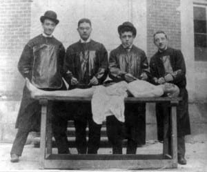
Nowadays, it’s commonplace for students studying anatomy to encounter human remains as part of their university degree, but this wasn’t always the case. In the early nineteenth century, there was a dire shortage in Britain of bodies for the purpose of medical research. For instance, the Edinburgh Medical College received fewer than five cadavers a year [1]. This was because only the remains of executed criminals could legally be used. The limitations put on scientific research because of this policy gave oxygen to the criminal business of ‘body-snatching’. When it began, the ‘snatchers’ invented a method to remove bodies from graves without detection: they used to dig holes, some distance away, and tunnel down into the graves before pulling bodies out by rope or hooks. Those who could afford it soon began to invest in mausoleums, vaults and table tombstones to ensure the safekeeping of their eternal resting places [2].

Medical students? Body-snatchers? Or both?

Medical students? Body-snatchers? Or both?

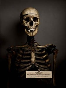
The business of bodysnatching, that fuelled medical research, soon turned even more sinister… In 1831 three men were arrested in London for the murder of vagrants, individuals whose deaths they thought would go unnoticed. On the day they were arrested, they had tried to sell the body of a fourteen year old boy to the lecturers of King’s College for twelve guineas [3]. There was also the famous case of William Burke and William Hare in Edinburgh, who murdered seventeen victims between 1827 and 1829, before selling the corpses to Dr Robert Knox at the Edinburgh Medical College. Unfortunately, this grisly business was inherently tied up in the advancement of medical knowledge.

The dissection of bodies was problematic, in both religious and moral terms, for contemporaries. In the first instance, many believed that their bodies had to remain intact for the afterlife, and dissection was also widely considered to be a punishment for the worst type of criminal. Take the fate of the Edinburgh bodysnatcher William Burke, for instance: he was executed by hanging in 1829 and his body was then publicly dissected at the Edinburgh Medical College [4]. And yet, in this period, recognition of the need for medical students to learn from human subjects was growing.

Courtesy of the Edinburgh City of Literature

Courtesy of the Edinburgh City of Literature

Public outcry, because of the black-market that had developed around medical research, helped the passing of a new bill: the 1832 Anatomy Act, which recognised that more bodies were needed for research and teaching. University College London’s Jeremy Bentham, who donated his own body to science (his auto-icon remains in the UCL South Cloisters), helped prepare the bill before his death in 1832. The act significantly extended access to cadavers, by allowing anatomists to dissect ‘unclaimed bodies’, individuals who died without anyone coming forward to pay for their burial. This was mostly people who died destitute in hospitals, workhouses and prisons. Dissection was no longer solely associated with individuals who were executed for murder, it was now also associated with the shame of dying in poverty [5].

It was really only in the mid-twentieth century that the donation of bodies to science became commonplace. Yet even now, we often feel squeamish about donating our bodies to science after we die. Attitudes certainly have changed, however, since 1832. From December 2015, individuals living in Wales will now have to opt-out if they don’t want their organs donated when they die, and legislation will certainly change soon in the rest of the United Kingdom.


[1] http://www.edinburgh-history.co.uk/burke-hare.html

[2] http://www.history.co.uk/study-topics/history-of-death/the-rise-of-the-body-snatchers

[3] http://www.exclassics.com/newgate/ng609.htm

[4] http://www.edinburgh-history.co.uk/burke-hare.html

[5] http://www.kingscollections.org/exhibitions/specialcollections/charles-dickens-2/italian-boy/anatomy-act

Further reading:

Colin Blakemore & Sheila Jennett, ‘body snatchers’, The Oxford Companion to the Body (2001). Encyclopedia.com. <http://www.encyclopedia.com>.

Food As Medicine

By Gemma Angel, on 24 June 2013

Sarah Savage by Sarah Savage


This blog post is dedicated to two of my favourite passions: medicine and food. As an historian of medicine examining epidemics, I am constantly fascinated by what past societies consumed for health and medicinal purposes. Today, most Londoners rely upon a trip to the local pharmacy for mass-produced pharmaceutical drugs to alleviate their symptoms or cure an illness. However, what did peoples consume before little engineered white pills? In my own research on the Spanish Influenza pandemic of 1918-1919, I discovered that American and English patients relied upon the use of herbal salves spread over the body and the consumption of soups, broths, milk, and chilled custards to reduce fevers and nourish the ill. One’s diet during a period of illness shifted from heavy meat and starch-based foods to items viewed as more acceptable to the feeble body such as clear vegetable-based broths, ground spices in warm water, and fresh fruits. Many of these natural foods do not seem so foreign to the present day reader. A warm bowl of soup and citrus fruit are commonplace today if someone is under the weather. On May 24th 2013 I attended the conference Spices and Medicine: From Historical Obsession to Research of the Future hosted by the UCL SoP Centre for Pharmacognosy and Phytotherapy, examining the use of spices and food for medicinal purposes. Archeologists, historians, and pharmacists discussed various different natural cures from those located in Roman ports in the 1st century CE ,to Southeast Asian missions in the 18th century CE. Some of the examined food items do not appear in our daily diets unless you are already fond of candied lark. Other foods and spices are still used today as cooking ingredients including black pepper, garlic, onions, limes, turmeric, ginger, and rice. As part of ancient and early modern medicinal treatments, the above listed ingredients had an intended medicinal purpose other than to simply add flavour to a dish. In Germany on the Rhine River, archeologists even discovered a military hospital that contained an ancient herb garden and spices in patients’ rooms for treatments. The Petrie Museum of Egyptian Archeology contains the remarkable remains of dried apricots, peaches, dates, and almonds that would have been part of the Egyptian diet.

Dried fruit in PetrieModern medicine acknowledges the benefits of foods rich in vitamin C as immunity boosters during flu and cold seasons. It is interesting to wonder whether the ancient people recognized that certain vitamin C rich fruits had inherent medicinal properties, or were these delicious fruits simply part of their diet for flavour reasons more so than preventative measures? One archeologist during the conference discussed the importance of trade routes to bring spices and fruits from the East, Middle East, and North Africa to Roman territories throughout the Mediterranean and Europe. During the Islamic period, there was a major increase in the range of spices imported into the empire. Since spices were expensive commodities, it is rare for archeologists to find spices in these ancient ports; however, letters from the Islamic period discuss what goods were traded and in what quantities. For those archeologists interested in food, it must be fascinating to find 2,000-year-old garlic cloves, squeezed limes, and dried aubergines, all buried under layers of sand. Although present day peoples consume pharmaceutical drugs for health, certain foods such as chicken noodle soup and herbal teas remain go-to sources of nourishment during times of illness.


Viruses of Mice and Men

By Gemma Angel, on 3 June 2013

Sarah Savage by Sarah Savage


Recently in the Grant Museum, I had the most exciting 35 minute engagement with a mother and son visiting London from Jersey in the Channel Islands.  Since her son was very interested in coming to UCL for undergraduate study, the mum thought the best idea would be to visit the campus and see all that UCL has to offer, including the museums on campus.  I caught this family on their first stop on the UCL museums trail.  After introducing myself and telling the boy’s mother a little bit about the UCL student engagers group, she quickly asked what my research is specifically about. I told her that I am an historical epidemiologist specializing in the Spanish Influenza Pandemic 1918-19, and the Encephalitis Lethargica Epidemic 1917-1930. Her eyes grew quite wide and she replied that her son had been hoping to meet someone doing research like mine, to find out more about pandemics. Her main reference point for Spanish Influenza was that the character Edward Cullen from the Twilight films had died from the pandemic! Alas, I encounter that response quite often. If anything, Twilight put the ‘forgotten pandemic’ on the radar of the general population and teenage girls everywhere.[1]

Spanish Influenza 1Although previously I’ve mainly engaged in the Petrie Museum next to objects of everyday Egyptian life that relate to disease, I found that amongst the great preserved animals of medical colleges past, many fascinating connections to my research topic presented themselves in conversation with visitors. The display of parasitic worms, although admittedly horrifying, can be used as a tool to demonstrate how a virus inhabits and travels through the body. A gentleman visitor later in the afternoon stood in shock when confronted with the incredible size of some of the parasitic worms that are able to live in the human body. 

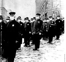
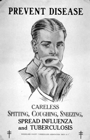
The brave visitor from Jersey further engaged with me to discuss exactly how viruses spread through the body, mutate, and ‘disappear’ after an outbreak. I put disappear in parenthesis, since some viruses can simply become dormant in the body.  During our conversation, she inquired as to what initially drew me to epidemics. “Most young students do not dream of studying viruses that wipe out entire populations for a living!” she told me. spanish Influenza 2Oh, but I was that student, fascinated by the plague, and how tiny organisms could exist in our bodies. Once I’d told her more about my academic background in the United States, she asked me how common it is for historians to examine medicine or epidemics. Although UCL previously had a Centre for the History of Medicine for postgraduate researchers, now we are divided amongst different disciplines including history, neurology, and psychology.  As an historian specialising in epidemics, I explained to her that I am not only interested in the physical side of how epidemics work, but also how societies react to an outbreak.  During the 1918-19 Spanish Influenza outbreak, governments in England and the United States quarantined areas of cities and closed all government buildings. Although these measures prevented the spread of the virus to some extent, many citizens became infected prior to the required quarantines and closures. There are many links between government measures and public behaviour during historical influenza epidemics during the early 20th century and the avian and swine flu outbreaks of present day.The visitor mentioned the 2009 Swine Flu outbreak, and how the fear of coming into contact with an infected person effected daily life and decisions to frequent public spaces. By the end of our lengthy conversation, we had discussed everything from 20th century epidemics to life on the Channel Islands and life as a UCL student. After her son had finished peering into every case in the Grant Museum, his mother expressed how enlightened and intellectually stimulated she felt to discuss such a specialised topic with a UCL researcher, before moving on to encounter another member of our team at one of UCL other museum spaces. As a new team member, this was a heartening conclusion to a very inspiring conversation, and I am thoroughly looking forward to future conversations with museums visitors from all over the world…


References

[1] Alfred W. Crosby: America’s Forgotten Pandemic: The Influenza of 1918, Cambridge: Cambridge University Press, (2003).


So Comfortable You Can’t Even Feel It! The Cocaine Tampon

By Gemma Angel, on 29 April 2013

Lisa Plotkin  by Lisa Plotkin


Last May, a Utah woman was in for a surprise when she purchased a $1.99 box of tampons from a local store in Salt Lake City. Instead of a cotton tampon inside the applicator, the woman discovered something else with a much steeper price tag: cocaine. At first she was completely astonished and didn’t realize it was cocaine – she thought that the cotton might have somehow disintegrated; so unlikely was the pairing of cocaine and tampons to her. Similarly, when the police were called in to collect the cocaine, they too expressed their surprise at this method of transporting drugs. Detective Carlie Wiechman, spokesperson for the Salt Lake City Police Department, said this in response to the crime: “It’s not every day we run across this. We run across different ways of packaging and distributing, but it never ceases to amaze us the different and creative ways of trying to move drugs around.”

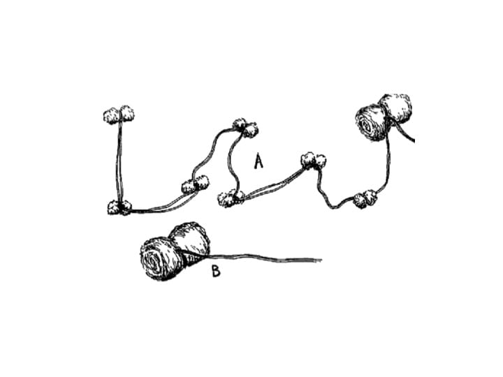
However, the marriage of cocaine and tampons is not as farfetched or creative as the Salt Lake City PD imagined, and for 19th century surgeons and gynecologists it was a regular – dare I say it, ‘everyday’ – medical sight. Throughout the 19th and early 20th centuries, cocaine was regularly used as a local anesthetic in surgery. It was often administered in the form of what doctors referred to as a tampon – a medical device invented in the 18th century primarily as an antiseptic to clog up bullet wounds. The tampon was traditionally soaked in whatever antiseptic or anesthetic drug was in general use, before being applied to a wound. These tampons were not particularly associated with women; at least not until later in the 19th century that is, when cocaine came to be  regarded as an especially effective treatment for gynecological diseases.


The medical tampon.

A: Kite-tail tampon; small wads of cotton tied together
on one string with a fairly large tampon on the end.
B: Ordinary rolled tampon.
Image from Practical Clinical Gynecology in:
American Journal of Surgery, vol. 39, issue 1 (1938).

Cocaine was believed effective against a whole range of women’s ailments: From painful intercourse; to uterine diseases; to cervical endometritis; to inflammation of the urethra; to dysmenorrhea – the list goes on and on. [1] In fact, cocaine was even believed to assure a ‘painless childbirth’ and according to Physician to the British Lying-in Hospital, John Philipps, could even cure the scourge of ‘sore nipples’. [2].

How would the cocaine be applied in these situations? A typical gynecological answer: by vaginally “inserting a tampon soaked in a freshly prepared solution of 2 % cocaine through a narrow Ferguson’s speculum.” [3]

Therefore, with regards to many women’s diseases, the question was not should cocaine be used- but how much. This was common until the interwar period. Of course, accidents do happen and sometimes these tampons were never removed, (most were). To read more about other ‘accidental’ foreign bodies left behind in women’s bodies, read my previous blog post here. Although many women were on board with the idea of being treated with cocaine, some did however refuse. So, the Utah woman who recently discovered cocaine in her tampon carton was by no means the first to say ‘thanks, but no thanks’ to tampons with coke on the side!

Learn more about our current UCL exhibition on all kinds of foreign bodies see.

References:

[1] Stephen R. Kandall: Women and Addiction in the United States (Cambridge, MA: Harvard University Press, 1996).

[2] John Philipps: ‘The Value of Cocaine in Obstetrics’, in The Lancet (26 November 1887), p. 1061

[3] ‘A Note on the Morphine-Hyoscine Method of Painless Childbirth’, in British Medical Journal (6 January 1917).


Of Foetuses & Fibroids: the Accidental Foreign Body

By Gemma Angel, on 8 April 2013

Lisa Plotkinby Lisa Plotkin


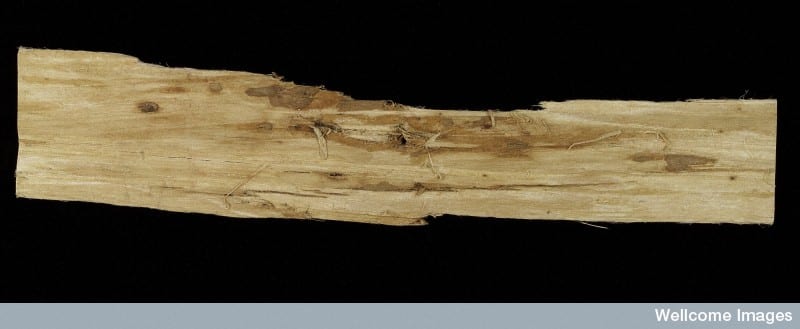
As our current exhibition in UCL’s north cloisters demonstrates, “foreign bodies” may take many forms, as well as being continually redefined throughout history. Putting it simply, the term “foreign body” in medicine usually refers to an external object introduced into the body that isn’t supposed to be there. As my colleague Dr. Sarah Chaney notes in her recent blog post, some of the most common foreign objects uncovered from the bodies of 19th and early 20th century patients were coins, safety pins, buttons and needles. These objects could enter the body accidentally or with purpose. Medical instruments or tools, for example, were on occasion accidentally lost inside the patient’s body during an operation. For those Seinfeld fans out there, think back to the “junior mint” episode. However, it was neither pins, mints or instruments which were the subject of a 1939 article in the British Medical Journal devoted entirely to foreign bodies. Rather, Dr. A. H. Charles, obstetric registrar at St. George’s Hospital, zeroed in on one particular foreign body that was with some frequency discovered in the female bladder: slippery elm bark. It may surprise some readers to discover (as it certainly did Dr. Charles), that slippery elm bark was commonly used as an abortifacient. In fact, it is still used by women to induce abortion today.[1] Writing on elm bark as a foreign body inside the bladder, Dr. Charles observed:

Five cases in which a piece of elm bark was used have been reported in detail previously, and in all of these the body has remained undiscovered for some time, until its removal suprapubically after calculus formation had taken place, causing symptoms leading to its discovery. Why the bark of this noble tree should be so popular is difficult to understand.[2]

Called “slippery” elm because when it gets wet it becomes slippery, this type of elm bark has traditionally been used to cause early uterine contractions and induce labor. The bark is inserted into the cervix where it then absorbs water and expands, dilating the cervix and triggering contractions. Needless to say, this procedure was not always successful and could cause life-threatening infections. Occasionally the bark could end up in the bladder by mistake, which is what Dr. Charles had observed. For many of the women who mistakenly inserted the slippery elm into their bladders, there the bark most likely stayed, unless a severe medical problem compelled them to seek medical attention. No doubt for many of the women attempting to self-abort, their experience with slippery elm was less than satisfactory and could have proven fatal.

Slippery Elm Bark, sold as an abortifacient

Slippery Elm Bark, sold as an abortifacient.

Accidental or intentional abortion occurred with a lot greater frequency in the 19th and early 20th centuries than some might imagine, and was inextricably wrapped up with both the idea and concrete reality of “foreign bodies.” For many, the coat hanger is the ultimate symbol of a foreign body inserted into the uterus to cause abortion. Still others may think of obstetric forceps as the foreign body which has caused thousands of fetal deaths during delivery. But what about uterine fibroids? How many abortions have they caused, and can they be regarded as foreign bodies if they are naturally occurring? A uterine fibroid is a benign tumour of the uterus, commonly found in women of reproductive age. Most fibroids are asymptomatic and therefore, most women are never aware that they even have one. However, on occasion these fibroids can cause health complications or interfere with pregnancy. Before the advances of late 19th century abdominal surgery and gynecology, uterine fibroids were not treatable. However, by 1916, obstetric surgeon Sir. John Bland-Sutton was able to boast that “uterine fibroids are common tumours; so common and troublesome that I have removed the uterus in 2,000 women.”[3]

From his experience performing hysterectomy on thousands of women a theme emerges: uterine fibroids closely mimic pregnancy in a variety of ways, and it is difficult – sometimes impossible – to distinguish between the two. Writing of this unfortunate similarity in 1913 Bland-Sutton observed, “A large sub-mucous fibroid produces similar changes in the uterus to those set up by the growth of the fetus […] Women with large sub-mucous fibroids are more or less in a condition resembling chronic pregnancy.”[4] What this similarity meant – and what gynecologists and obstetricians of the time openly acknowledged – was that sometimes a hysterectomy was performed to remove a fibroid that either never existed in the first place, or was also sitting alongside a feotus, masking a pregnancy. Either way, an abortion was performed.

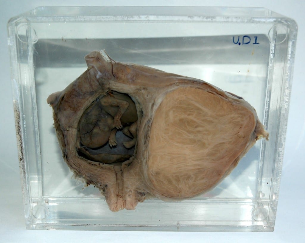
The photograph below demonstrates the reality of such surgeries. This particular specimen belongs to UCL Pathology Collections, and is currently on display in the Foreign Bodies exhibition. The anonymous woman patient underwent a hysterectomy most likely sometime in the early 20th century, in order to remove the sizable uterine fibroid, which can be seen on the right side of the image. However, on closer examination of the image, we see on the left side a preserved feotus, frozen in development, somewhere between 8-11 weeks.  It is unclear whether this woman or her doctor even knew she was pregnant.

Feotus in uterus, with large fibroid tumour. UCL Pathology Collections. Photograph Gemma Angel.

Feotus in utero, with large fibroid tumour.
UCL Pathology Collections. Photograph Gemma Angel.

Such examples abound in medical literature, and Victorian and Edwardian gynaecologists, obstetricians, and surgeons spoke of them with little or no censure. It was all a part of the surgical trial and error that they were practicing. The feotus was sometimes viewed as a necessary casualty in removing a potentially life-threatening fibroid. Either way, be it by slippery elm, by accident, or with purposeful intent, the feotus was removed as a foreign body, like any other. By examining the medical establishment’s attitudes towards fibroid removal we catch a glimpse into one way the feotus, and the experience of pregnancy in general, was understood in the past.


References:

[1] David A Grimes, Janie Benson, Susheela Singh, Mariana Romero, Bela Ganatra, Friday E Okonofua, Iqbal H Shah. “Unsafe abortion: the preventable pandemic.” The Lancet Sexual and Reproductive Health Series, October 2006.

[2] British Medical Journal, 29 July 1939.

[3] Sir John Bland-Sutton, “A Clinical Lecture on 200 Consecutive Hysterectomies for Fibroids Attended With Recovery” reprinted British Medical Journal, 4 July 1916.

[4] Sir John Bland-Sutton, “The Visceral Complications Met With Hysterectomy for Fibroids and the Best Methods for Dealing With Them” British Medical Journal, 1 November 1913.

Diagnosing Foreign Bodies

By Gemma Angel, on 25 March 2013

Sarah Chaneyby Sarah Chaney


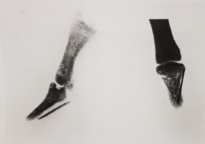
One of the most important diagnostic tools to assist in foreign body removal was the development of the x-ray. In 1895, Wilhelm Röntgen, a German physics professor, developed the x-ray photograph, which enabled the interior of the body to be made visible using electromagnetic radiation.[1]  In the late 1890s and early 1900s, medical reports on foreign bodies frequently focused on the use of the “Röntgen Rays” or “skiagraphs” (as x-rays were then widely called) to locate such objects. A few weeks after Röntgen published the first X-ray photograph, Norman Collie at UCL made his own x-ray tube in order to locate a broken needle in the thumb of a female patient.

World’s first diagnostic x-ray, by Norman Collie at UCL. UCL Special Collections, on display in the Octagon Gallery until 30 April: http://www.ucl.ac.uk/museums/whats-on

World’s first diagnostic x-ray, by Norman Collie at UCL.
UCL Special Collections, on display in the Octagon Gallery
until 30 April: http://www.ucl.ac.uk/museums/whats-on


Many of the foreign bodies case histories in hospital records of the period focus on the use of this new diagnostic technique. However, x-rays could also lead to tension between patients and clinicians, when the photographs contradicted stories told by patients. One German publication of 1899 reported a soldier’s claim to have been bitten by a horse as “malingering” (a serious military crime) when broken needles were found in the wound, suggesting that the injury was self-inflicted.[2]

Image showing self-inflicted burns in a “hysterical” patient, from John Collie’s Malingering and Feigned Sickness (1913)

Image showing self-inflicted burns in a
“hysterical” patient, from John Collie’s
Malingering and Feigned Sickness (1913)

Yet, surprisingly (given the widespread publicity given to so-called malingering in civilian populations in the decades preceding the National Insurance Act of 1911), these stories seem to have been of less interest to many surgeons than the diagnostic procedure itself. At the Royal London Hospital in 1898, for example, little interest was shown in the fact that the x-ray photographs of 38-year-old domestic servant Elizabeth Quaife did not tally with the history she gave.[3] Elizabeth claimed that she had suffered pain in the knee joint ever since a long hat pin ran into her leg while she was sweeping under a bed: in hospital, however, five separate needles were discovered in the joint. Unlike in published cases, the surgeon made no reference to the potential use of x-ray imagery to detect fraud, but instead used the case to evaluate the usefulness of the technique itself. This, it was thought, had been successful in locating and removing four needles but “the fifth needle shewed by the skiagraph was … not found. It is probable that the figure shewed in the skiagraph was due to a shadow of the other needles. This, once more, shews that the skiagraph may be deceptive.”

This emphasis on diagnosis and removal certainly tallies with the lack of interest surgeons tended to show in the cause of foreign bodies. Foreign Body in this period was a diagnosis, not an exploration of a patient’s state of mind. When Rachel Taylor was admitted to the Royal London in 1900 – after swallowing a pin and a tin tack – and again in 1906 having swallowed two nails “the night before last”, it was not noted whether either instance was accidental or intentional.[4] Despite published concern over the potential abuse of charitable treatment, in practice this does not seem to have been a significant issue for either surgeons or physicians at the Royal London Hospital: cases of “artefact injury” were treated without question whether or not the patient paid for their treatment.


References:

[1] Lisa Cartwight, Screening the Body: Tracing Medicine’s Visual Culture (Minneapolis: University of Minnesota Press, 1995)

[2] “Self-Inflicted Injuries Diagnosed by the Roentgen Rays,” The Lancet, 153, no. 3955 (1899): 1668.

[3] Elizabeth Quaife, RLHA Microfilm Case Records (Surgical), DEAN F1898, pt. no. 29 & 511.

[4] Rachel Taylor, RLHA Microfilm Case Records (Surgical), TAY F1900, pt. no. 2326 & FENWICK 1906, pt. no. 906.


Sword Swallowing & Surgical Performance

By Gemma Angel, on 11 March 2013

Sarah Chaneyby Sarah Chaney


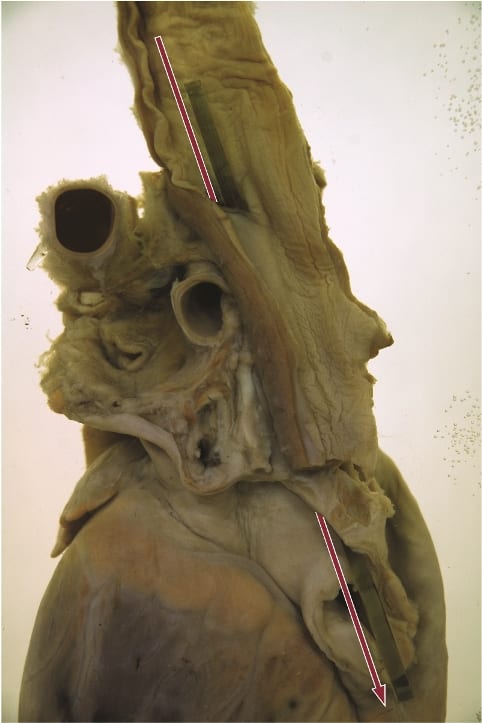
We know sadly little about the sword swallower’s sword that resides in the UCL Pathology Collection: not even how long it has been here. What we do know is that this performer was very unlucky. Perhaps he (or, indeed, she) didn’t tilt his head back far enough. Perhaps he moved during the process of insertion. Whatever the case, the sword pierced the flexible tube of the oesophagus, leading to the performer’s death. The heart and oesophagus were preserved – perhaps as a warning of the dangers of such feats – alongside the weapon that led to his demise.

Fatally ruptured oesophagus, caused by the sword swallower's sword. Photograph Gemma Angel, UCL Pathology Collections.

Fatally ruptured oesophagus, caused by the sword swallower’s sword. Photograph Gemma Angel, UCL Pathology Collections.

Sword swallowing seemingly originated in India some 4,000 years ago, but reached the western world of Ancient Greece and Rome in the first century AD. The performer tilts his or her head back, extending the neck, and learning to relax muscles that usually move involuntarily. A rigid weapon can then be passed down as far as the stomach, usually for just a few seconds, before removal. It is dangerous, certainly, but few performers suffer the fate of the individual preserved in the UCL collections. According to one recent article in the British Medical Journal, most serious incidents occur owing to distraction or attempts at exceedingly complex feats:

For example, one swallower lacerated his pharynx when trying to swallow a curved sabre, a second lacerated his oesophagus and developed pleurisy after being distracted by a misbehaving macaw on his shoulder, and a belly dancer suffered a major haemorrhage when a bystander pushed dollar bills into her belt causing three blades in her oesophagus to scissor. [1]

In many ways, sword swallowing is the opposite of the ingestion of other foreign bodies: rather than swallowing, the performer maintains absolute control over the process of consumption, taming the body’s reflexes and realigning the organs. As Mary Cappello notes in her fascinating literary biography of surgeon Chevalier Jackson (1865 – 1968), who was an expert in foreign body removal, sword swallowing was recognised by doctors as inspirational to their own techniques. Jackson took his lead from German professors Alfred Kirstein and Gustav Killian, who lectured that sword swallowing proved the possibility of passing a rigid tube into the oesophagus, in order to remove lodged objects. Jackson, who developed his own oesophagoscope in 1890, admitted that the abilities of circus performers had opened his eyes to the opportunity of removing foreign objects without dangerous surgery. He even taught his children how to “scope” themselves.[2]

In an intriguing parallel, the insertion of some foreign objects into the human body thus assisted with the removal of others. At the turn of the 20th century, the removal of foreign bodies lodged in the throat and airways frequently required an incision to be made into the trachea or oesophagus, an operation which could prove fatal. In the records of the Royal London Hospital, from 1890 to 1910, we find no mention of oesophagoscopy or bronchoscopy: instead, surgery or the probang or “coin-catcher” was the norm. This latter instrument was generally a simple hook, inserted without any kind of viewing device or illumination. The practitioner would feel blindly for the object, and either attempt to hook it out, or push it into the stomach. This might lead to numerous complications. In 1903, surgeons at the Royal London attempted to remove a halfpenny from the throat of a five-year-old boy by pushing it into the stomach. However, it was subsequently reported that the coin catcher broke off in the boy’s throat, necessitating a major operation from which the child did not survive.[3] Small wonder that, less than a decade later, Jackson declared such objects “rough, unjustifiable, brutal”.[4]

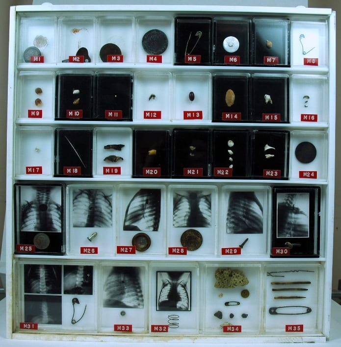
foreignbodies

UCL Pathology Collections contains many examples of foreign objects removed from the
human body: this purpose built display showcases many such objects, some with
small x-rays of the objects prior to removal.

X-ray imaging techniques aided the removal of foreign objects by instruments, and foreign body specimens are often accompanied by photographs showing the item’s location in the human body. The above set of items is found in the UCL Pathology Collection, the objects having been gathered by several surgeons in the 1920s – ‘50s. At some point, the individual boxes made for each specimen were mounted together, in a specially designed plastic surround. Fittings on the back indicate that the case was made to hang on a wall. But why? To decorate the office of a surgeon, showing off his achievements? To offer a warning to others to take care (particularly parents, for all these objects were removed from children and infants)?

Chevalier Jackson claimed that his collection of more than two thousand foreign bodies (now housed in Philadelphia’s Mutter Museum) was not a curiosity, but indicative of the everyday nature of foreign body ingestion and inspiration. Yet many of these specimens are not everyday. The two boxes of multiple objects in the bottom right, for example, were removed from the vaginas of young girls (six and eight years old respectively). The case notes do not indicate how these objects arrived in their location. Did the girls insert them themselves, or might it be a sign of sexual abuse? In her research into the medical histories of Jewish immigrants to the East End of London in the late nineteenth and early twentieth centuries, Carole Reeves came across a case of multiple foreign body insertion in a young woman, whose vagina was found to be tightly packed with pins. Reeves speculated that Leah G. might have inserted these items in an effort to ward off potential (and actual) abusers.[5]

In most instances, we can uncover little about the motivations of those in the late 19th and early 20th centuries whose foreign bodies are recorded in medical records: surgeons were often little interested in how the object came to be in its current location, but only in its removal. Yet this may often make such displays still more intriguing than otherwise. As Mary Cappello put it, in a video discussion of the UCL artefact pictured above for the Damaging the Body website: “What is the border or boundary between human flesh, between human life and the object world?”


References:

[1] Brian Witcombe and Dan Meyer, “Sword Swallowing and its Side Effects”, in British Medical Journal, 333 (2006), 1285-7, p. 1287.

[2] Mary Cappello, Swallow: Foreign Bodies, Their Ingestion, Inspiration and the Curious Doctor who Extracted Them, New York, London: The New Press (2011). Website: http://www.swallowthebook.com/

[3] Royal London Hospital Archives, Surgical Index 1903, LH/M/2/9, patient no. 4086.

[4] Chevalier Jackson, Lecture to the Kings County Medical Society, December 19 1911, quoted in Cappello, p. 208.

[5] Carole Anne Reeves, Insanity and Nervous Diseases Amongst Jewish Immigrants to the East End of London, 1880 – 1920 (Unpublished PhD thesis, University of London, 2001), p. 213.

Toxic Tattoos: Mercury Based Pigments in the 19th and 20th Centuries

By Gemma Angel, on 4 February 2013

  by Gemma Angel


In January this year, myself and fellow Research Engager Sarah Chaney went to visit the UCL Geology Collections, to see if there were any mineral or rock samples in the collection that would fit in with our upcoming cross-collections exhibition, Foreign Bodies. Neither of us being geologists, we didn’t have particularly high expectations – how interesting can rocks be, really? As it turned out, the answer to that question is – very! We spent a fascinating hour in the Rock Room, where we quickly realised that there were many specimens that could be interpreted as foreign bodies in one way or another: The fossilised forms of plants and animals in rock; a rusted nail fused into a lump of lava; and perhaps the ultimate foreign body, a beautifully patterned fragment of meteorite.

One particular sample drew my attention – a surprisingly heavy lump of purplish-red rock with pretty pink and bright red veins (pictured below). When I asked if I could have a closer look, I was told that I would have to wear gloves to handle this piece of rock, as it was in fact toxic. The rock sample was cinnabar, the common ore of mercury. I am well aware of the toxicity of mercury from my own research – gloves are also required when I’m handling preserved tattooed human skins as part of my work at the Science Museum archives. It is speculated that one of the substances used in the dry-preservation process of human skin is mercuric sulphide, and many of the specimens betray the typical orange-red staining that this chemical causes. But there is another unexpected connection between mercury and my research. Cinnabar has been used to make bold red pigments since antiquity – and this pigment was also historically used in European tattooing.

Cinnabar ore and powder (8.5% Hg) sample, in the UCL Rock Room.
UCL Geology Collections.


Red mercuric sulphide occurs naturally, and has been manufactured for use as a pigment since the early Middle Ages. The pigment was referred to interchangeably as vermilion or cinnabar, although vermilion became the more commonly used term by the 17th century. [1] Vermilion is now the standard English name given to red artists’ pigment based on artificially produced mercuric sulphide. [2] Since the toxic effects of mercury were historically well known, it might seem strange that cinnabar was used in tattooing at all. In fact, mercury has been used in medicine to treat a range of ailments throughout history, most notably syphilis. In European tattooing, red pigments were not commonly used pre-20th century, with red inks tending to be used sparingly for small areas of embellishment.

Most cinnabar was mined in China and by the mid 19th century, Chinese vermilion was generally considered to be the purest form, producing a superior hue to the European variety. The cinnabar ore on which vermillion production depended was costly; as a result, European vermilion was often mixed with inexpensive materials including brick, orpiment, iron oxide, Persian red, iodine scarlet, and minium (red lead). Whilst these additives also produced a bright red pigment, their relative impermanence made it an inferior choice for artists’ colours.

This may explain why there is marked variability amongst preserved tattoos containing red inks, in terms of both permanence and vibrancy of colour: The more commonly available and cheaper European variety of vermilion used by some 19th century tattooists likely contained additives which reduced colour saturation, and made the pigment more susceptible to light-degradation over time. The Wellcome Collection possesses only a handful of tattoos containing red dye, and most of these are very degraded, such that little colour is visible. In these cases, the red has often faded far more dramatically than the black ink used in the same tattoos. However, there are one or two preserved specimens containing exceptionally bright ink, which has lost none of its vivid red colour, an example of which can be seen below.

Tattooed human skin with bold red pigment, likely cinnabar.
Science Museum object no. A687. Photograph © Gemma Angel,
courtesy of the Science Museum London.


Since heavy mineral pigments do not generally lose saturation over time, it is possible to speculate that the bold red ink seen here very likely contains a high concentration of cinnabar, although it is impossible to know for certain without physical testing. There are, however, historical references to the use of mercury-based pigments in tattooing, most of which can be found in 20th century medical journals. As may be expected, these sources focus on the toxic effects of cinnabar-based tattoo pigments. In particular, mercury dermatitis in tattoos was sometimes reported during the early-mid 20th century, often many years after the tattoo was acquired by the patient.

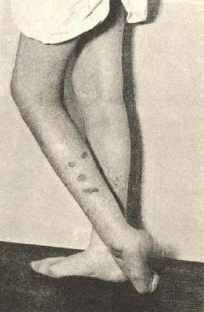
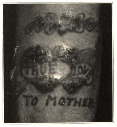
In 1930, one such case appeared in the Archives of Dermatology and Syphilology, written by Dr. Paul Gerson Unna. His patient, a 63-year-old man who had been tattooed in his youth, suddenly developed itching, swelling and blistering in the red portions of the tattoo, following a mercury-based treatment for haemorrhoids. Three years later, Dr. D. B. Ballin reported a case in which a young male patient had developed itching, swelling and oozing in the red portions of a tattoo, 2 years after he had been tattooed. The patient was treated by the removal of the affected areas using a dermal punch, and the tattooed skin samples were sent for histological testing; however, the resultant scar tissue in the punched areas later developed the same reaction.

Photograph from Ballin’s 1933 report,
Cutaneous Hypersensitivity to Mercury from Tattooing
Caption reads: “Forearm of patient showing sensitivity
to mercury as a result of tattooing.”

Throughout the 1940s and 50s, cases of mercurial sensitivity and dermatitis in red tattoos appear sporadically in the medical literature, [4] though the apparent causes of the onset of symptoms vary. According to Keiller and Warin:

In some cases the use of mercurial applications elsewhere has led to the development of sensitivity and the red areas of the tattoo have subsequently become swollen. Other cases are reported in which the sensitivity has developed spontaneously. [5]

Interestingly, there were also reports of the apparent ‘positive’ effects of cinnabar tattoo pigments in cases of cutaneous syphilis during the early 20th century. It was observed that the red portions of a tattoo were seldom effected by syphilis sores – even in cases where adjacent areas of skin tattooed in black ink were engulfed by the infection.


References:

[1] R. D. Harley: Artists’ Pigments c.1600-1835: A Study in English Documentary Sources, (1982) Butterworth Scientific, p.125.

[2] Rutherford J. Gettens et. al. : ‘Vermilion and Cinnabar’, in Studies in Conservation, Vol. 17 No. 2. (May 1972), p.45. Available on JSTOR: http://www.jstor.org/stable/1505572

[3]  D. B. Ballin: ‘Cutaneous Hypersenistivity to Mercury From Tattooing’, in Archives of Dermatology and Syphilology, Vol. 27, No.2 (February 1933), pp.292-294.

[4] See, for example: Howard I. Goldberg: ‘Mercurial Reaction in a Tattoo’, in Canadian Medical Association Journal, Vol. 80 (Feb. 1 1959), pp.203-204. Available online: http://www.ncbi.nlm.nih.gov/pmc/articles/PMC1830587/ ; also R. A. G. Lane et. al.: ‘Mercurial Granuloma in a Tattoo’, in Canadian Medical Association Journal, Vol. 70 (May 1954), pp.546-548. Available online: http://www.ncbi.nlm.nih.gov/pmc/articles/PMC1825326/

[5] F. E. S. Keiller & R.P. Warin: ‘Mercury Dermatitis in a Tattoo: Treated With Dimercaprol’, in The British Medical Journal, Vol. 1, 5020 (Mar. 23, 1957), p.678. Available on JSTOR: http://www.jstor.org/stable/20361174

[6] For more on the history of tattooing and skin disease, see Gemma Angel: ‘Atavistic Marks and Risky Practices: the Tattoo in Medico-Legal Debate 1850~1950’, in J. Reinarz & K. Siena (eds.) A Medical History of Skin: Scratching The Surface, Pickering Chatto, (2013) pp.165-179.

[analytics-counter]

Doctors, Dissection & UCL

By Gemma Angel, on 21 January 2013

  by Sarah Chaney


A visit to the current Museum of London exhibition, Doctors, Dissection and Resurrection Men (on until 14 April 2013), brought to mind the recent Buried on Campus exhibition in the Grant Museum. Several of us have previously blogged about reinstating the stories of the forgotten dead, as well as the issues around the display and interpretation of human remains in a museum context. As I myself wrote, the disinterrment of human remains is not unusual during building work: the Museum of London exhibition focuses on the excavation of the former Royal London Hospital burial site, during recent improvement works. The bones found showed traces of a variety of practices, including dissection for autopsy, as well as marks made during surgical practice and articulation for the creation of teaching specimens.

Dissection, particularly in the case of medical teaching, was often linked to artistic practice. Doctors, Dissection and Resurrection Men opens with the grisly plaster cast of James Legg, hanged for murder in 1801. Legg was subsequently flayed and posed as if crucified: a collaborative project between artists Benjamin West and Richard Crossway, and sculptor Thomas Banks, who believed that most depictions of Christ’s crucifixion were anatomically incorrect (for more on the Anatomical Crucifixion see Gemma Angel’s post). Rather less theatrically, anatomical drawings and textbooks were also created directly from dissection practice. During a recent session in the Art Museum, I discussed with visitors the way in which anatomy textbooks create stylised images, removing certain body parts in order to emphasise others. Students re-created these images for themselves: first with the corpse, then in their own sketches, re-interpreting the body in a way that made sense for their practice.

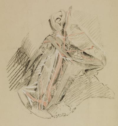
Joseph Lister – Side of the Neck and Floor of the Mouth (1850), UCL Art Museum #4801

Amongst the UCL Art Collections are a number of student sketches of the famous surgeon Joseph Lister (1827 – 1912), well-known for his introduction of antiseptic techniques into surgical practice. Born in Essex, Lister came to UCL in 1844, initially as a student of the arts. After graduating, however, he subsequently turned his attention to medical studies, continuing at UCL until he gained his M.B. in 1852. The sketches in the collection mainly date from 1849-50, produced as part of Lister’s studies. The techniques used indicate some of the interesting artistic choices available to anatomical illustrators: perhaps also the influence of Lister’s varied education and interests. The sketch above, for example, was made on tinted paper, which enabled the young Lister to highlight structures using white chalk. This emphasis, along with the effective use of colour (in this instance, major blood vessels are depicted in red, standing out clearly in an otherwise monochrome drawing), enables quick and easy recognition of bodily structures, adding depth to the sketch. For an un-trained eye, the mass of tissues within the human body could not be read in such a manner. The ability to render the three-dimensional body in a series of recognisable images – and then understand the physical body through such images – was as important as surgical skill.

Box Viewer from the UCL
Physiology Collections (080:RFH)

The huge variety of techniques for anatomically representing the human body is also evident elsewhere in the UCL Collections. The Physiology Collection includes a volume of the Edinburgh Stereoscopic Atlas of Anatomy, published in 1905. Stereoscopy became a popular technique of representing three-dimensional structures from its inception in the 1840s. Two offset photographs or other images are presented to the viewer which, when viewed through the stereoscope, are seen separately by the left and right eye. As occurs in ordinary vision, the brain combines the images perceived by both eyes; in the case of stereoscopy, giving the illusion of three-dimensional depth. The Edinburgh Atlas aimed to use this technique to represent photographs of dissections in a manner closer to that seen in the three-dimensional human body than simple sketches. Bulky and expensive, the success of the Atlas was relatively limited. It still serves, however, as an unusual reminder of the way in which the human body has continued to require anatomical translation.

Art and Psychiatry: Henry Scott Tuke

By Gemma Angel, on 15 October 2012

by Sarah Chaney


Henry Scott Tuke was a student at the Slade School of Fine Art in the 1870s, winning a three-year-scholarship in 1877: sadly, this was twenty years too early for his prize-winning work to have made it into the UCL Art Museum. The Tuke family had a number of connections with UCL, however: Henry’s elder brother was a medical student at University College Hospital.

Although biographies of the Newlyn painter often mention his doctor father, little is made of this connection – Henry remains a figure of interest for art and cultural historians, and his father for historians of medicine. Yet parallels between art and psychiatry were often emphasised in the late nineteenth century. Daniel Hack Tuke, Henry’s father, was a governor at Bethlem Royal Hospital from the 1870s until his death in 1897, and art was an important topic at the asylum, for patients and staff alike. Daniel’s obituary in the Hospital Magazine, Under the Dome, concentrated on his well-known son, noting that:

The early death of his eldest son, who was a brilliant student of University College Hospital, was a painful blow to Dr. Tuke, but no doubt he found some amount of solace under this loss in the successful career as a painter of his other son, Mr. H.S. Tuke. The latter has been a foremost member of the Newlyn School, and like most of his brother artists of that school of painters, has lived a good deal on his boat on the coast of Cornwall, and, we remember, that about three seasons since, Dr. Tuke, upon his first visit to the Hospital, after his autumn holiday, said to the present writer that he had much enjoyed it, having in good part spent it with his son upon the latter’s studio-boat. [1]

From this remark, it would seem that Henry and his father were close. It may be interesting to pay closer attention to the fact that Henry Scott Tuke is best remembered today for his Impressionist style paintings of male nudes, becoming a cult figure in gay cultural circles. Was this connection also made in his life? Certainly, homosexuality (or ‘sexual inversion’ as it was more commonly known at this time) was a topic of interest for many psychiatrists, among whom Daniel Hack Tuke was extremely prominent. In Austria, for example, forensic psychiatrist Richard von Krafft-Ebing devoted much of his magnum opus, Psychopathia Sexualis (first published in 1886) to the topic, later becoming a fervent opponent of Paragraph 26, which outlawed homosexual acts in German and Austrian law.

English sexologist, Havelock Ellis, meanwhile, had been mentored by Hack Tuke during his own days as a medical student. In the early 1890s Ellis and writer (and self-confessed ‘invert’) John Addington Symonds corresponded about a book they wished to write together on the topic. Ellis complained about the lack of interest from his medical colleagues, in particular that Daniel Hack Tuke himself “wrap[ped] a wet blanket around it [the topic], with averted eyes”. Symonds had similarly been annoyed in a letter to his friend, Edmund Gosse, that when he tried to ‘draw’ Tuke on the topic of ‘sexual inversion’ he “found that he preferred to discourse on ‘hypnotism’.”[2]

Symonds and Gosse were both certain of the ‘character’ of Henry Scott Tuke’s art (i.e. homosexual). Perhaps this was also why Symonds felt that Daniel would be a natural ally, in addition to the doctor being an old friend of his father’s. In 1891, he sent the psychiatrist his philosophical text on homosexuality, A Problem in Modern Ethics, but informed Havelock Ellis that Tuke “shrinks from entertaining the question in any practical way.”[3] A year later, Symonds was made ‘angry’ by Daniel’s attitude, seeing it as evidence of the refusal of English Medical Psychologists to discuss the topic at all. Tuke, Symonds claimed, was “unscientifically prejudiced to the last degree.”[4] Today, we may well feel that Symonds’ anger was justified, and that Daniel Hack Tuke should have lent his well-known name to a project, in support of his son. Or we may feel that the “sentimental” psychiatrist (as his colleagues described him) was the very worst person for Symonds and Ellis to approach, and that the topic of sexual inversion might, to him, have appeared personally painful. He may have worried that his involvement might reflect badly on his son’s career or, alternatively, he might not have regarded the topic as falling into the field of pathology at all. We can conclude, however, that Ellis and Symonds felt that Daniel Hack Tuke’s personal connections should encourage a commitment to exploring homosexuality both medically and politically, and that the quiet, serious doctor did not.

He did, however, keenly support his son’s career, attending exhibitions at the Royal Academy of Art – and, perhaps, at the Slade before that. While there is no work by Henry Scott Tuke himself in the UCL Collection, the Art Museum includes work by his teachers. The artist studied under Sir Edward Poynter, depicted here in a portrait by Alphonse Legros, another of Henry’s teachers.

Sir Edward Poynter by Alphonse Legros (1837 – 1911) from the UCL Art Museum


[1] Anon. “Daniel Hack Tuke, M.D., F.R.C.P., LL.D.” Under the Dome, vol. 4, no. 14 (June 1895)

[2] Havelock Ellis, John Addington Symonds, and Ivan Crozier, Sexual Inversion: A Critical Edition, (Basingstoke: Palgrave Macmillan, 2008) , p. 39; Symonds to Gosse, 15 Nov 1890 in John Addington Symonds, The Letters of John Addington Symonds: Volume III 1885-1893, eds. Herbert M. Schueller and Robert L. Peters, ed.(Detroit: Wayne State University Press, 1969) , p. 518

[3] Symonds to Ellis, July 1891, Symonds, John Addington, 1969, p. 587

[4] Symonds to Ellis, July 7 1892, Symonds, John Addington, 1969, p. 710