X Close

Researchers in Museums

Home

Engaging the public with research & collections

Menu

Of Foetuses & Fibroids: the Accidental Foreign Body

By Gemma Angel, on 8 April 2013

Lisa Plotkinby Lisa Plotkin


As our current exhibition in UCL’s north cloisters demonstrates, “foreign bodies” may take many forms, as well as being continually redefined throughout history. Putting it simply, the term “foreign body” in medicine usually refers to an external object introduced into the body that isn’t supposed to be there. As my colleague Dr. Sarah Chaney notes in her recent blog post, some of the most common foreign objects uncovered from the bodies of 19th and early 20th century patients were coins, safety pins, buttons and needles. These objects could enter the body accidentally or with purpose. Medical instruments or tools, for example, were on occasion accidentally lost inside the patient’s body during an operation. For those Seinfeld fans out there, think back to the “junior mint” episode. However, it was neither pins, mints or instruments which were the subject of a 1939 article in the British Medical Journal devoted entirely to foreign bodies. Rather, Dr. A. H. Charles, obstetric registrar at St. George’s Hospital, zeroed in on one particular foreign body that was with some frequency discovered in the female bladder: slippery elm bark. It may surprise some readers to discover (as it certainly did Dr. Charles), that slippery elm bark was commonly used as an abortifacient. In fact, it is still used by women to induce abortion today.[1] Writing on elm bark as a foreign body inside the bladder, Dr. Charles observed:

Five cases in which a piece of elm bark was used have been reported in detail previously, and in all of these the body has remained undiscovered for some time, until its removal suprapubically after calculus formation had taken place, causing symptoms leading to its discovery. Why the bark of this noble tree should be so popular is difficult to understand.[2]

Called “slippery” elm because when it gets wet it becomes slippery, this type of elm bark has traditionally been used to cause early uterine contractions and induce labor. The bark is inserted into the cervix where it then absorbs water and expands, dilating the cervix and triggering contractions. Needless to say, this procedure was not always successful and could cause life-threatening infections. Occasionally the bark could end up in the bladder by mistake, which is what Dr. Charles had observed. For many of the women who mistakenly inserted the slippery elm into their bladders, there the bark most likely stayed, unless a severe medical problem compelled them to seek medical attention. No doubt for many of the women attempting to self-abort, their experience with slippery elm was less than satisfactory and could have proven fatal.

Slippery Elm Bark, sold as an abortifacient

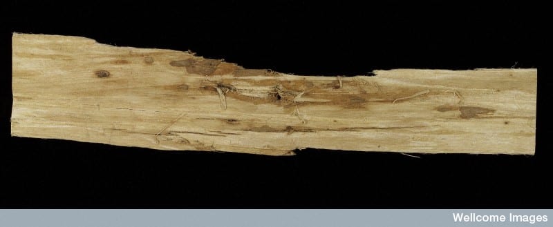
Slippery Elm Bark, sold as an abortifacient.

Accidental or intentional abortion occurred with a lot greater frequency in the 19th and early 20th centuries than some might imagine, and was inextricably wrapped up with both the idea and concrete reality of “foreign bodies.” For many, the coat hanger is the ultimate symbol of a foreign body inserted into the uterus to cause abortion. Still others may think of obstetric forceps as the foreign body which has caused thousands of fetal deaths during delivery. But what about uterine fibroids? How many abortions have they caused, and can they be regarded as foreign bodies if they are naturally occurring? A uterine fibroid is a benign tumour of the uterus, commonly found in women of reproductive age. Most fibroids are asymptomatic and therefore, most women are never aware that they even have one. However, on occasion these fibroids can cause health complications or interfere with pregnancy. Before the advances of late 19th century abdominal surgery and gynecology, uterine fibroids were not treatable. However, by 1916, obstetric surgeon Sir. John Bland-Sutton was able to boast that “uterine fibroids are common tumours; so common and troublesome that I have removed the uterus in 2,000 women.”[3]

From his experience performing hysterectomy on thousands of women a theme emerges: uterine fibroids closely mimic pregnancy in a variety of ways, and it is difficult – sometimes impossible – to distinguish between the two. Writing of this unfortunate similarity in 1913 Bland-Sutton observed, “A large sub-mucous fibroid produces similar changes in the uterus to those set up by the growth of the fetus […] Women with large sub-mucous fibroids are more or less in a condition resembling chronic pregnancy.”[4] What this similarity meant – and what gynecologists and obstetricians of the time openly acknowledged – was that sometimes a hysterectomy was performed to remove a fibroid that either never existed in the first place, or was also sitting alongside a feotus, masking a pregnancy. Either way, an abortion was performed.

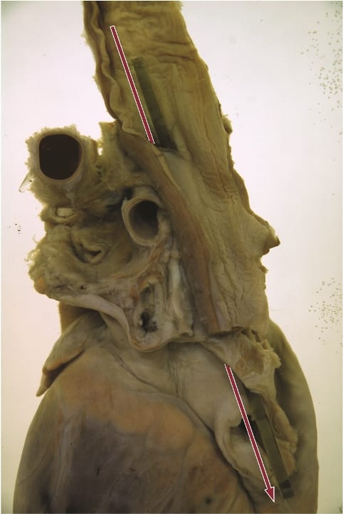
The photograph below demonstrates the reality of such surgeries. This particular specimen belongs to UCL Pathology Collections, and is currently on display in the Foreign Bodies exhibition. The anonymous woman patient underwent a hysterectomy most likely sometime in the early 20th century, in order to remove the sizable uterine fibroid, which can be seen on the right side of the image. However, on closer examination of the image, we see on the left side a preserved feotus, frozen in development, somewhere between 8-11 weeks.  It is unclear whether this woman or her doctor even knew she was pregnant.

Feotus in uterus, with large fibroid tumour. UCL Pathology Collections. Photograph Gemma Angel.

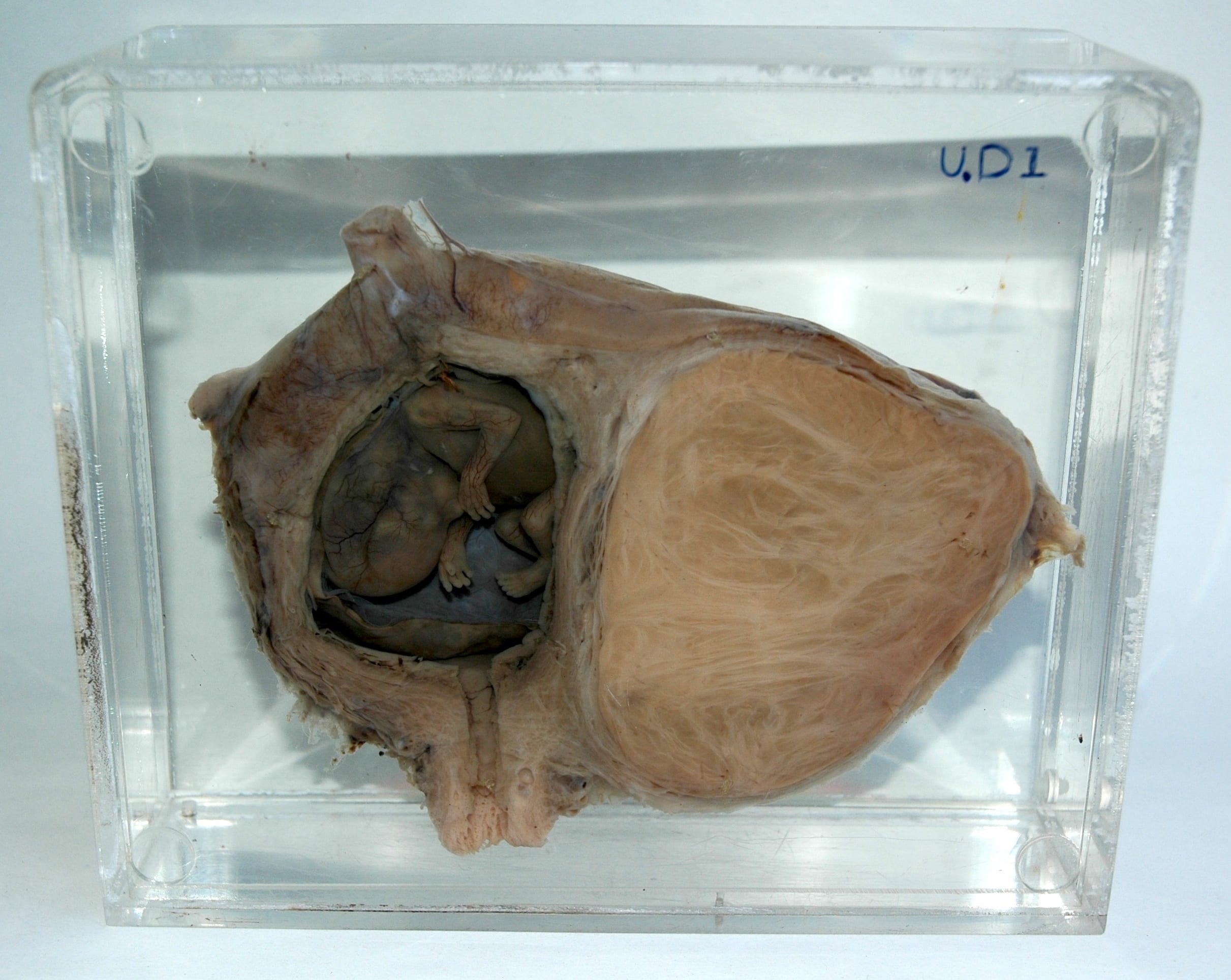
Feotus in utero, with large fibroid tumour.
UCL Pathology Collections. Photograph Gemma Angel.

Such examples abound in medical literature, and Victorian and Edwardian gynaecologists, obstetricians, and surgeons spoke of them with little or no censure. It was all a part of the surgical trial and error that they were practicing. The feotus was sometimes viewed as a necessary casualty in removing a potentially life-threatening fibroid. Either way, be it by slippery elm, by accident, or with purposeful intent, the feotus was removed as a foreign body, like any other. By examining the medical establishment’s attitudes towards fibroid removal we catch a glimpse into one way the feotus, and the experience of pregnancy in general, was understood in the past.


References:

[1] David A Grimes, Janie Benson, Susheela Singh, Mariana Romero, Bela Ganatra, Friday E Okonofua, Iqbal H Shah. “Unsafe abortion: the preventable pandemic.” The Lancet Sexual and Reproductive Health Series, October 2006.

[2] British Medical Journal, 29 July 1939.

[3] Sir John Bland-Sutton, “A Clinical Lecture on 200 Consecutive Hysterectomies for Fibroids Attended With Recovery” reprinted British Medical Journal, 4 July 1916.

[4] Sir John Bland-Sutton, “The Visceral Complications Met With Hysterectomy for Fibroids and the Best Methods for Dealing With Them” British Medical Journal, 1 November 1913.

Painted Skins & Butterfly Wings

By Gemma Angel, on 1 April 2013

Gemma Angelby Gemma Angel


When I first began my doctoral research into tattoo preservation three and a half years ago, I assumed that tattoo collections such as those held by the Science Museum in London were rare. Whilst collections of inked human skin are most definitely unusual, I was soon surprised and intrigued to discover that such objects exist in almost every museum archive, university anatomy department, or pathology collection that I have visited over the course of my research. The largest collections, of which the Wellcome Collection is the major exemplar, are dry-preserved and date from the 19th century – similar collections can be found across Europe, and the MNHN in Paris has a collection of 56 tattoos which are very similar to those in the Wellcome collection. Historically, these collections may be medical, anthropological, or criminological in origin.

In anatomy and pathology collections, tattoo specimens tend to be wet-preserved and date more recently, usually from the early part of the 20th century anywhere up to around the 1980s. But why are these objects preserved in anatomy and pathology departments at all? There is of course nothing pathological about tattooed skin in itself – so it seems strange that specimens like the one pictured below are displayed alongside other pathological skin specimens such as cutaneous anthrax, fibromas, keloids and glanders. What, if anything, can be learned from these tattoos in medical terms? Or are these striking collections of decorated human skin merely objects of curiosity? Often, the simple answer is that they are a little bit of both…

Tattooed human skin specimen. UCL Pathology Collections. Photograph © Gemma Angel.

Tattooed human skin specimen. UCL Pathology Collections.
Photograph © Gemma Angel.

The collection of tattoos pictured here are a case in point. These particular tattoos belonged to one individual, whose very brief case notes have been recorded and retained along with the specimen in UCL Pathology Collections. The notes provide an intriguing glimpse into the life of the individual to whom the tattoos belonged, as well as revealing something of the clinical interests and collecting practices of the doctor who preserved them:

From a man aged 79 years who had earned his living for many years as the Tattooed Man in a circus. His entire body, except for the head and neck, hands and soles of his feet, was covered with elaborate tattoo designs. He died of peritonitis due to a perforation of an anastomatic ulcer … In tattooing, fine particles of pigment are introduced through the skin, taken up by histiocytes and become lodged in the tissue spaces of the dermis. Pigment also passes to the regional lymph glands via the lymphatics. In this case, all the superficial lymph nodes were heavily pigmented.

It is clear from these brief comments that the nature and extent of this man’s tattoos were indeed of anatomical interest to the medical practitioner: The tattooed man had been so extensively tattooed that gradual migration of ink particles resulted in the collection of pigment in the lymph glands. This demonstrates that although tattoo ink is trapped permanently under the skin following healing, it does actually travel within the body over time, filtering into the body’s tissue drainage system, and collecting in the lymph glands. Whilst this is certainly an interesting anatomical observation, it is not the pigmented lymph glands that the doctor has chosen to preserve, but rather the tattooed skin itself. Without these accompanying case notes, we would never have known that this man’s tattoos had exerted any effect on another of the body’s organs and systems at all.

Reverse panel of tattooed human skin specimen Z6,  showing tattoos of a butterfly and a flying fish.UCL Pathology Collections.Photograph © Gemma Angel.

Reverse panel of tattooed human skin
specimen Z6, showing tattoos of a
butterfly and a flying fish.
UCL Pathology Collections.
Photograph © Gemma Angel.

It would be equally impossible to know whether or not these were the only tattoos he possessed – or indeed, if they all necessarily belonged to the same person. There are strong stylistic similarities between the butterfly motifs, suggesting the work of a single tattooist, or perhaps that the individual motifs were part of a larger design. But just how large or complex the design may have been, we certainly cannot tell just by looking at these 5 small tattoos. We know that they belonged to a 79-year-old man, who made his living as a Tattooed Man, only because the doctor tells us so. He or she also tells us that his body was covered in tattoos – yet only 5 small pieces have been preserved. Five carefully selected motifs, chosen by the doctor from an already complete collection, which provided the livelihood and told the life story of one unnamed man. What selection criteria did the pathologist adopt when deciding which tattoos to preserve, and which to consign to the grave? The manner in which the specimens have been excised and mounted are strikingly reminiscent of a lepidopterist‘s collection of butterflies – could this reflect the personal collecting interests of the pathologist, or perhaps even the Tattooed Man himself? Both the pathologist and Tattooed Man alike chose these butterflies – did they also share a passion for lepidoptery?

Many people will be familiar with the kind of insect specimen displays that are a staple of natural history collections – the old 19th century museum cases containing neat rows of pinned and mounted moths and butterflies, neatly organised according to subspecies and morphological characteristics. The tattooed butterflies share some remarkable similarities with these entomology collections; they are arranged one above another, and “pinned” to a support with small surgical stitches. Unusually for specimens found in pathology collections, this support is a slightly translucent black. This appears to be a deliberate choice on the part of the pathologist – the black perspex provides a contrasting ground for the display of tattoos on opposite sides of the vitrine, such that they do not visually detract from one another. These aesthetic choices suggest a nuanced interest in the collection and display of these specimens, which goes far beyond a straightforward medical interest in the anatomy of the tattoo. From the limited case notes and analysis of the specimen itself, we can learn something about the pathologist’s interest in the tattoo, but we are still no closer to being able to answer the fundamental question – why collect tattoos at all?

Butterfly display at UCL's Grant Museum. Photograph © UCL, Grant Museum of Zoology.

Lepidoptera display at UCL’s Grant Museum. Photograph © UCL, Grant Museum of Zoology.

There is no part of the body able to register the history of a life lived so much as the skin: wrinkles, scars and lines all map out our lives on the surface of our bodies as we age. The tattoo reinforces this unique capacity of the skin to record the traces of our experience, in the conscious act of permanently inscribing memory in skin. The pathologist, uniquely acquainted with death by virtue of their specialism, is perhaps best positioned amongst medical professionals to appreciate the peculiar relationship of the tattoo with mortality. It is a trace of the subjectivity of the deceased that is capable of outliving them, akin to a photograph or written memoir. From this point of view, it no longer seems surprising that tattoos are so often found in pathology collections; perhaps the pathologist who collected the Tattooed Man’s butterflies simply wished to preserve a small part of a colourful and remarkable life.


[analytics-counter]

Sword Swallowing & Surgical Performance

By Gemma Angel, on 11 March 2013

Sarah Chaneyby Sarah Chaney


We know sadly little about the sword swallower’s sword that resides in the UCL Pathology Collection: not even how long it has been here. What we do know is that this performer was very unlucky. Perhaps he (or, indeed, she) didn’t tilt his head back far enough. Perhaps he moved during the process of insertion. Whatever the case, the sword pierced the flexible tube of the oesophagus, leading to the performer’s death. The heart and oesophagus were preserved – perhaps as a warning of the dangers of such feats – alongside the weapon that led to his demise.

Fatally ruptured oesophagus, caused by the sword swallower's sword. Photograph Gemma Angel, UCL Pathology Collections.

Fatally ruptured oesophagus, caused by the sword swallower’s sword. Photograph Gemma Angel, UCL Pathology Collections.

Sword swallowing seemingly originated in India some 4,000 years ago, but reached the western world of Ancient Greece and Rome in the first century AD. The performer tilts his or her head back, extending the neck, and learning to relax muscles that usually move involuntarily. A rigid weapon can then be passed down as far as the stomach, usually for just a few seconds, before removal. It is dangerous, certainly, but few performers suffer the fate of the individual preserved in the UCL collections. According to one recent article in the British Medical Journal, most serious incidents occur owing to distraction or attempts at exceedingly complex feats:

For example, one swallower lacerated his pharynx when trying to swallow a curved sabre, a second lacerated his oesophagus and developed pleurisy after being distracted by a misbehaving macaw on his shoulder, and a belly dancer suffered a major haemorrhage when a bystander pushed dollar bills into her belt causing three blades in her oesophagus to scissor. [1]

In many ways, sword swallowing is the opposite of the ingestion of other foreign bodies: rather than swallowing, the performer maintains absolute control over the process of consumption, taming the body’s reflexes and realigning the organs. As Mary Cappello notes in her fascinating literary biography of surgeon Chevalier Jackson (1865 – 1968), who was an expert in foreign body removal, sword swallowing was recognised by doctors as inspirational to their own techniques. Jackson took his lead from German professors Alfred Kirstein and Gustav Killian, who lectured that sword swallowing proved the possibility of passing a rigid tube into the oesophagus, in order to remove lodged objects. Jackson, who developed his own oesophagoscope in 1890, admitted that the abilities of circus performers had opened his eyes to the opportunity of removing foreign objects without dangerous surgery. He even taught his children how to “scope” themselves.[2]

In an intriguing parallel, the insertion of some foreign objects into the human body thus assisted with the removal of others. At the turn of the 20th century, the removal of foreign bodies lodged in the throat and airways frequently required an incision to be made into the trachea or oesophagus, an operation which could prove fatal. In the records of the Royal London Hospital, from 1890 to 1910, we find no mention of oesophagoscopy or bronchoscopy: instead, surgery or the probang or “coin-catcher” was the norm. This latter instrument was generally a simple hook, inserted without any kind of viewing device or illumination. The practitioner would feel blindly for the object, and either attempt to hook it out, or push it into the stomach. This might lead to numerous complications. In 1903, surgeons at the Royal London attempted to remove a halfpenny from the throat of a five-year-old boy by pushing it into the stomach. However, it was subsequently reported that the coin catcher broke off in the boy’s throat, necessitating a major operation from which the child did not survive.[3] Small wonder that, less than a decade later, Jackson declared such objects “rough, unjustifiable, brutal”.[4]

foreignbodies

UCL Pathology Collections contains many examples of foreign objects removed from the
human body: this purpose built display showcases many such objects, some with
small x-rays of the objects prior to removal.

X-ray imaging techniques aided the removal of foreign objects by instruments, and foreign body specimens are often accompanied by photographs showing the item’s location in the human body. The above set of items is found in the UCL Pathology Collection, the objects having been gathered by several surgeons in the 1920s – ‘50s. At some point, the individual boxes made for each specimen were mounted together, in a specially designed plastic surround. Fittings on the back indicate that the case was made to hang on a wall. But why? To decorate the office of a surgeon, showing off his achievements? To offer a warning to others to take care (particularly parents, for all these objects were removed from children and infants)?

Chevalier Jackson claimed that his collection of more than two thousand foreign bodies (now housed in Philadelphia’s Mutter Museum) was not a curiosity, but indicative of the everyday nature of foreign body ingestion and inspiration. Yet many of these specimens are not everyday. The two boxes of multiple objects in the bottom right, for example, were removed from the vaginas of young girls (six and eight years old respectively). The case notes do not indicate how these objects arrived in their location. Did the girls insert them themselves, or might it be a sign of sexual abuse? In her research into the medical histories of Jewish immigrants to the East End of London in the late nineteenth and early twentieth centuries, Carole Reeves came across a case of multiple foreign body insertion in a young woman, whose vagina was found to be tightly packed with pins. Reeves speculated that Leah G. might have inserted these items in an effort to ward off potential (and actual) abusers.[5]

In most instances, we can uncover little about the motivations of those in the late 19th and early 20th centuries whose foreign bodies are recorded in medical records: surgeons were often little interested in how the object came to be in its current location, but only in its removal. Yet this may often make such displays still more intriguing than otherwise. As Mary Cappello put it, in a video discussion of the UCL artefact pictured above for the Damaging the Body website: “What is the border or boundary between human flesh, between human life and the object world?”


References:

[1] Brian Witcombe and Dan Meyer, “Sword Swallowing and its Side Effects”, in British Medical Journal, 333 (2006), 1285-7, p. 1287.

[2] Mary Cappello, Swallow: Foreign Bodies, Their Ingestion, Inspiration and the Curious Doctor who Extracted Them, New York, London: The New Press (2011). Website: http://www.swallowthebook.com/

[3] Royal London Hospital Archives, Surgical Index 1903, LH/M/2/9, patient no. 4086.

[4] Chevalier Jackson, Lecture to the Kings County Medical Society, December 19 1911, quoted in Cappello, p. 208.

[5] Carole Anne Reeves, Insanity and Nervous Diseases Amongst Jewish Immigrants to the East End of London, 1880 – 1920 (Unpublished PhD thesis, University of London, 2001), p. 213.

Pulling Teeth: Ovarian Teratomas & the Myth of Vagina Dentata

By Gemma Angel, on 4 March 2013

  by Gemma Angel


In preparation for our upcoming exhibition, Foreign Bodies, several members of the engagement team went to visit UCL Pathology Collections, to have a look at a collection of foreign objects removed from the human body. We soon encountered a number of other specimens which resonated with the exhibition theme in various ways: From a liver infected with syphilis, to a ruptured oesophagus and the sword swallower’s sword that caused the fatal injury; to a feotus inadvertently discovered during a hysterectomy, which was performed to extract a large tumour on the uterus.

The UCL Pathology Collections comprise over 6,000 specimens dating back to around 1850, many of which have been absorbed from other London medical institutions over the past 25 years, and these are currently in the process of being re-catalogued and conserved. It is a fascinating, not to mention an educationally invaluable collection – not least because it contains many specimens that demonstrate gross clinical manifestations of diseases which are now very rare in the Western world. Some of these diseases, such as syphilis, are unfortunately making a comeback, so it seems more important than ever that medical students are able to recognise the clinical signs of these infections. Pathology collections are a highly valuable medical teaching resource; particularly since these kinds of collections are now unlikely to be expanded in the wake of the 2004 Human Tissue Act.

As with many historical pathology collections, UCL possesses its share of medical anomalies or curiosities. Fragments of preserved skin belonging to a tattooed man certainly seem to fall into the category of the anatomically curious – there is certainly nothing pathological about this specimen. One of the biggest surprises I encountered during my visit to the collections, was the revelation that the female reproductive anatomy can, and occasionally does, grow teeth.

Teratoma with Tooth and Hair

Dermoid cyst (cystic teratoma) with fully developed
tooth and hair. UCL Pathology Collections.

The specimen shown here (right) is a dermoid cyst, or cystic teratoma, which has formed inside an ovary. When I first came across it, I experienced a strong visceral reaction: I didn’t have to be a medical student to recognise that this tooth, entwined in long hair drifting in the liquid-filled vitrine, was out of place – so much so, that the sight of it provoked an immediate and simultaneous sense of revulsion and fascination. The term teratoma is derived from the Greek, tera, meaning monster, and literally means “monstrous growth”; it was easy for me to see how such biological anomalies could become the stuff of nightmares. Despite the ominous name, however, ovarian teratomas are usually benign, and arise from totipotent stem cells which are capable of developing into any type of body cell. One 1941 pathology text describes these tumours as follows:

Dermoid cysts are usually globular in shape and dull white in color. They contain structures associated with epidermal tissues, such as hair, teeth, bone, sebaceous material resembling fat … The following is a partial list of tissues which have been found in dermoids: Skin and its derivatives, sebaceous glands, hair, sweat glands, and bone, especially the maxillae containing teeth. Up to 300 teeth have been found in one cyst … Long bones, digits, fingernails, and skull have been found. Brain tissue and its derivatives, intestinal loops, thyroid tissue, eyes, salivary glands, may occasionally be found. Even rudimentary fetuses have been described, such as a pelvis with hairy pubes and a vulva and clitoris. Brains with ventricles, spinal cords and a few complete extremities, have been observed. [1]

Although teratomas can develop in almost any part of the body – including the brain, neck, bladder, and the testes in men – being confronted with a toothy tumour in the female reproductive organs brought to mind mythic archetypes of the sexually devouring and deadly woman. I was immediately struck by the parallels between this specimen and the image of the vagina dentata. I am not the first to make such an observation,[2] and whilst I am not suggesting that there is any explanatory relationship to be found between the biological phenomena and the myths, it is certainly an intriguing association. The toothed vagina appears in the creation myths and folk stories of many cultures, from Native America, Russia and Japan (amongst the Ainu), to India, Samoa and New Zealand. [3] Funk and Wagnalls Standard Dictionary of Folklore, Mythology and Legend records this entry concerning vagina dentata:

The toothed vagina motif, so prominent in North American Indian mythology, is also found in the Chaco and the Guianas. The first men in the world were unable to have sexual relationships with their wives until the culture hero broke the teeth of the women’s vaginas (Chaco). According to the Waspishiana and Taruma Indians the first woman had a carnivorous fish inside her vagina. [4]

Many 19th and 20th century European interpretations linked the motif to Freudian concepts of castration anxiety, in which young males are said to experience an unconscious fear of castration upon seeing female genitalia. Whilst a Freudian analysis is undoubtedly culturally and historically specific, many vagina dentata legends explicitly articulate male fears of castration in the act of normal sexual intercourse, and warn of the necessity of removing the teeth from women’s vaginas, in order to transform her into a nonthreatening and marriageable sexual partner. A particularly telling collection of stories comes from India, in which the ferocious sexual appetites of beautiful young women are tamed and ‘made safe’ to men through the violent breaking of the teeth hidden inside their vaginas. [5]

Lloyd, Charles Augustus, d 1930. Lloyd, Charles A fl 1880s-1912 (Photographer) : Maori wood carving of the goddess Hine-nui-te-po, and Maui. Original photographic prints and postcards from file print collection, Box 14. Ref: PAColl-6585-10. Alexander Turnbull Library, Wellington, New Zealand. http://natlib.govt.nz/records/22708288

Māori wood carving of the goddess Hine-nui-te-pō and Māui.
Photograph by Charles Augustus Lloyd, c.1880s-1912.
Alexander Turnbull Library, Wellington, New Zealand.

The toothed vagina motif is not exclusively associated with male fears of the ‘castrating female’, however. In some traditions, the terrible power of the vagina dentata lies principally not in fears of the sexual act, but in its associations with death. The Māori legend of Māui and Hine-nui-te-pō is particularly interesting in this respect. Hine-nui-te-pō was the goddess of death and gatekeeper of the underworld, whom the trickster demigod Māui sought to kill in order to win immortality for humankind. When Māui asks his father what his ancestress Hine-nui-te-pō is like, he responds by pointing to the icy mountains beneath the fiery clouds of sunset. He explains:

What you see there is Hine-nui, flashing where the sky meets the earth. Her body is like a woman’s, but the pupils of her eyes are greenstone and her hair is kelp. Her mouth is that of a barracuda, and in the place where men enter her she has sharp teeth of obsidian and greenstone. [6]

Undeterred by his father’s grave warnings, Māui sets off on his quest with a gathering of bird companions. He proposes to kill Hine-nui-te-pō by entering her vagina and exiting through her mouth whilst she is sleeping, thus reversing the natural passage into life via birth. Māui finds the great goddess sleeping “with her legs apart” such that they can clearly see “those flints that were set between her thighs”, and he transforms himself into a caterpillar in order to crawl through her body. But his bird companions are so struck by the absurdity of his actions, that they laugh out loud and wake Hine-nui-te-pō from her slumber. Angry at Māui’s impiety, she crushes him with the obsidian teeth in her vagina; thus Māui becomes the first man to die and seals the fate of all humankind, who were ever after destined to die and be welcomed into the underworld by Hine-nui-te-pō. In this version of the myth, the vagina dentata appears as an inverse manifestation of the generative, life-giving powers of woman, which Māui attempts to subvert – he endeavours to overcome the forces of life and death, and therefore “by the way of rebirth he met his end.” [7]

Ovarian Dermoid Cyst

X ray of a dermoid cyst, showing a cluster of teeth in the pelvic cavity.

The mythical theme of the vagina-with-teeth can in most cases be read as an attempt to render the potentially dangerous sexuality of women nonthreatening to patriarchal power, through heroic acts of “pulling the teeth”. Some authors have even suggested a correspondence between this mythic construct and practices of clitoridectomy and ‘female circumcision’ in some cultures. [8] Whilst there can be little correlation between ancient stories and the observation of biological phenomena such as dermoid cysts, the removal of these peculiar tumours and their retention in pathology collections nevertheless reminds us of the remarkable complexity and diversity of human understandings of the body, and their wider cultural significance. For those readers interested in the practical removal of teratomas such as those discussed here, a demonstration of the surgical procedure can be viewed in this educational film (contains scenes of graphic live surgery).


References:

[1] Harry Sturgeon Cross and Robert James Crossen: Diseases of Women, St. Louis (1941), p.685.

[2] See, for example, Bruce Jackson: ‘Vagina Dentata and Cystic Teratoma’, in The Journal of American Folklore, Vol. 84 No. 333 (July-Sept 1971), pp.341-342. Available on JSTOR: http://www.jstor.org/stable/539812

[3] Verrier Elwin: ‘The Vagina Dentata Legend’, in British Journal of Medical Psychology, (1943) Vol. 19, pp. 439-453.

[4] Maria Leach (ed): Funk and Wagnalls Standard Dictionary of Folklore Mythology and Legend, Volume 2 J-Z (1950), p.1152.

[5]  Verrier Elwin: ‘The Vagina Dentata Legend’, in British Journal of Medical Psychology, (1943), Vol. 19, pp.439-453. A particularly illustrative example of one of these stories is recounted by Elwin on pp.439-440:

There was a Baiga girl who looked so fierce and angry, as if there was magic in her, that for all her beauty, no one dared to marry her. But she was full of passion and longed for men. She had many lovers, but – though she did not know it – she had three teeth in her vagina, and whenever she went to a man she cut his penis into three pieces. After a time she grew so beautiful that the landlord of the village determined to marry her on the condition that she allowed four of his servants to have intercourse with her first. To this she agreed, and the landlord first sent a Brahmin to her  – and he lost his penis. Then he sent a Gond, but the Gond said, “I am only a poor man and I am too shy to do this while you are looking at me.” He covered the girl’s face with a cloth. The two other servants, a Baiga and an Agaria, crept quietly into the room. The Gond held the girl down, and the Baiga thrust his flint into her vagina and knocked out one of the teeth. The Agaria inserted his tongs and pulled out the other two. The girl wept with the pain, but she was consoled when the landlord came in and said he would now marry her immediately.

[6] Antony Alpers: Maori Myths and Tribal Legends, Pearson Education, New Zealand (1964), p.67.

[7] Ibid, p.70.

[8] See for example, Jill Raitt: ‘The “Vagina Dentata” and the “Immaculatus Uterus Divini Fontis”‘, in Journal of the American Academy of Religion, Vol. 48 No. 3 (Sept. 1980), pp.415-431. Available on JSTOR: http://www.jstor.org/stable/1462869

[analytics-counter]

A Dark Chapter in Tattoo History: Nazi Prisoner Tattoos

By Gemma Angel, on 28 January 2013

  by Gemma Angel


Edit note: This post was amended on 25 August 2023 in order to remove an image of the tattoo, remove broken links, provide further detail on the UCL Pathology Collections and update the pejorative use of ‘inmates’ to describe interned people.

Over the course of the past 3 years working on the history of preserved tattooed human skin, I have frequently met with difficult questions: Material and conservation concerns pose the question how were they preserved? Historical and anthropological approaches lead me to ask why were they collected? – which of course raises the inevitable consideration of who were these people? Who did the tattoos belong to in life, and who collected them postmortem? This has proven to be the most thorny question of all – identifying both the tattooees and the collectors of tattooed skin is challenging in itself – but quite often it is the relationship between these two groups that raises the most controversial issues.

Frequently, a relationship of power and domination emerges, in which one group has the authority to study, scrutinize, classify – and ultimately, to dissect and preserve – the bodies of others under their institutional control. This is certainly the case for tattoos collected during the late 19th century by physicians and criminologists, who studied the tattoos of criminals and military personnel in prisons, barracks and hospitals. The criminals, soldiers and common men in these institutions very likely did not give consent for their tattoos to be excised and preserved after death; a practice that was rarely questioned during the late 19th century.

Ilse Koch was the wife of Karl Otto Koch,
Kommandant of the Buchenwald and Madjanek
concentration camps. She was convicted of war
crimes in 1947 and sentenced to life imprisonment.

The collecting of tattooed human skin – both for research purposes and as a kind of fetishized collector’s item – tailed off dramatically after the end of the second World War in the wake of revelations of Nazi concentration camp atrocities. Reports emerged from Buchenwald of the manufacture of everyday items such as gloves, knife sheaths, book-bindings and lampshades from the skins of murdered inmates.[1] In particular, stories of the collection of tattooed human skin, removed from the bodies of inmates at the behest of Ilse Koch, the wife of Kommandant Karl Otto Koch, caused a scandal in the Allied press. Although photographs documenting some of these objects were taken when the camps were liberated, no other material evidence of them was recovered to be entered into Koch’s trial at Dachau in 1947, or later at her second trial at Augsburg in 1950. Without material proof, Koch could not be convicted of the charges relating to the human skin objects.

The Nazis did not just collect the tattoos of prisoners as grotesque trophies of war, however – they also used tattooing as a weapon of dehumanization and control. From May 1940, prisoner numbers were introduced for all concentration camp prisoners deemed capable of work at the Auschwitz concentration camp complex – those sent directly to the gas chambers were not registered and did not receive numbers. These numbers were initially sewn onto prisoner uniforms. However, as the daily mortality rate increased and clothes were removed, this soon proved impractical as a way of identifying the dead. Tattooing of prisoner numbers was thus introduced at Auschwitz in the autumn of 1941. Tattoos were applied to either the inner or outer side of the left forearm on registration at the camp. More than 400,000 inmates were forcibly tattooed in this way at Auschwitz.

The SS introduced number sequences beginning with ‘A’ in mid-May 1944 – 20,000 men and 30,000 women were assigned numbers in this series. One of these women was Holocaust survivor Henia Bryer, who was liberated from Bergen-Belsen concentration camp in 1945. Henia still bears the number ‘A26188’ inscribed on her inner arm. She described her reasons for keeping her prisoner tattoo in a recent BBC programme:

I was offered by various surgeons to remove it, and they were very glad to do it, but I wanted to keep it on. So that when people say that it didn’t exist – these days, that the Holocaust didn’t exist, it’s a figment of your imagination – I wanted to show them. And many people don’t even know what this number means, still today. I wanted to keep it as a witness … as a sign that it really happened.

Tattoos carry with them a powerful capacity to evoke memory, and this quality is often a motivating factor for many people who choose to become tattooed with marks commemorating important life events or rites of passage. For those, like Henia, who have been forcibly tattooed, the mark may come to stand as testament to a personal and collective history of suffering and survival. For other Holocaust survivors, these marks became unwelcome reminders of trauma, and were removed by surgeons after the war. Some of these tattoos were retained in pathology collections, perhaps for reasons similar to those described by Henia above – ‘to stand witness’ to Nazi war crimes that should never be excised from the historical memory.

Original documentation record for object no. A.5.2. UCL Pathology Collections.


Whilst working with the UCL Pathology Collections, I came across such a tattoo: a small skin specimen in a perspex vitrine, catalogued as A.5.2 (pictured above). The original documentation associated with this object is the brief catalogue entry: “A tattooed identification number from the forearm of an inmate of Belsen concentration camp during World War II.”* No other information is known about this individual – since only people interned at Auschwitz were tattooed, it is very likely that this person, like Henia, endured the “death marches” from Auschwitz to Bergen-Belsen, where they were later liberated. Many Nazi records were destroyed at the end of the war, and the many thousands of files that do remain are scattered across Europe – even today, it is difficult to trace the identities of interned people based on their tattooed prisoner numbers.

Nor is it known exactly where this specimen was acquired, or who preserved it. Over the past 25 years, UCL Pathology Collections have absorbed a number of collections originating from other London medical institutions as a result of the creation of the Bloomsbury Health Authority in 1982 and subsequent UCLH NHS Trust established in 1994. A large number of the pathology specimens received by UCL arrived in a state of neglect, requiring intensive conservation and re-cataloguing – a task made all the more difficult for the lack of associated documentation. Specimens such as A.5.2 may make us uneasy, particularly when they are unprovenanced and inherited. Their histories are lost and fraught with ethical entanglements – but they cannot simply be discarded, and perhaps should not be hidden away. Henia reminds us of the need to remember past trauma, and of the role that material culture can play in this process. Nazi prisoner tattoos are a powerful reminder of the lived experience of war and genocide. As objects, their presence in pathology collections is undoubtedly troubling; yet they remain as an important testament to the horrors of the Holocaust – they are fragments of lives that should never be forgotten.


References:

[1] Flint Whitlock: The Beasts of Buchenwald, (2011) Cable Publishing, p.81. See also, Alexandra Przyrembel: ‘Transfixed by an Image: Ilse Koch, the ‘Komandeuse of Buchenwald’, in German History, Vol. 19 No. 3, (2001).

*The current catalogue entry for this specimen reflects recent research and has been updated to amend the pejorative term ‘inmate’. A.5.2

[analytics-counter]