X Close

Researchers in Museums

Home

Engaging the public with research & collections

Menu

If a salamander can grow back a limb, why can’t I?

By Julia R Deathridge, on 29 August 2017

Regeneration has been the deemed the “Holy Grail” of scientific and medical research: the ability to regrow a limb, replace damaged tissue and heal without scars would completely change the face of modern medicine. Whilst regeneration, on a large scale, still precludes us Homo sapiens, other members of the animal kingdom are light years ahead.
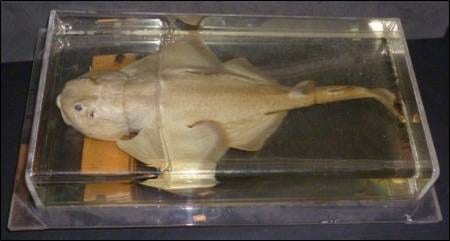
W584

Accession no. W584

Not wanting to completely disregard humans, it is important so say that we are capable of a small degree of regeneration. We can heal the upper layer of our skin without scarring, regenerate parts of our gut lining, expand our liver to replace damaged parts, and can even regenerate the very tips of our fingers. However, if you go to the far left corner of the Grant museum you will come face to face with an axolotl salamander, whose regenerative powers are worthy of a science fiction character.

Like many salamander species, the axolotl has the remarkable ability of re-growing lost limbs. An axolotl’s leg can be amputated hundreds of times and it will continue to grow back perfectly! However, what makes axolotls so unique is that as well as regenerating their limbs they can also regenerate their jaw, tail, heart and spinal cord, all without scarring. If you want to find out more about the extraordinary features of the axolotl, read this post from the Grant Museums’ Specimen of the Week blog.

To recreate the limb the axolotl, and other salamander species, need to be able to regrow everything – bone, muscle, blood vessels, nerves – and all of this needs to be built from scratch. So, how do they do this?

Regeneration relies on the formation of highly pluripotent cells that have the capacity to develop into multiple cell types. These types of cells are known as stem cells, and they are the building blocks of all embryo development. As stem cells mature during development, they become restricted to a defined cell fate and lose the ability to become multiple cell types. However, salamanders, and other creatures that can regenerate, are able to revert defined cells, at the site of the lost limb, back to this immature stem-like state by a process known as de-differentiation. Once in an immature state these cells congregate together to form a blastema, which is capable of growing into all the different cell types of the missing limb

CC: Wikipedia

Salamander. CC: Wikipedia

Despite our understanding the basic steps of regeneration, the intricacies of each step are extremely complicated and a lot of questions still remain unanswered. Imagine having to reconstruct an entirely new building from previously used disassembled building materials, without any blueprint or instructions. How would you know what to make and where each building material goes? This is the exact dilemma facing the cells of the regenerating limb, and how exactly they overcome this hurdle continues to puzzle scientists.

OK there are a lot of complex mechanisms involved – but surely if a simple salamander can regrow a limb we should be able to as well?

Well actually our increased complexity could be what’s stopping us from regenerating. All our cell behaviours need to be tightly regulated in order to maintain the function of our complex organ systems and prevent aberrant growth. If cells are reverted back to their immature state they will be more difficult to control and thus more likely to misbehave, which could result in the formation of a tumour or loss of organ function. With less complex animals, such as the salamander, these misbehaving cells are unlikely to inflict as much damage and cause as much of a problem.

Some researchers have proposed that we have the capacity to regenerate but, for unknown reasons, this mechanism was switched off during evolution. The theory is that if our genetic code allowed us to develop entire limbs and structures in the womb, then that information must still exist inside of us. It could be as simple as switching on a few genes and – hey presto – you’ve built yourself a limb! Furthermore, recent discoveries have identified genes involved in the regeneration process of the axolotl that are turned off in humans. Could these hold the key to regeneration?
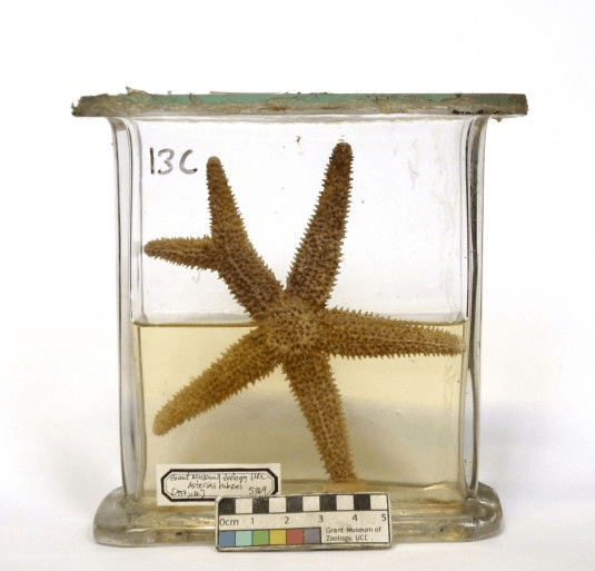
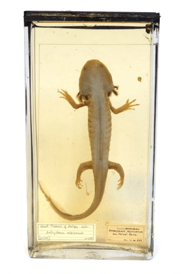
Accession no. S369

Accession no. S369

Another theory is that it is easier for amphibians to regenerate, as they are cold blooded and therefore have fewer metabolic requirements. A salamander can hide away for months without eating, waiting for their limb to grow back. However, this would be a death sentence for mammals that would need to heal much faster in order to keep up with their metabolic demands. Moreover, the regeneration time of the axolotl is far from rapid, if humans were to regenerate at the same rate it would take up to 15-20 years to regrow a full limb! Therefore, alternative medical interventions are likely to provide a more pragmatic solution.

The axolotl and other salamanders are not the only animals that can regenerate. Hydras, starfish, flat worms, zebrafish, and even the African spiny mouse all have the capacity to regenerate themselves to a certain degree. So next time you encounter one of these creatures whilst exploring the Grant Museum, remember that although they may look simple these animals have science fiction powers that, right now, humans can only dream about.


Question of the Week: Why do brains have wrinkles?

By ucbtch1, on 27 April 2017

The brains displayed at the entrance of the Grant Museum are mostly mammal’s brains but we can observe differences in sizes and in how smooth or wrinkly they are. The folds of a brain are called gyri and the grooves are called sulci. These morphological features are produced by the folding of the cortex, the part of our brain responsible for higher cognitive processes like memories, language and consciousness. During development, all brains start off with a smooth surface and as they grow, gyrification (the development of the gyri and sulci) occurs. It is interesting to note that the major folds are very consistent amongst individuals, meaning that development is similar sometimes even amongst species.

c_ucl_gmz_matt_clayton020

The brain collection on display at the Grant Museum of Zoology (Image credit Grant Museum of Zoology).


It has been assumed that the wrinkles in brains correlate with an animal’s intelligence. The reasoning behind this is that a bigger brain, and hence more neurons, need more space. The folds allow the cortex to increase its area while being packed in a confined space like our cranium. There are several factors and hypothesis of how gyrification occurs. Recently, researchers at Harvard developed a 3D gel model based on MRI (magnetic resonance imaging) images to understand how this process occurs. They found out that it all boils down to the mechanical properties of the cortex. While neuronal cells grow and divide, the increasingly bigger brain leads to a compression of the cortex and to the formation of the folds. The researchers were able to mimic the folds of the cortex and were stunned at how similar their gel model looked to a real human brain.

tumblr_o21mv85Nt01t5fphqo1_1280

Gel model of a foetal brain (Image credit: Mahadevan Lab/Harvard SEAS).


Even though most of the brains on display in the Grant museum have gyri and sulci, in nature, most animals have smooth brains. In general, larger brains have folds while smaller brains do not, even small mammals like rats or mice have smooth brains. In humans, a lissencephalic brain is one without gyri and sulci and is a result of a rare disorder that is characterised by mental abnormalities. From the collection of brains in the Grant Museum, there is only one lissencephalic brain—next time you visit the museum see if you can spot it. Additionally, try to find the brain coral. Because of its intricate maze–like pattern, Diploria labyrinthiformis has very similar ridges and grooves as a brain, and so is referred to as brain coral. Overall, I find looking at brains and their grooves fascinating, each species with their own pattern and each groove in a specific place. Makes me wonder how brain coral gets its patterns.

brain coral 3

Diploria labyrinthiformis also known as brain coral(Grant Museum C1084).


References:

Roth, G. and Dicke, U., 2005. Evolution of the brain and intelligence. Trends in cognitive sciences9(5), pp.250-257.

Ronan, L. and Fletcher, P.C., 2015. From genes to folds: a review of cortical gyrification theory. Brain Structure and Function220(5), pp.2475-2483.

Manger, P.R., Prowse, M., Haagensen, M. and Hemingway, J., 2012. Quantitative analysis of neocortical gyrencephaly in African elephants (Loxodonta africana) and six species of cetaceans: comparison with other mammals. Journal of Comparative Neurology520(11), pp.2430-2439.


Question of the Week: What’s that zigzag on your skull?

By Arendse I Lund, on 25 April 2017

Stan hangs out in a corner of the Grant Museum amid cases filled with insect exoskeletons and bisected animal heads. Standing at around two meters, he keeps watch through empty sockets over the animal bones, taxidermy, and jar specimens.

“Can I hold his hand?” I’ve been asked more than once. “Is he real?” comes the hesitant question. As a matter of fact, Stan is a model skeleton, the likes of which you’ve probably seen in any biology classroom. Although he’s resin and missing a joint or two he’s still a remarkably good way to explain what we’re made of once you strip all our clothes, skin, and muscles away.

One of Stan’s characteristics is a zigzagging line arching its way across his skull. Surprised by the mark, a visitor wanted to know why Stan bears this line. She might have been surprised to know that she has one too. It’s actually a feature all human skulls have. Known as the coronal suture, it’s an immovable joint that runs transverse across the skull, separating the frontal bone from the parietal bones.

Top view of a skull with coronal suture extending from ear to ear (Image: Stanford's Children Health Hospital)

Top view of a skull with coronal suture extending from ear to ear (Image: Stanford’s Children Health Hospital)

At birth, the various bones of the skull don’t quite join up, making it easier for the infant to fit through the birth canal; following the birth, the gap persists for a while and the coronal suture reflects where that separation once was. There can be “premature closing” of the suture if the bones fuse too soon and people will develop conditions such as oxycephaly—where the skull is lengthened—or plagiocephaly—where the skull is flattened.

Top view of skull casts, the left found in Beijing and commonly referred to as the "Peking man" but is actually thought to be female (Grant Museum Z2681); and the right of a Rhodesian Man found in Kabwe and known as the Broken Hill 1 skull (Grant Museum Z2684).

Top view of skull casts, the left found in Beijing and commonly referred to as the “Peking man” but is actually thought to be female (Grant Museum Z2681); and the right of a Rhodesian Man found in Kabwe and known as the Broken Hill 1 skull (Grant Museum Z2684).

If you take an “exploded skull” view then you can see how the various parts of your head all join up. We can see these sutures in other skulls than just modern humans as these skulls are formed in similar ways.

Chimpanzee skull (left, Grant Museum Z461) and Neanderthal skull (right, Grant Museum Z2020) both showing coronal sutures.

Chimpanzee skull (left, Grant Museum Z461) and Neanderthal skull (right, Grant Museum Z2020) both showing coronal sutures.

Stan has a few friends at the Grant Museum. There’s a Neanderthal skull alongside Homo erectus, Homo habilis, and Australopithecus afarensis. There’s also a human skeleton that oversees the museum up on a balcony accompanied by an orangutan, gorilla, and chimpanzee—all bearing these sutures.

Next time you see a human skull in a museum, see if you can spot the coronal suture. While knowing its name may not win you any prizes in a pub quiz, it’ll certainly impress Stan. He’ll be waiting to say hi.

Follow @Arendse on Twitter or read more of her blog posts here.

Question of the Week: How do sharks hear?

By Stacy Hackner, on 23 April 2014

by Stacy Hackner

“Sharks have eyes and mouths, and we hear all about their ability to smell blood. How do they hear?” Once again, a visitor had me stumped. Despite their having only tiny holes for external ears, sharks actually have very acute hearing, I later learned. Like in humans and other mammals, the shark’s inner ear has tiny hairs called stereocilia that vibrate, which is interpreted by the brain as sound. The stereocilia are arranged in three fluid-filled tubes, allowing the shark to hear in multiple directions. (These tubes are also responsible for the shark’s sense of balance.)

Sharks can hear low frequencies much better than humans, ranging from 10-800 Hertz (for reference, humans can hear between 25-16,000 Hertz), and can hear prey up to 800 feet away. In combination with their formidable sense of smell and speed, this makes them fearsome predators. (The big ones, at least.)

The-angel-shark-at-the-Grant-Museum-of-Zoology

The angel shark, with ears visible just behind the eyes.
Courtesy Grant Museum.

Sources:

Shark Trust

Sharks Interactive 

Foreign Bodies: Attack of the Clones

By Gemma Angel, on 18 February 2013

Profile  by Felicity Winkley


One of the most controversial specimens amongst the Grant Museum’s encyclopaedic collection is a preserved domestic cat; in fact, on one occasion, I was standing quite close to this object at the exact moment when a small child laid eyes upon it and promptly burst into tears. The fact that the sight of preserved animals, particularly domesticated or fluffy ones, provokes such a response would be ample topic for a debate in its own right, however in this instance I am more interested in the way the Grant have developed the subject in their museum signage. Beside the exhibit, they point out that in 2004 the first domesticated cat was cloned for $50,000 – a kitten called ‘Little Nicky’, commissioned by a Texan woman called Julie after the original cat ‘Nicky’ had died [1] – and ask whether or not this was a good thing to do? nickyAt the time of the cloning in 2004, the response from the scientific community was negative: it was thought a fatuous use of the technology to reproduce a domestic pet, as well as inhumane given the animal’s short life-expectancy (roughly a third of cloned cats did not survive beyond 60 days).[2] Today, expanding the subject beyond the cloning of domestic animals, as part of the successful QRator scheme (in which visitors are invited to record their responses to topical questions relating to the collections), the Grant Museum asks the public to contribute to a wider debate: Should we clone extinct animals?

The argument is a complex one. For one thing, extinct animals may have died out because of their own comparative weaknesses, and therefore any attempts to reintroduce them may prove futile. The journalist Chris Packham, for example, has famously lambasted attempts to conserve the Giant Panda, criticising the huge amounts of money spent on attempting to breed an animal which is so reluctant to reproduce itself. He suggests that the Giant Panda is “a species that of its own accord has gone down an evolutionary cul-de-sac” and therefore should be allowed to die out, not least because any attempts to reintroduce it into the wild will be limited by the increasingly diminishing area of its potential habitat anyway.[3] Where cloning animals and reintroducing them is concerned, habitat is also an issue in terms of preempting any potential environmental changes that might have occurred since the species was last present in the wild. The repercussions of reintroducing clones despite drastic ecosystem change are fairly clearly (if not necessarily realistically!) laid out for us to see in Jurassic Park. Although the author accepts this is an extreme example, it is nevertheless an effective visualisation of what can occur when we tamper with complicated systems of which we have limited understanding.

Jurassic Park III


But what of those species made extinct by human influence, and through no fault of their own? The quagga, hunted to extinction in 1883, and the thylacine, in 1936, are both on display in exhibition cases at the Grant Museum. If we accept, then, the fault of human oversight, perhaps these two could justifiably be cloned and reintroduced into the wild – but given the cost of the procedure and the potentially limited life-span of the animal subjects, wouldn’t the enormous investment be better applied to conserving those species still alive today but in dire need of assistance? The Amur leopard population, for example, is currently at a critical low, with just 7-12 thought to remain in the wild in China and 20-25 in Russia.[4]

Taking into account all of these conflicting arguments where cloning is concerned, it was with some interest, therefore, that I read a few weeks ago about a Harvard professor’s hopes for recruiting a female volunteer willing to surrogate a baby created with Neanderthal DNA.[5] Geneticist Professor George Church has recently completed enough Neanderthal bone-sample analysis to accurately isolate the genetic code that would enable him to create artificial Neanderthal DNA, according to his publication Regenesis: How Synthetic Biology Will Reinvent Nature and Ourselves.[6] Having previously been involved in the Human Genome Project, which successfully mapped human DNA, Professor Church would insert artificial Neanderthal DNA into stem cells and inject these into a human embryo in the earliest stages, allowing this to develop in the laboratory before implanting it into the womb of a potential surrogate mother. He believes that Neanderthals, whose population flourished in Europe and extended throughout the Middle East and into China between 70,000 and 30,000 years ago, were highly intelligent. This impression is certainly supported by archaeological evidence: the skulls of Neanderthals held large brains, “in the range of and exceeding the cranial capacity of modern humans” state Lewin and Foley.[7] As such, Professor Church proposes that a cloning and reintroduction of Neanderthals could be useful to increase diversity, and introduce an alternative way of thinking into society:

When the time comes to deal with an epidemic or getting off the planet or whatever, it’s conceivable that their way of thinking could be beneficial. They could maybe even create a new neo-Neanderthal culture and become a political force. The main goal is to increase diversity. The one thing that is bad for society is low diversity.[8]

Aside from the obvious concerns about the potential risks to the surrogate mother of a baby created via this method, critics have also challenged the ethics of the proposed experiment. Whilst the Charter of Fundamental Rights of the European Union prohibits reproductive human cloning in member states of the EU, and it is likewise illegal in the UK under the Human Fertilisation and Embryology Act 2008 – because the project proposes the cloning of a Neanderthal rather than a Homo Sapiens, there are fears that current legislation may not apply. In any case, there is no uniform guideline agreed for the United States of America on human cloning, whether reproductive or therapeutic. But were Professor Church to have his way, how would a new Neanderthal cope in modern-day society? Physically, could their immune system withstand it? Emotionally, would they successfully integrate, or be outcast as a monster? Whatever the answer – and luckily at the moment our concerns are purely speculative – there is no denying that a neo-Neanderthal person would be the ultimate foreign body.


Does Size Matter? Evolution and the Primate Penis

By Gemma Angel, on 17 September 2012

Suzanne Harvey #2by Suzanne Harvey


Anatomy is destiny … The genitals themselves have not taken part in the development of the human body in the direction of beauty: they have remained animal, and thus love, too, has remained in essence just as animal as it ever was.

When Sigmund Freud wrote this in 1912, he may have been surprised to hear that some hundred years later, evolutionary theory would come to the same conclusions. Despite the frequently discussed individual variation in human penis size, the shaft of an average human penis is around twice the length and width of the shaft of an average chimpanzee penis. It is also useful to mention some more unusual facts: firstly, while chimpanzees have penises half the size of humans, they have testicles three times as large. Moreover, while silverbacks are formidable looking creatures, gorillas in fact have the smallest penis to body size ratio of any mammal. So, what causes these seemingly contradictory differences among the great apes, and how can evolutionary theory make sense of all creatures great and small?

Sperm Competition

As Freud’s quote suggests, the clue to the evolution of the penis is not just in their physical appearance but also in the social aspects of sex. In fact, generally speaking, the mating system of a species can be used to predict penis size. Chimpanzees live in large multi-male, multi-female groups, where females are able to mate with many males. Sperm can live for up to 4 days after ejaculation, and consequently when females mate with two males in close succession, sperm from two males can be in direct competition. The male who produces more sperm will have the best chance of fertilizing an egg. This evolutionary advantage of producing large amounts of sperm can explain the relatively large testicle size of chimpanzees. Correspondingly, the male gorilla’s huge stature is in fact the reason why he has such a small penis: when competition between males occurs through physical aggression, an alpha male may fight off rivals and control his own mating success without the need for sperm competition. Other physically smaller males have little access to females in the group.

Understanding the Human Penis

The mystery of the human penis is that ancestral hominids lived in similarly large and promiscuous social groups, but did not evolve the large testicles seemingly necessary to compete via sperm competition. One might be forgiven for thinking that larger penises evolved as a result of sexual selection; the theory that a preference for larger penises in females has led to greater reproductive success for males with larger penises, with these males passing on the trait to their offspring. However, the latest research shows that penis size may also be the result of sperm competition and natural selection.

The Semen Displacement Theory (Gallup and Burch, 2004) essentially explains the advantages of the size and shape of the human penis in terms of a device evolved to remove another male’s semen before fertilization.

As well as being larger and wider than other primate penises, the human penis has the unique shape of a shaft with a ridge leading to a wider tip, known as the coronal ridge. This is more pronounced than in other species. All of these elements are important in terms of semen displacement: the coronal ridge removes semen by ‘scooping it out’ as it passes over the tip, is trapped behind the ridge and pulled out during intercourse. Recent research shows that (using artificial genitalia) a penis with a coronal ridge will displace 91% of semen, while one without will displace only 35% (Gallup et al. 2003). Thrusting during sex creates a vacuum that aids this process, as the width of the shaft provides a plug in the vagina. In Gallup’s experiment, the same penis removed 90% of semen when fully inserted and only 39% when inserted three quarters of the way. Therefore, the length of the shaft simply improves reach and maximizes the amount of semen that can be removed.

So yes, when it comes to penises, size – and shape – matters when it comes to natural selection!


Suzanne Harvey is a PhD student in Biological Anthropology, working on social interactions and communication in wild olive baboons. She is also a teaching assistant on the UCL Arts and Sciences BASc, a new interdisciplinary degree, and can be found on twitter @suzemonkey.


References:

Freud, S. (1912). On the Universal Tendency to Debasement in the Sphere of Love. Oxford Literary Review 30: 109-146 DOI 10.3366/E0305149808000199, ISSN 0305-1498

Gallup, G. G. & Burch, R. L. (2004). Semen Displacement as a Sperm Competition Strategy in Humans. Evolutionary Psychology 2: 12-23

Gallup, G. G., Burch, R. L., Zappieri, M. L., Parvez, R. A., Stockwell, M. L. & Davis, J. A. (2003). The human penis as a semen displacement device. Evolution and Human Behavior 24: 277–289