X Close

Researchers in Museums

Home

Engaging the public with research & collections

Menu

Archive for the 'Question of the Week' Category

Question of the Week: How do dogs recognise other dogs?

By Cerys R Jones, on 30 April 2019

From Great Danes and Dogue de Bordeauxs to miniature Dachshunds and Chihuahuas, man’s best friend comes in a variety of shapes and sizes, so how can they recognise fellow dogs even when they all look so different?

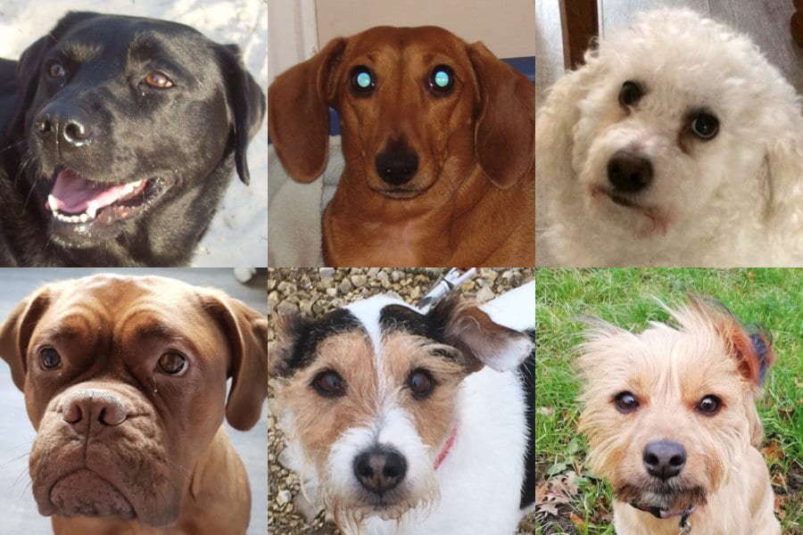
Dogs come in a variety of different shapes and sizes, featuring Jess the black Labrador, Jewell the miniature Dachshund, Percy the Bichon Frise, Luna the Dogue de Bordeaux, Scratch the Jack Russell Terrier, and Spud the mixed-breed. (Engager’s own photos)

The Kennel Club recognises 211 different breeds of dogs but with different coats and mixed-breeds, there are by no means 211 dog-shaped moulds. Despite this, your dog can decipher between a Bichon Frise and a lamb instantly. This is in part due to their impressive sense of smell which they use to smell the hormones secreted by other dogs. Not only do they have a large nose cavity, which contains a folded surface covered by the sensing organ that is up to 23 times larger than in humans, they also have a vomeronasal organ in the roof of their mouth for detecting smells [1]. This means dogs can smell up to 10,000 times better than humans [1].

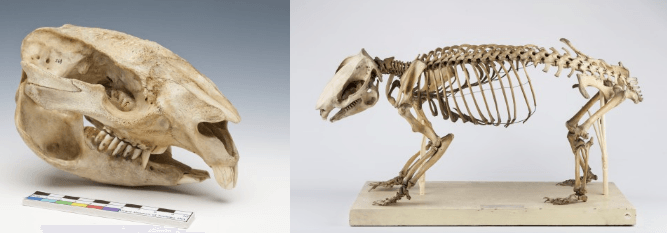
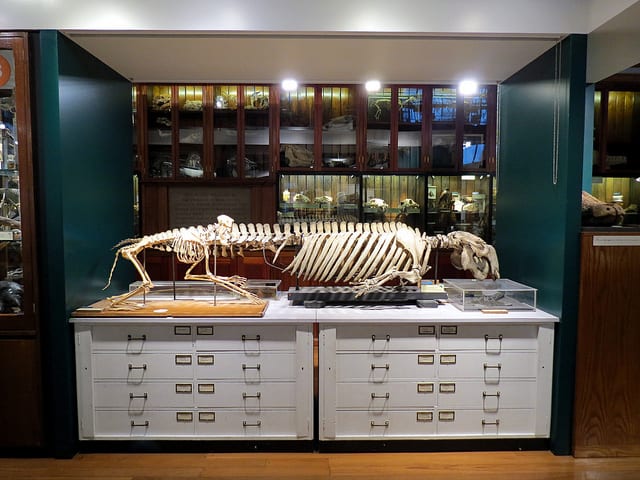
Seven domestic dog skulls on display in the Grant Museum (Accession number: Z2909)

Dogs’ ability to recognise different chemicals through their sense of smell has been used by humans to sniff out drugs, explosives and even illnesses such as cancer and diabetes. But is this the only sense dogs rely on to recognise other canines? A study from 2013 tested nine dogs’ ability to correctly identify other dogs from pictures [2]. The dogs were shown two images: one of a dog (from a set of 3000 pictures of different breeds, including mixed-breeds) and one of a non-dog animal, which included cats, cows, rabbits, birds, reptiles and even humans. On command, the dog participant had to correctly distinguish between the images and place their paw on the picture of the dog. All nine dogs successfully chose the images of the dogs over the images of non-dogs the required 10 times out of 12. The study concluded that dogs could “form a visual category of “dog pattern”” ([2] page 647); however, it did not allow the researchers “to determine which dog morphotypes or which species were easier to discriminate” ([2] page 648). As the dogs were successful at distinguishing between dogs and other animals from photographs alone, it is clear that they don’t solely rely on a sense of smell.

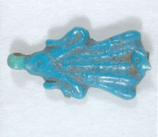
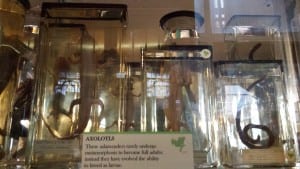
Hair curlers with a hunting dog on from the Petrie Museum (Accession number: UC8529)

Although varying highly in appearance, from the colour of their coat to the length of their snout, dogs use both their senses of smell and sight to identify others. Exactly which visual cues are required is still unknown. One thing we know for certain is, regardless of how they look, they’re all good dogs!


Bibliography

[1] Miklosi, A., (2018) “The Dog: A Natural History” Ivy Press, Brighton

[2] Autier-Derian, D., Deputte, B.L., Chalvet-Monfray, K., Coulon, M., and Mounier, L., (2013) “Visual discrimination of species in dogs (Canis familiaris)” Anim Cong, 16, pp 637-651.

Question of the Week: Why are sea sponges considered animals?

By Sarah M Gibbs, on 29 May 2018

By Sarah Gibbs

In that big comparative anatomy lecture hall in the sky, Robert Grant, founder of UCL’s Grant Museum of Zoology, must be smiling. This week’s featured question focuses on Grant’s favourite animal: the sea sponge. Grant’s work definitively proved that sponges are animals, not plants or simple celled organisms.

So, why is a sea sponge more closely related to a dog that a cactus? Read on to find out!

lue Barrel Sponge (Scientific American; Creative Commons Chris Coccaro)

The ever-sage Encyclopedia Britannica informs us that early naturalists classed sponges as plants because, you know, they lack organs, don’t move, and often have branches. Understandable, to be sure. In the eighteenth century, however, scientists began to notice animal characteristics of sponges, including the changes in diameter of their central cavity, and their creation of distinct water currents. Zoologists imagined that sponges occupied an isolated position in the animal kingdom, but molecular testing has since proved that sponges and more complex animals (like humans) developed from a common ancestor; sponges also possess many of the qualities biologists use to distinguish people from plants. For example, bodily composition: the elastic skeletons of sponges are made from collagen, the same protein found in human tendons and skin. Prevailing theories suggest that sponges are early animals which produced no subsequent evolutionary line.

The Venus Flower Basket Sponge (Scientific American; Creative Commons Ryan Somma)

The folks over at Scientific American note that sponges’ specialized cells differentiate them from multicellular protists, creatures which are not animals, plants, or fungus, and which form no tissues. It is the thinness of the sponge body and the fact that its cells are exposed to circulating water—which supplies food and oxygen, and removes waste—that make organs unnecessary. Sponges may have been the first multicellular animals. Multicellularity (which means that cells adhere to one another, communicate, are mutually dependent for survival, and specialize to perform different tasks) is the key to producing more complex organisms. Scientists speculate that sponges emerged, flexing their multicellular muscles*, at least 543 million years ago (*as sponges lack arms, they are sadly ineligible for body building contests). According to Scientific American, sponges were the first filter feeders, tiny Brita jugs of the sea** (**mixed metaphor alert).

So, sponges are in fact the original animal hipster; they were multicellular before it was cool. Let’s close with a few fun sponge facts.

Absorbing (!) Facts:

  1. Sponges can range in height from less than one centimeter to two metres tall.
  2. Most sponges are hermaphroditic (male and female cells exist in one animal) and reproduce sexually by releasing spermatozoan into the water current to be carried to other sponges, where they interact with eggs. Sponges can also reproduce asexually.
  3. Some deep-water sponges are carnivorous. Animals like the ping-pong tree sponge lie in wait for small crustaceans and other hapless sea dwellers to alight on their branches, the hook-like spicules on which prevent escape. Digestive cells migrate to the site of capture and the feast begins. Bet you’ll never look at a loofah the same way again.


Sources:

Frazer, Jennifer. “Sponges: The Original Animal House.” Scientific American, 17 Nov. 2011, https://blogs.scientificamerican.com/artful-amoeba/sponges-the-original-animal-house/

Sarà, Michele. “Sponge.” Encyclopedia Britannica. Britannica Academic, Britannica Digital Learning, https://academic-eb-com.libproxy.ucl.ac.uk/levels/collegiate/article/sponge/110257

Question of the Week: Why do box jellyfish have eyes?

By tcrnkl0, on 10 May 2018

If you meet me during one of my shifts as a PhD Student Engager in the Grant Museum, you’ll find me next to the Micrarium, facing a case packed full of jellyfish and their ghostly relatives. I’ve never had an interest in jellyfish before, but hours and hours of staring at them over the time I’ve been an Engager has inspired my admiration (as well as a previous blog post).

Box jellyfish specimen at the Grant Museum, photo by author.

In recent weeks, I’ve had a number of visitors ask me about box jellyfish eyes, because it’s surprising to find out that something which often looks and moves like a floating plastic bag has eyes. And not just one or two eyes, but 24 in total. Their eyes are bundled into four structures called rhopalia, which sit around the bottom of its bell. Two of the eye types have the capability to form images, while the other two types help with swimming navigation, avoiding obstacles, and responding to light. Fun fact: Box jellyfish can regenerate their eye bundles (rhopalia) in as quickly as two weeks’ time.

On the specimen in the Grant Museum, you can only see two of it’s eyes because it’s been carefully bisected to reveal its internal anatomy.

Grant Museum specimen with eyes highlighted by author.

Like other jellies, box jellyfish have no brain, perceiving the world only through their nervous systems. Most jellyfish catch their prey without having either brains or eyes, just by floating transparently through the sea until prey run into their tentacles. So, our question should actually be: why do box jellyfish even need eyes?

There are at two main possibilities:

1)  Habitat: Unlike most jellies, which live on the open sea, box jellyfish tend to live in shallow water, which has many obstacles. Scientists have shown that box jellies near Puerto Rico can navigate around the dense mangrove swamps where they live, and also make sure that they don’t drift away to where there is less prey. Their upper lens eye can actually peer through the water’s surface to navigate from landmarks above the water, and perhaps celestial ones as well! Some scientists think these kinds of jellies actively hunt rather than passively encounter prey.

2) Reproduction: Among jellyfish, box jellies also have unusual mating practices, involving the precise transfer of sperm, which might involve the use of their complex eyes to identify mates.

Many things about jellyfish biology and behaviour are still a mystery to scientists, so keep a lookout for ongoing discoveries.


Bonus fact: box jellyfish also need to rest their eyes

Scientists have only recently discovered that jellyfish appear to sleep at night—an activity usually only associated with vertebrates. Some reasons why they might do this include is because of their reliance on vision for hunting (they don’t see well enough to hunt in the dark) or because they jellies simply need to take a break from the neural processing their eyes require.


Question of the Week: Why Do Wombats Poop Cubes?

By Arendse I Lund, on 14 February 2018


A wombat waddling along (Image: © Jack Ashby)

With pudgy little legs and a determined waddle, wombats are amongst Australia’s cutest marsupials. I mean, have you ever seen a wombatlet (not the technical term, unfortunately) sneeze? There’s lots to love about wombats—including their cube-shaped poop.

Wombat faeces—not a snack treat (Image: Bjørn Christian Tørrissen)

This odd wombat feature has sparked a lot of gleeful speculation. The prevailing thought is that these six-sided excrements are caused by a combination of the digestion time, the shape of the large intestine, and the dryness of the resulting fecal matter.

Wombats have a slow digestive system—it takes up to 2.5 weeks for food eaten to make its way down the alimentary canal, through the stomach, small intestine, and finally out the anus as fecal matter. (On the scale of animal defecation time, wombats aren’t even in the running. One snake was recorded as “holding it” for 420 days.)

A common wombat, or Vombatus ursinus, skull with large teeth for masticating grasses and roots, and a skeleton with large front claws for digging (Images: Grant Museum of Zoology, Z68 and Z67)

After being processed by the stomach, the digested matter transverses the large intestine, which is a long tube-like organ with ridged sides. These ridges may help to break the matter into compact sections. Since the final part of the intestine is much smoother, these cubed sections retain their shape all the way to the anus.

A wombat’s long digestive time means that this matter becomes condensed and, ultimately, dry as the nutrients are extracted. Wombats have some of the driest faeces amongst mammals and, it turns out, it’s a handy evolutionary trait. Wombats use their droppings to mark territory; with a propensity to defecate on logs and other elevated objects, cubes won’t roll off, unlike cylindrical droppings. As wombats drop between 80 and 100 scats a day, it would be a pain if they, well, scattered.


According to Jack Ashby, Manager of the Grant Museum of Zoology, “Another thing to note about wombat poo is that since wombats have backwards-facing pouches, larger wombatlets end up spending a lot of time with their faces in poo. It has been suggested that this is an important way that they gain helpful gut bacteria that they need to digest the wombat diet of tough Australian grasses.”

If you want to see fake wombat faeces in action, Robyn Lawrence created a video demonstrating a wombat’s digestive system. She uses Jell-O to illustrate the forming and squeezing of the food into cube shapes, which then passes unchanged through the colon and out the fake anus.

So no, the wombat rectum isn’t square.

———

Further Reading:

Menkhorst, P. A Field Guide to the Mammals of Australia. South Melbourne: Oxford University Press, 2001.

Triggs, Barbara. The Wombat: Common Wombats in Australia. University of New South Wales Press, 2002.

Question of the Week: Why do brains have wrinkles?

By ucbtch1, on 27 April 2017

The brains displayed at the entrance of the Grant Museum are mostly mammal’s brains but we can observe differences in sizes and in how smooth or wrinkly they are. The folds of a brain are called gyri and the grooves are called sulci. These morphological features are produced by the folding of the cortex, the part of our brain responsible for higher cognitive processes like memories, language and consciousness. During development, all brains start off with a smooth surface and as they grow, gyrification (the development of the gyri and sulci) occurs. It is interesting to note that the major folds are very consistent amongst individuals, meaning that development is similar sometimes even amongst species.

c_ucl_gmz_matt_clayton020

The brain collection on display at the Grant Museum of Zoology (Image credit Grant Museum of Zoology).


It has been assumed that the wrinkles in brains correlate with an animal’s intelligence. The reasoning behind this is that a bigger brain, and hence more neurons, need more space. The folds allow the cortex to increase its area while being packed in a confined space like our cranium. There are several factors and hypothesis of how gyrification occurs. Recently, researchers at Harvard developed a 3D gel model based on MRI (magnetic resonance imaging) images to understand how this process occurs. They found out that it all boils down to the mechanical properties of the cortex. While neuronal cells grow and divide, the increasingly bigger brain leads to a compression of the cortex and to the formation of the folds. The researchers were able to mimic the folds of the cortex and were stunned at how similar their gel model looked to a real human brain.

tumblr_o21mv85Nt01t5fphqo1_1280

Gel model of a foetal brain (Image credit: Mahadevan Lab/Harvard SEAS).


Even though most of the brains on display in the Grant museum have gyri and sulci, in nature, most animals have smooth brains. In general, larger brains have folds while smaller brains do not, even small mammals like rats or mice have smooth brains. In humans, a lissencephalic brain is one without gyri and sulci and is a result of a rare disorder that is characterised by mental abnormalities. From the collection of brains in the Grant Museum, there is only one lissencephalic brain—next time you visit the museum see if you can spot it. Additionally, try to find the brain coral. Because of its intricate maze–like pattern, Diploria labyrinthiformis has very similar ridges and grooves as a brain, and so is referred to as brain coral. Overall, I find looking at brains and their grooves fascinating, each species with their own pattern and each groove in a specific place. Makes me wonder how brain coral gets its patterns.

brain coral 3

Diploria labyrinthiformis also known as brain coral(Grant Museum C1084).


References:

Roth, G. and Dicke, U., 2005. Evolution of the brain and intelligence. Trends in cognitive sciences9(5), pp.250-257.

Ronan, L. and Fletcher, P.C., 2015. From genes to folds: a review of cortical gyrification theory. Brain Structure and Function220(5), pp.2475-2483.

Manger, P.R., Prowse, M., Haagensen, M. and Hemingway, J., 2012. Quantitative analysis of neocortical gyrencephaly in African elephants (Loxodonta africana) and six species of cetaceans: comparison with other mammals. Journal of Comparative Neurology520(11), pp.2430-2439.


Question of the Week: What’s that zigzag on your skull?

By Arendse I Lund, on 25 April 2017

Stan hangs out in a corner of the Grant Museum amid cases filled with insect exoskeletons and bisected animal heads. Standing at around two meters, he keeps watch through empty sockets over the animal bones, taxidermy, and jar specimens.

“Can I hold his hand?” I’ve been asked more than once. “Is he real?” comes the hesitant question. As a matter of fact, Stan is a model skeleton, the likes of which you’ve probably seen in any biology classroom. Although he’s resin and missing a joint or two he’s still a remarkably good way to explain what we’re made of once you strip all our clothes, skin, and muscles away.

One of Stan’s characteristics is a zigzagging line arching its way across his skull. Surprised by the mark, a visitor wanted to know why Stan bears this line. She might have been surprised to know that she has one too. It’s actually a feature all human skulls have. Known as the coronal suture, it’s an immovable joint that runs transverse across the skull, separating the frontal bone from the parietal bones.

Top view of a skull with coronal suture extending from ear to ear (Image: Stanford's Children Health Hospital)

Top view of a skull with coronal suture extending from ear to ear (Image: Stanford’s Children Health Hospital)

At birth, the various bones of the skull don’t quite join up, making it easier for the infant to fit through the birth canal; following the birth, the gap persists for a while and the coronal suture reflects where that separation once was. There can be “premature closing” of the suture if the bones fuse too soon and people will develop conditions such as oxycephaly—where the skull is lengthened—or plagiocephaly—where the skull is flattened.

Top view of skull casts, the left found in Beijing and commonly referred to as the "Peking man" but is actually thought to be female (Grant Museum Z2681); and the right of a Rhodesian Man found in Kabwe and known as the Broken Hill 1 skull (Grant Museum Z2684).

Top view of skull casts, the left found in Beijing and commonly referred to as the “Peking man” but is actually thought to be female (Grant Museum Z2681); and the right of a Rhodesian Man found in Kabwe and known as the Broken Hill 1 skull (Grant Museum Z2684).

If you take an “exploded skull” view then you can see how the various parts of your head all join up. We can see these sutures in other skulls than just modern humans as these skulls are formed in similar ways.

Chimpanzee skull (left, Grant Museum Z461) and Neanderthal skull (right, Grant Museum Z2020) both showing coronal sutures.

Chimpanzee skull (left, Grant Museum Z461) and Neanderthal skull (right, Grant Museum Z2020) both showing coronal sutures.

Stan has a few friends at the Grant Museum. There’s a Neanderthal skull alongside Homo erectus, Homo habilis, and Australopithecus afarensis. There’s also a human skeleton that oversees the museum up on a balcony accompanied by an orangutan, gorilla, and chimpanzee—all bearing these sutures.

Next time you see a human skull in a museum, see if you can spot the coronal suture. While knowing its name may not win you any prizes in a pub quiz, it’ll certainly impress Stan. He’ll be waiting to say hi.

Follow @Arendse on Twitter or read more of her blog posts here.

A Physicist’s Guide to Zoology

By ucbtcwi, on 21 February 2017

As any lover of Attenborough will, I’m sure, understand, the idea that someone is not naturally interested in nature and zoology is something that I, as a researcher of primates (specifically, their gut bacteria), had never really considered before. Aware as I am that the fascinating but visually underwhelming (I’m sorry!) sea squirt might take a bit of effort to enthuse people I sort of assumed a general underlying love of at least all the four-legged, big-eyed, furry, woolly things of the world.

This wholly unreasonable assumption of mine was proven wrong during last week’s shift at the Grant Museum by one simple question from a very enthusiastic and lovely retired physicist:

“What would a group of physicists find interesting in a Zoology museum?”

What follow here are just two examples of nature seen through a different lens, which I hope go some way towards enthusing those not naturally curious about zoology.

All that glitters isn’t gold, all that shimmers isn’t green

Most of the green birds you see are pretenders.  Rather than truly being green, they’re a beautiful example of something called structural colouring.

When you use paint to colour a surface, what you are applying are coloured molecules, called pigments.  These produce colour through absorption of different wavelengths of light; to produce green, for example, red and blue light are absorbed whilst green light is reflected into your eyes.

Honeycreeper

The Green Honeycreeper, not a green bird. Photo credit: CC Image courtesy of Lip Kee on Flickr

First observed by Robert Hooke and Sir Isaac Newton and explained by Thomas Young a century later, structural colouring, however, is the production of colour through the interference of white light by microscopic surfaces, rather than absorption of certain wavelengths.  This can work in conjunction with pigments — for example, a peacock feather is pigmented brown, but microscopically structured so that they reflect blue and green light, and also making them iridescent, showing different colours depending on the angle from which you view them.

Structural colouring in animals, particularly birds, can be a big evolutionary advantage.  Creating pigments can be very energy-costly, and often requires rare elements that are difficult to extract from food during digestion, such as metals like cadmium, cobalt or chromium for green pigments.  Structural colouring is an ingenious way to create these brilliant colours through feather shape alone, hugely useful when trying to attract a mate or hide from predators in the trees.

Turacos are the interesting exception to these structural colourists.  Found in forests and woodlands in sub-Saharan Africa, these birds actually produce their own unique red and green pigments, called turacin and turacoverdin respectively, using an unusually high amount of copper.  Just why they make this pigment is still a mystery.  Their habitat coincides with the world’s richest copperbelt, leading some to speculate that this pigment production might’ve evolved to detoxify the large amount of copper these birds ingest through their food.  Whatever the reason, this unique ability to use copper in this way makes turacos some of the only truly green birds.

A truly green Angolan Turaco. Photo credit: C. P. Ewing

A truly green Angolan Turaco. Photo credit: CC Image courtesy of C. P. Ewing on Flickr

There are many examples of structural colouring in the Grant Museum, from the peacock’s feather to the wings of iridescent butterflies and the gold sheen of some beetles.  I highly recommend seeing how many you can spot next time you’re there.


A (constructal) theory of everything


It might not be the unified theory that Stephen Hawking is searching for, but the Constructal Law is a physics theory that can be used to explain the shapes of all the bones, limbs and preserved animal specimens that you see around you in the Grant Museum.

In its simplest form, Constructal Law states that systems naturally evolve over time to minimise energy waste.  Substitute the word “animals” for “systems”, and you have its application to zoology.  This seems like an obvious benefit; wasting less energy allows animals to get the most out of the food they eat, allowing them to flee from predators faster, spend less time gathering food and more time chatting each other up, and produce better-fed offspring. Where this rule becomes most interesting though is when you consider animal locomotion.

Even though running, flying and swimming have all evolved as separate methods of locomotion, they’re all linked by this simple physics principle.  Despite involving very different body mechanics, it turns out that there is a universal relationship between animals’ mass and speed, as well as the frequency and force of limb or tail movement, whether those are legs, wings or fins.  The relationship between a winged animal’s mass and the frequency of their wing beats shows the same relationship as between mass and rate of swimming in fish, as well as mass and stride frequency in running animals, and has all evolved to move the animal at optimal speed, reducing energy wastage whilst maintaining quick movement.  No other factors, such as type of creature, limb length, wingspan or otherwise, seem to factor in to this, only body mass and limb or tail movement.

Grant Museum

Paddling and running on display at the Grant Museum. Photo credit: CC Image courtesy of Justin Pickard on Flickr

This principle helps determine how animals move around and is a brilliant example of how the great diversity of life still converges to fit fundamental physics principles.  Next time you’re in the Grant Museum, have a think about how all the animals around you have been shaped in part by this universal law.

The physicist I met got me to consider the animal specimens in the museum from a whole new angle, making me think about what different people would find interesting about zoology and, importantly, why, rather than just assuming everyone has an inbuilt love.  Just like the iridescent wings of certain animals, looking at a familiar collection from a different angle can offer a whole new view on zoology.  And seriously, give the sea squirt a chance.

Question of the Week: What is Egyptian Faience?

By Arendse I Lund, on 2 February 2017

Many of the most noticeable objects in the Petrie Museum’s collection are a striking blue. Visitors are often surprised by their brilliance and ask me whether these objects, thousands of years old, have been recently repainted. They haven’t; they’re part of an ancient Egyptian material called faience.

Shabti with hieroglyphs of the reverse: "the god's father beloved of the god, ruler of the goddess Bat Amunireru (?)" (Petrie Museum, UC13211)

Shabti with hieroglyphs of the reverse: “the god’s father beloved of the god, ruler of the goddess Bat Amunireru (?)” (Petrie Museum, UC13211)

Faience was commonly used for small objects to be worn—such as amulets and beads—as it is smooth to touch. In many cases, these objects are quite similar to glass: the technique involves crushing quartz or sand and applying a soda-lime silica glaze. While faience is often studied and discussed in relation to pottery, in actuality it’s a type of ceramic, most popularly glazed in blue.

string of beads

String of beads: gold, lapis lazuli, glazed steatite (Petrie Museum, UC5432)

I’m often asked if the amulets are made of lapis lazuli, an intensely blue semi-precious stone favored throughout the ancient and medieval worlds. (Ground-up lapis is the source of the color ultramarine.) Blue faience was viewed as a substitute of sorts for the more precious lapis and the objects in the Petrie collection are more frequently faience.

pendant

Pendant (Petrie Museum, UC1231)

Faience may have been produced in Armana, the short-lived capital city built by the Pharaoh Akhenaten and the site of one of William Flinders Petrie’s most famous excavations. While Petrie did not find the remnants of any actual faience kilns, he did find a multitude of artifacts which are now on display in his namesake museum. Unsurprisingly, faience is popular in museum displays due to its shockingly blue hue; you can stop by the V&A and spot this famous sceptre or pop into the Met and say hi to “William” the blue faience hippopotamus—other artifacts that also use the faience technique. Once you start noticing all the faience, you just can’t stop.

Further Reading:

  • Nicholson, Paul T., and Ian Shaw. Ancient Egyptian Materials and Technology. Cambridge: Cambridge UP, 2000.
  • Stevenson, Alice. The Petrie Museum of Egyptian Archaeology: Characters and Collections. London: UCL, 2015.


Question of the Week: How do snakes poop?

By Arendse I Lund, on 9 November 2016

Sometimes kids ask the darndest things, and this week in the Grant Museum one kid asked me how do snakes poop? I didn’t know, and none of the books we consulted seemed to get to the bottom of this.

carpet snake eating toad

Juvenile carpet snake eating a cane toad (Photo: Andrew Mercer)

It turns out that a snake’s excretion process is highly variable from species to species. When a snake eats something—be it a mouse, deer, or hippo—it’s digested, and the gut extracts the nutrients. Poop consists of everything that couldn’t, for whatever reason, be extracted. Rat snakes defecate approximately every two days; bush vipers defecate every 3-7 days. A good rule of thumb is that if a snake eats frequently, it will defecate frequently. If a snake eats infrequently, it will defecate infrequently. Simple in theory, this means that a snake may defecate only a few times a year. (One snake was recorded holding it for 420 days!)

Because of this, up to 5-20% of a snake’s body weight at any given time may be fecal matter. In a human of 130 lbs, that would be 6.5-26 lbs of feces. If you think that number is incredible, it turns out that a snake eating a particularly large meal will potentially experience its body mass more than double!

Alazarin stained snake

Grass snake stained with Alazarin Red to show the skeletal structure (Grant Museum, X50)

How big a snake is and where it lives matters too. Scientists have found there to be a positive correlation between the ingestion to defecation period and the relative body mass of snake species. An arboreal snake will defecate soon after eating to maximize mobility; a terrestrial snake such as the Gaboon Viper, which lies still for days on end, doesn’t require the same speediness and doesn’t defecate as frequently. One theory suggests that holding onto its feces may help a snake as the increased size and weight could anchor it in attacks on larger and heavier animals. If the added weight becomes cumbersome, then the snake can simply dispel it. (Please don’t try this at home.)

rock python eating antelope

An African Rock Python eating an antelope (Photo: Alex Griffiths)

So in the end, where does it all go? Once the meal is reduced to poop, the snake can get rid of it through an anal opening, or cloaca, which is Latin for ‘sewer.’ This opening can be found at the end of a snake’s belly and beginning of its tail; unsurprisingly, the feces are the same width as the snake’s body. A snake will use the same opening to defecate, urinate, mate, and lay eggs—now that’s multi-purpose!

Follow Arendse Lund on Twitter

Question of the Week:

What is an axolotl and why is it so unique?

By ucbtch1, on 4 May 2016

By Citlali Helenes Gonzalez

From the many specimens that are on exhibit at the Grant Museum of Zoology, it is hard to choose one that stands out. For me, it has to be the axolotl, a small little creature that passes by unnoticeable. It is not the biggest or the strangest but it has a unique feature that we humans desire to achieve: tissue regeneration. Although newts, salamanders and starfish can also regenerate tissue, the axolotl is probably the most interesting of these animals because of the extent of its capacity.

20160210_134444When we humans have an injury, our body starts forming scar tissue over the injury. With just a few exceptions, like the skin that is regenerating constantly and the liver that does something similar to regeneration, the rest of our body has very limited regeneration capacity.

The axolotl, on the other hand, after an injury, the tissue at the wounding site starts to regrow new healthy tissue instead of scar tissue. It does so to the extent that it can regrow a whole limb, jaw, tail, spinal cord and even some parts of its brain. Scientists have even transplanted organs from one axolotl to another with no rejection issues. And if all of this is not enough for you to be amazed, the axolotl is over 1000 times more resistant to cancer than mammals. This is why it has been used as a model animal for the study of regeneration and development.

Studying the axolotl has huge implications for medical research because if we can learn how the mechanisms of regeneration in this little animal work, then maybe we can simulate it in humans. And in the long run, if we learn how to regenerate and repair tissue, this could mean no more need for transplants, no more prosthesis of arms or legs, helping burn victims just to name a few benefits.

Unfortunately, this little animal native to Mexico has been listed as an endangered species due to the destruction of its natural habitat in the lakes and canals of Mexico City. But scientists across the globe are still studying them and looking closer into their genome to try to unlock the secrets of tissue regeneration. So next time you stop by the Grant Museum take a closer look at this fascinating animal that even though its small size has a lot to offer to human medical research.


Further reading:

Roy, S. and Lévesque, M., 2006. Limb regeneration in axolotl: is it superhealing? The Scientific World Journal6, pp.12-25.

http://blogs.scientificamerican.com/guest-blog/regeneration-the-axolotl-story/