X Close

Researchers in Museums

Home

Engaging the public with research & collections

Menu

Archive for the 'Foreign Bodies Exhibition' Category

Tattoos That Repel Venomous Creatures! The Tragic Tale of Prince Giolo

By Gemma Angel, on 27 May 2013

Gemma Angel by Gemma Angel


The tattooed body has been an object of spectacle and a source of fascination in Europe for at least 4 hundred years. Tattooed natives captured by European explorers were transported to Europe and put on display as curiosities or ‘sights’ from as early as the middle of the 16th century. In 1566, a tattooed Inuit woman and her child were kidnapped by French sailors and put on display in a tavern in Antwerp, The Netherlands. 10 years later, the sometime pirate and seaman Martin Frobisher returned to England from his voyage to Baffin Island in northeastern Canada with a native man whom he had abducted; this unfortunate individual caused such a stir in London, that Frobisher returned from his second voyage to the region with 3 more Inuit captives, who drew equally fascinated crowds when he landed in Bristol. Sadly, all 3 of his human cargo died shortly after their arrival on British shores, succumbing to common European illnesses against which they had no natural immunity.

A similar fate befell the Miangas islander named Jeoly, who became popularly known as ‘Prince Giolo’ when he arrived in England in 1691. Perhaps the most famous of all the tattooed ‘curiosities’ exhibited in Britain, Jeoly was purchased as a slave by the buccaneer-adventurer William Dampier in Mindanao, the Philippines, in 1690. Having failed in his ambitions to discover unexploited spice and gold wealth in the Spice Islands, Dampier returned to England broke, with only his diaries and his ‘Painted Prince’ to show for travels. On his arrival home, Dampier sold Jeoly on to business interests, and later published his journals under the title A New Voyage Around the World, in 1697. In these diaries, Dampier describes Jeoly’s elaborate tattoos in some detail:

He was painted all down the Breast, between his Shoulders behind; on his Thighs (mostly) before; and the Form of several broad Rings, or Bracelets around his Arms and Legs. I cannot liken the Drawings to any Figure of Animals, or the like; but they were very curious, full of great variety of Lines, Flourishes, Chequered-Work, &c. keeping a very graceful Proportion, and appearing very artificial, even to Wonder, especially that upon and between his Shoulder-blades […] I understood that the Painting was done in the same manner, as the Jerusalem Cross is made in Mens Arms, by pricking the Skin, and rubbing in a Pigment. [1]

Prince Giolo, 1692

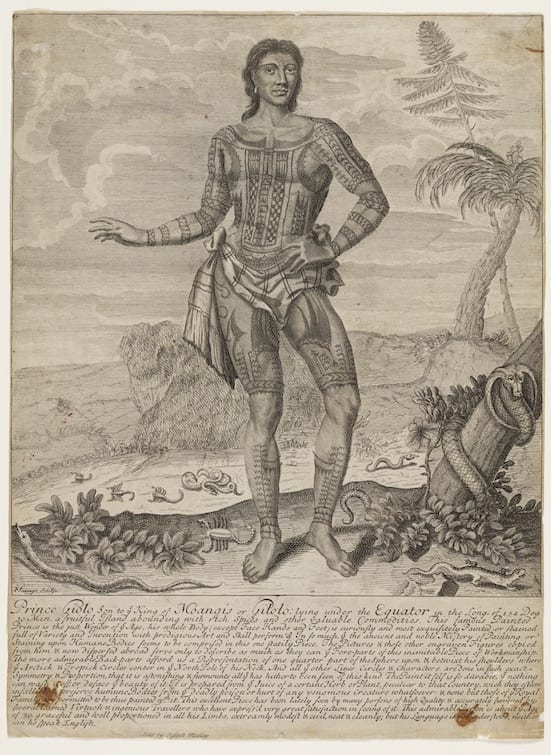
Playbill advertising ‘Prince Giolo’ in London, 1692.
Etching by John Savage.
Image courtesy of the Mitchell Library, State Library of New South Wales, Australia.

Jeoly was put on display ‘as a sight’ at the Blue Boar’s Head Inn in Fleet Street in June 1692. A number of copies of the playbill advertising his public appearances survive (pictured above). The original advertisement includes a detailed etching of Jeoly by John Savage, showing the tattoos over the front of his body, arms and legs, which resemble traditional Micronesian tattoos of the Caroline and Palau Islands. [2] As well as this striking image, a somewhat embellished story of his life was printed beneath the illustration. Interestingly, this accompanying text ascribes potent protective and healing powers to Jeoly’s tattoos, claiming that his people believed them to be a defense against ‘venomous creatures’:

The Paint it self is so durable, that nothing can wash it off, or deface the beauty of it: It is prepared from the Juice of a certain Herb or Plant, peculiar to that Country, which they esteem infallible to preserve humane Bodies from the deadly poison or hurt of any venomous Creatures whatsoever.

Whilst tattooing was considered to possess magical, protective and medicinal properties in many cultures, it is more than likely that the stories claiming that Jeoly’s tattoos repelled venomous creatures were dreamed up by his exhibitors, rather than having any genuine basis in his own native belief system. Dampier himself remarked upon the ‘Romantick stories’ which circulated in England about Jeoly’s origins, openly ridiculing the marketing campaign:

In the little printed Relation that was made of him when he was shown for a Sight in England, there was a romantick Story of a beautiful Sister of his a Slave with them at Mindanao; and of the Sultan’s falling in Love with her; but they were Stories indeed. They reported also that this Paint was of such Virtue, that Serpents, and venomous Creatures would flee from him, for which reason, I suppose, they represented so many Serpents scampering about in the printed Picture that was made of him. But I never knew of any Paint of such Virtue: and as for Jeoly, I have seen him as much afraid of Snakes, Scorpions, or Centapees, as my self. [3]

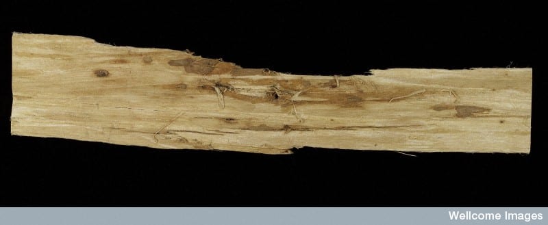
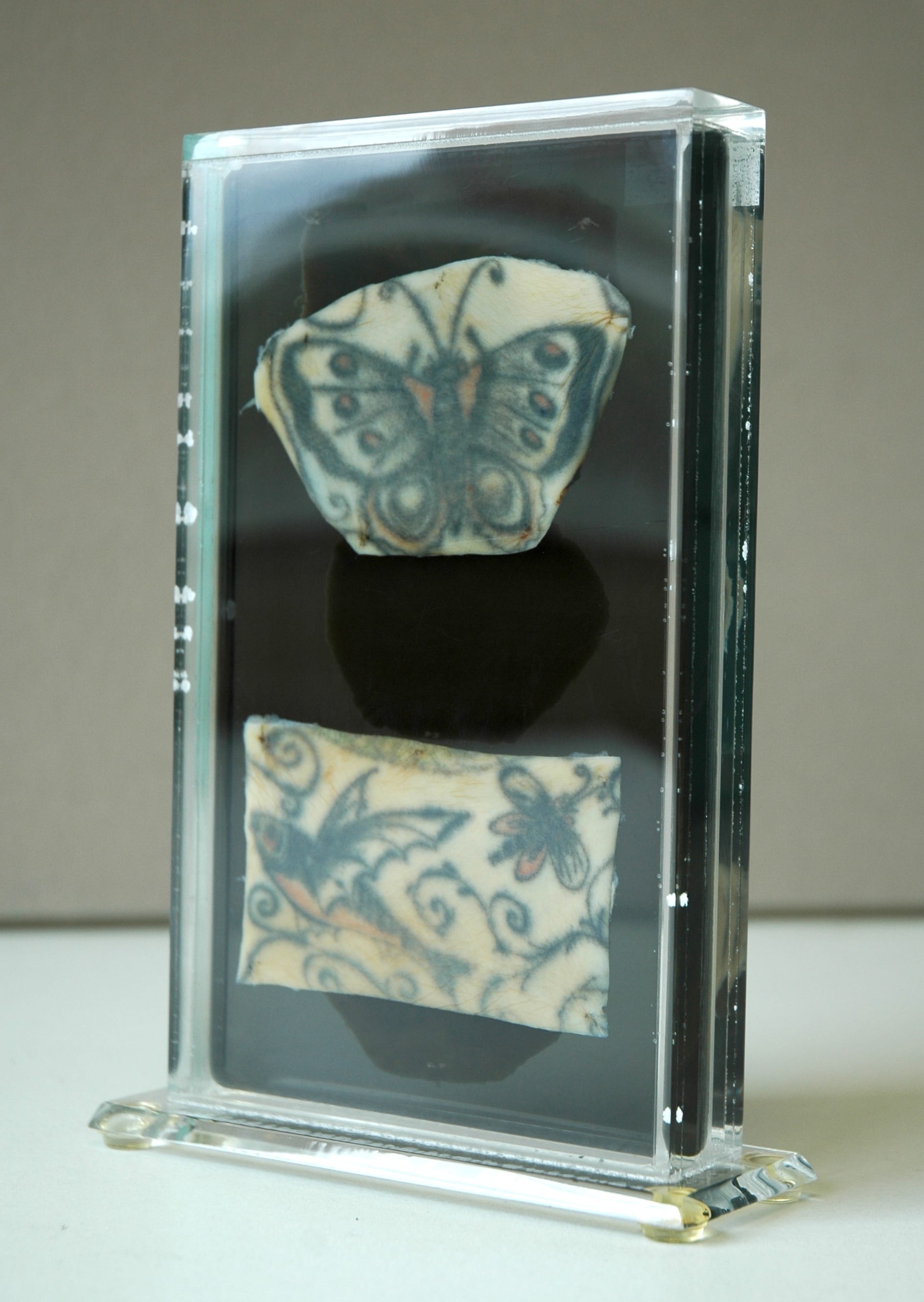
In the lower foreground of the illustration, a variety of reptiles and scorpions can be seen fleeing from Jeoly’s feet, his tattoos apparently acting as some kind of aposematic deterrent. Tragically however, Jeoly’s tattoos could not protect him from the foreign infections that he was exposed to in England; he died of smallpox in Oxford sometime in 1693. Although his grave is not marked, and his name does not appear in the Parish register, Jeoly is thought to be buried in St Ebbe’s Churchyard. After his death, a fragment of his tattooed skin was removed and preserved for the Anatomy School collections at Oxford University by the surgeon Theophilius Poynter. This skin fragment was recorded in a list of ‘Anatomical Rarities’ in the Appendix of John Pointer’s 4 volume catalogue for his Musaeum Pointerianum, the cabinet of curiosities he left to St. John’s College Oxford in 1740. [4] Although the skin did not survive, having been lost by the early 20th century, this appears to be the first documented instance of the collection and preservation of tattooed human skin as an anatomical curiosity in England.

Jeoly’s tragic story of enslavement, forced re-location to Europe, public exhibition for profit, fatal illness, and the preservation of his tattooed skin for display as an anatomical rarity, speaks of the foreign body on multiple levels. From the 16th century onwards, the tattooed body of the native became a powerful symbol of foreignness, that could reliably draw curious European crowds and turn a profit for unscrupulous entrepreneurs; but the consequences for displaced foreigners like Frobisher’s Inuits and Dampier’s ‘Painted Prince’ were grave indeed. Exposed to invisible and deadly foreign bodies such as measles and smallpox, they died far from home, unable to fight off common European illnesses against which they had no natural defences.


References:

[1] William Dampier, A New Voyage Around the World, ed. N. M. Penzer (London: Adam & Charles Black), 1937, p. 344.

[2] See Tricia Allen, “European Explorers and Marquesan Tattooing: The Wildest Island Style” in D.E. Hardy (ed) Tattootime Volume V: Art from the Heart, (1991) pp. 86-101; also Kotondo Hasebe, “The Tattooing of the Western Micronesians” in The Journal of the Anthropological Society of Tokyo Vol. XLIII No.s 483-494 (1928), pp. 129-152 (in Japanese).

[3] Dampier, A New Voyage Around the World, p.346.

[4] Geraldine Barnes “Curiosity, Wonder and William Dampier’s Painted Prince“, Journal for Early Modern Cultural Studies, Vol. 6, No. 1 (2006), p. 32 & 43.

[analytics-counter]

Mythical Hybrids and Fantastic Beasts

By Gemma Angel, on 13 May 2013

Gemma Angelby Gemma Angel


I’m going to describe a creature, and you have to try and guess what it is, based on the following three clues: 1) it lays eggs; 2) it has venomous claws; and 3) it uses electroreception to assist it in catching prey under water. You probably guessed some sort of reptile, right? Wrong. Ok, so those questions were a bit tricky. I’ll give you another three clues: 4) it’s semi-aquatic; 5) it has thick fur; and 6) despite laying eggs, it suckles its young on milk. Some of you will probably have worked out what this mysterious animal is by now. I am, of course, describing Ornithorhynchus anatinus, or as it is more commonly known, the platypus.

Growing up in Australia, I was fascinated by the native wildlife. As a curious 7-year-old recently emigrated from England, I tried to assimilate the unfamiliar Antipodean fauna into my limited understanding of the animal kingdom, largely through approximations: To me, the wombat was like a kind of stout, snub-nosed badger; sugar gliders were reminiscent of squirrels; and the echidna was a larger and longer-nosed version of the hedgehog. Kangaroos were a more difficult species to accommodate, with a face similar to a deer, and the hind legs of some sort of giant Alice-in-Wonderlandesque rabbit. But my system completely fell down when it came to the platypus. This creature was truly weird, a kind of animal cut-and-paste that defied all of the categories that I tried to fit it into. As it turned out, I wasn’t alone in my estimations of this remarkable and unique creature.

Platypus-sketch

Ornithornhynchus anatinus, John Gould (1863).

As an Australian native, the platypus has been known in Aboriginal culture for millennia – but it was not until 1797 that Europeans first encountered them. Captain John Hunter of the Royal Navy sent a pelt and a sketch back to Britain in 1798, [1] but the bizarre appearance of the creature baffled European naturalists. Some considered it to be an elaborate hoax, and Scottish zoologist Robert Knox believed the creature to be the work of an inventive Asian taxidermist. Even George Shaw, the first man to scientifically describe the platypus, admitted that “a degree of scepticism is not only pardonable, but laudable … I almost doubt the testimony of my own eyes.” [2]

Whilst it makes perfect sense that European observers would find the platypus strange, having never encountered anything like it in the Northern hemisphere outside of the bizarre chimerical creatures of mythology, it is perhaps more surprising that Aboriginal Dreamtime legends also describe the platypus as a peculiar exception within the animal realm. Known as the ‘mallangong’, tambreet’ or ‘duliawarung’ to local indigenous peoples, Aboriginal story-telling traditions use myth to explain the unique appearance and behavioural characteristics of the platypus. The platypus was believed to be the offspring of a mother duck and a father water rat, accounting for its unusual characteristics – inheriting the duck-bill, webbed feet and egg-laying abilities of their mother, and the thick fur, claws and four legs of their father. In an origin story of the platypus from Northern New South Wales, their poor mother Gaygar is ostracized by the other ducks because of her bizarre-looking hatchlings, and is forced to leave her home on Narran Lake. She takes her babies up into the Warrumbungle mountains, thereby accounting for why platypus are only found in particular regions. In another story from the New South Wales Central Coast, the animals argue amongst themselves about who is the most important creature. They form three exclusive groups, all convinced of their superiority: The animals with fur who can run across land, the birds who lay eggs, and the water creatures who can swim. All of the groups want the platypus to join them, since he shares characteristics with all of them, and each faction invites him to be part of their group. After thinking about this for some days, the platypus gathers all the animals to tell them his decision:

I don’t have to join anyone’s group to be special because I am special in my own way. Because I have fur and love to run across the land, I have a little bit of animal in me. I also have a little bit of bird in me because of my bill and the fact that my wife lays eggs. As well, I also have a bit of water creature in me because I love to swim and explore the underwater world. […] I don’t know why the ancestors have made us all different, but we must learn to accept these differences and live with each other. [3]

All of the animals listening, including people, agreed that the platypus was very wise; and the people decided that they would not hunt the platypus because he was so special. Non-human animal hybrids of Eurasian mythology have also often been considered special, such as the Griffin, which combined features of the lion and eagle, both of which were regarded as especially regal animals.

Animal-hybrids from diverse mythological traditions demonstrate the significance of animals within human culture, playing an important role in origin stories and cosmology, as well as in defining what it is to be human. In the Aboriginal story above for instance, the strange ‘hybrid’ character of the platypus reminds us to accept and learn from our differences. To early European observers, the platypus must have seemed like the ultimate foreign creature, an almost perfect embodiment of mythical animal-assemblages such as the Chimera, a fire-breathing, androgynous, composite creature of ancient Greek legend that had the head and body of a lion, a snake for a tail and the head of a goat emerging from its back. But the platypus does not merely look like an odd melding of different species; recent scientific research has revealed that the platypus also has a very complex genetic lineage. Studies on platypus venom, which is secreted from a gland in the male’s hind legs and delivered by a ‘spur’, or hollow claw-like structure, have shown that their venom contains 80 different toxins, which share genetic similarities to poisons produced by snakes, lizards, spiders, starfish and sea anenomes, as well as containing 3 proteins that are unique to the platypus. [4] Despite these genetic similarities, this research suggests that platypus venom is an example of convergent evolution, whereby similar traits in different genetic lineages can arise independently due to similar environmental pressures. The eye, wings and fins are all examples of convergent evolution. Thus it seems that whilst the platypus appears to closely resemble a range of other species – both on the surface and genetically – it is nevertheless a uniquely adapted and very special creature indeed.

Platypus taxidermy specimen at the Grant Museum of Zoology. © The Grant Museum, UCL.

Platypus taxidermy specimen at the Grant Museum of Zoology.
Photograph © The Grant Museum, UCL.


References:

[1] Brian K. Hall, The Paradoxical Platypus in BioScience, Vol. 49 No. 3 (March 1999), p. 211. 

[2] George Shaw, The naturalist’s miscellany – Platypus Anatinus, June 1799, Vol. 10, published by Frederick P. Nodder, (London 1813/14). Available online from the Library of NSW.

[3] Helen F. McKay, Pauline E. Jones, F. Francis & June E. Barber: Gadi Mirrabooka: Australian Aboriginal Tales from the Dreaming. Libraries Unlimited (2001), pp. 57-60 & 83-85.

[4] Ewen Callaway, Poisonous Platypuses Confirm Convergent Evolution in Nature, (October 12th 2012).

[analytics-counter]

Was Helen of Troy a Natural Blonde?

By Gemma Angel, on 6 May 2013

Tzu-i Liaoby Tzu-i Liao


Petrie Museum holds an extraordinary sample of pale yellow human hair, which is attached to a patch of scalp and entangled with darker curls that are most likely hair from a wig. It is very tempting to assume that the wig was used to conceal – even in the afterlife – the natural blonde hair colour of the wearer. On discovering this artefact in a tomb at Gurob, Petrie himself wrote that “the person was light-haired and wore a wig of black, hiding the foreign token.” Petrie based his argument on traditional studies of the ancient Mediterranean world. It is very likely that the vast majority of ancient Egyptians probably did have darker, coarser hair, and blonde hair in this context would have been very unusual. The use of wigs was not an uncommon practice amongst the ancient Egyptians, as many Egyptologists of Petrie’s era recognised. Indeed, bodily features were definitely a strong point of reference in recognising foreigners, and having different hair colour was surely one of them. Consider, for example, the common epithets (nick-names) for different ethnic groups used in ancient Greek literature: the hairy-headed Achaeans (kare komoontes), Abantes (Thracians), known for their long hair (opithen komoontes), and the bright-haired (likely golden, or blonde) Menelaus (xanthos) – including Helen of Troy (described as having bostrychous xanthes komes).

The example of Helen is particularly interesting for our discussion, not only because she might have been blonde, like the owner of the black wig in Petrie Museum, but also because she may have been to ancient Egypt as well. In Euripides’ tragedy Helen, the beautiful wife of Menelaus was not abducted to Troy; instead, she was sent by a god to Egypt to avoid this terrible fate. If Petrie’s theory about the purpose of the black wig was correct, Helen, as a refugee with conspicuous blonde hair, would probably have tried to ‘hide her foreign token’ with a similar black wig.

British Museum.

Ancient Egyptian mutli-tonal wig. From the British Museum collections.


While this all sounds very intriguing to a classicist like me, there is no textual or archaeological evidence which can confirm the theory that wigs were used to conceal particular hair types denoting unfavourable ethnic features. On the contrary, as the hairstyles represented in Egyptian art are often described by Egyptologists as wigs, it appears that wigs of this kind were more likely worn more commonly for other aesthetic reasons. Another wig from ancient Egypt (Thebes) in the British Museum is a good example of this counterargument. This piece consists of hair of two colours; a lighter shade arranged into looser curls on the top, and darker long braids underneath. The two colours are presented in different styles in one hairpiece, the lighter colour appearing more prominently over the darker hair. Having lighter hair colour does not seem to be too much of a concern here. The important thing is to have the style on display – or even, to distinguish the special status of the wearer. It is less likely that our blonde woman wore the black wig to make herself look just like everyone else. This wig was probably worn on special occasions, or to denote her high social status. J. Stevens Cox even suggests that the prevalent use of wigs as fashion and status markers in the Roman empire was in fact a result of contact with Egypt, where it was already a popular practice.[1]

The reason that Petrie so quickly assumed that the black wig was worn for purposes of concealment of ethnic identity, probably owes more to his interest in eugenics. Since the time of Aristotle, many people have made often spurious connections between appearance and character. In the era of eugenics, bodily features supposedly denoting psychological or moral character were not only categorised but also ranked in a hierarchy. Petrie’s interest in these theories are apparent in his archaeological work as well as in his writing; he extensively documented Egyptian monuments that displayed “racial types”; he collected glass and terracotta figurines or heads that depict ethnic phenotypes and labeled them somewhat arbitrarily (a selection of these are presented in our recent Foreign Bodies exhibition). As late as 1934, Petrie sent skulls of “racial types” to Francis Galton, the father of eugenics, for further research.[2] In the preface of his Janus in Modern Life [3], Petrie clarifies the purpose of such studies, describing them as “physiological research of the obscure causes of [present] troubles” (to use a medical metaphor), since “the present time seems to most people so infinitely more important…than the past or future,” and “[t]hey forget that it is only a fiction to speak of the present…and every such present has been entirely conditioned by its past…” Looking at his work retrospectively, it is clear that much of his own reading of the past was actually largely conditioned by his present. He made some of the very mistakes he set out to help the world to avoid.


References:

[1] J. Stevens Cox, “The construction of an Ancient Egyptian wig (c. 1400 B.C.) in the British Museum”, in Journal of Egyptian Archaeology Vol. 5 No. 63 (1977).

[2] For further information, please see the website of Petrie Museum exhibition Typecast.

[3] W. M. F. Petrie, Janus of Modern Life. G.P. Putnams’ Sons (1907).

So Comfortable You Can’t Even Feel It! The Cocaine Tampon

By Gemma Angel, on 29 April 2013

Lisa Plotkin  by Lisa Plotkin


Last May, a Utah woman was in for a surprise when she purchased a $1.99 box of tampons from a local store in Salt Lake City. Instead of a cotton tampon inside the applicator, the woman discovered something else with a much steeper price tag: cocaine. At first she was completely astonished and didn’t realize it was cocaine – she thought that the cotton might have somehow disintegrated; so unlikely was the pairing of cocaine and tampons to her. Similarly, when the police were called in to collect the cocaine, they too expressed their surprise at this method of transporting drugs. Detective Carlie Wiechman, spokesperson for the Salt Lake City Police Department, said this in response to the crime: “It’s not every day we run across this. We run across different ways of packaging and distributing, but it never ceases to amaze us the different and creative ways of trying to move drugs around.”

However, the marriage of cocaine and tampons is not as farfetched or creative as the Salt Lake City PD imagined, and for 19th century surgeons and gynecologists it was a regular – dare I say it, ‘everyday’ – medical sight. Throughout the 19th and early 20th centuries, cocaine was regularly used as a local anesthetic in surgery. It was often administered in the form of what doctors referred to as a tampon – a medical device invented in the 18th century primarily as an antiseptic to clog up bullet wounds. The tampon was traditionally soaked in whatever antiseptic or anesthetic drug was in general use, before being applied to a wound. These tampons were not particularly associated with women; at least not until later in the 19th century that is, when cocaine came to be  regarded as an especially effective treatment for gynecological diseases.


The medical tampon.

A: Kite-tail tampon; small wads of cotton tied together
on one string with a fairly large tampon on the end.
B: Ordinary rolled tampon.
Image from Practical Clinical Gynecology in:
American Journal of Surgery, vol. 39, issue 1 (1938).

Cocaine was believed effective against a whole range of women’s ailments: From painful intercourse; to uterine diseases; to cervical endometritis; to inflammation of the urethra; to dysmenorrhea – the list goes on and on. [1] In fact, cocaine was even believed to assure a ‘painless childbirth’ and according to Physician to the British Lying-in Hospital, John Philipps, could even cure the scourge of ‘sore nipples’. [2].

How would the cocaine be applied in these situations? A typical gynecological answer: by vaginally “inserting a tampon soaked in a freshly prepared solution of 2 % cocaine through a narrow Ferguson’s speculum.” [3]

Therefore, with regards to many women’s diseases, the question was not should cocaine be used- but how much. This was common until the interwar period. Of course, accidents do happen and sometimes these tampons were never removed, (most were). To read more about other ‘accidental’ foreign bodies left behind in women’s bodies, read my previous blog post here. Although many women were on board with the idea of being treated with cocaine, some did however refuse. So, the Utah woman who recently discovered cocaine in her tampon carton was by no means the first to say ‘thanks, but no thanks’ to tampons with coke on the side!

Learn more about our current UCL exhibition on all kinds of foreign bodies see.

References:

[1] Stephen R. Kandall: Women and Addiction in the United States (Cambridge, MA: Harvard University Press, 1996).

[2] John Philipps: ‘The Value of Cocaine in Obstetrics’, in The Lancet (26 November 1887), p. 1061

[3] ‘A Note on the Morphine-Hyoscine Method of Painless Childbirth’, in British Medical Journal (6 January 1917).


Extraordinary Eaters: Swallowing Foreign Objects for a Living

By Gemma Angel, on 22 April 2013

Sarah Chaneyby Sarah Chaney


In 1935, Dr Isaac Lloyd Johnstone decided to publish a “case of unusual surgical and psychological interest” in the British Medical Journal. This concerned a patient he had encountered over a decade before, while he was a surgical dresser at the Middlesex Hospital. This man was operated on for the removal of several nails and, as Lloyd Johnstone took the patient’s medical history, he discovered that his abdomen was “a mass of scars”; the patient described having had more than a dozen similar operations in the past. Before leaving the hospital, the patient had handed the dresser his memoirs, intriguingly titled “Things I have swallowed since 1905”.[1]

Foreign bodies removed from the stomach of a 26-year-old woman in 1915, The Museums at the Royal College of Surgeons.

Foreign bodies removed from the stomach of a 26-year-old woman in 1915, The Museums at the Royal College of Surgeons.

The patient’s account described around a dozen operations for the removal of items, predominantly nails, screws and cutlery, but also hairpins, safety pins and, once, a tin whistle. According to Lloyd Johnstone, his informant would make money from his ability to swallow unusual items, by showing a group of objects to his companions in various public houses, and taking wagers against his ability to swallow them. The patient stated that he had always been able to pass objects up to two and a half inches in length, and Johnstone considered that larger objects found their way into the patient’s abdomen when “a drink or two and an intimate knowledge of the hospitals of London made him reckless”.

Indeed, the wagers did not necessarily end with the initial swallowing. In 1912, the patient reported having wound up in Guy’s Hospital after swallowing a 6 ½ inch nail, which took three operations to remove. In his own words:

Before being operated on there was a bet between the Night Nurse and the Student as were the 6 ½ inch lied, one said it was in the transfered Coln, and the Night Nurse said it layed in the Coln, to make sure Mr. John Dunn had me X Rayed and Skiagraphed and the Skiagraph showed that it was in the Coln, and then I was operated on straightaway. The Night Nurse won the Bet which was £5 0, 0 which my Dresser Mr. Taylor had to pay up. [sic]

In Mr XYZ’s account (as Lloyd Johnstone called him), the bet is emphasised over and above his own recovery, indicating the importance he laid on this aspect of his swallowing. The patient remained proud of his abilities, despite the painful and dangerous nature of his career. On at least one occasion, his actions had been thought fatal by hospital staff (after “6 Larg Safty Pins and 5 Ladies Hair Pins … they gave me up for Dead”), yet his account ends proudly with the words “I defy contradiction”. His composure and purpose, as Lloyd Johnstone noted, made XYZ very different from the “usual” hysterical or suicidal cases of foreign body ingestion.

Yet swallowing objects for money or notoriety has a lengthy history, bound up in the notions of performance and risk covered in a previous blog post on sword swallowing. Historian Emma Spary has researched the connections between the medical profession and the swallowers often referred to as “extraordinary eaters”. Her recent book – Eating the Enlightenment – includes a chapter on the involvement of the medical profession in cases of the consumption of non-nutritive items in 18th century Paris.[2]

The contents of the stomach of a knife eater, Gordon Museum (King’s College London)

The contents of the stomach of a knife eater, Gordon Museum (King’s College London)

One such case which features in a London museum is the “contents of the stomach of a knife eater”, housed in the Gordon Museum. This collection of rusty blades, buttons and medallions was removed by surgeons at Guy’s Hospital from the stomach of an American seaman named Cummings, known to the British medical profession from 1799. This unfortunate individual (much like the sword swallower in the UCL collections) ended up being dissected by surgeons a decade later. Like Mr XYZ, Cummings reminds us that the swallowing of foreign items is not necessarily an irrational pursuit, and might be carried out for a wide variety of reasons.

You can watch a video of Dr Spary discussing “extraordinary eaters” on the Damaging the Body website here.


References:

[1] I. Lloyd Johnstone: “Swallowing Foreign Bodies for a Livelihood” British Medical Journal, 21 Sept 1935, p. 546.

[2] Emma Spary: Eating the Enlightenment: French Food and the Sciences, 1670-1760, Chicago: University of Chicago Press, (2012).

I have sung and praised the sun disc, I have joined the baboons

By Gemma Angel, on 15 April 2013

Suzanne Harvey #2  by Suzanne Harvey


What links the evolution of language to the collection of baboon figurines at the Petrie Museum of Egyptology? I have previously speculated on the reasons why Ancient Egyptians might create figures of baboons performing acrobatics, playing the harp and even drinking beer. After months of sporadic research and conversations with museum visitors on the subject, I have finally chosen a favourite theory (without a hint of bias) that just happens to link directly to my own research on baboon communication.

Monkey with beer potThis post was inspired by an essay entitled Some Remarks on the Mysterious Language of the Baboons, [1] which mentions this quote from the Egyptian Book of the Dead, Chapter 100:

I have sung and praised the sun disc, I have joined the baboons.

The reason that Egyptians considered baboons to be sacred is actually quite straightforward. When baboons wake in the morning, like many primates (humans included), they tend to stretch and produce vocalisations. To some, the pose baboons adopt while stretching – sometimes raising their front legs in the air – resembles worship. As they stretch more often at sunrise, this action together with their ‘chattering’ noises when moving from sleeping sites, was interpreted as singing and dancing to praise the Sun-god, Ra. [2]


Caption for baboon chatterThis only explains the role of language in making baboons sacred. Of several Gods to whom they are sacred, the deity who links baboons unequivocally with language is Thoth. Thoth is often depicted as a baboon scribe who not only spoke and wrote, but who actually gave the gift of language to the Egyptians, rather than simply understanding it. [3]

The voice of the baboon is the voice of God

This title might seem a somewhat unusual interpretation of the famous vox populi, vox Dei maxim, but it is in fact the Ancient Egyptian variation on this theme. Their belief was that whoever understood the language of the baboons had access to religious knowledge that was usually hidden. This is very good news indeed for modern primatologists – though I’ve yet to decipher any religious revelations while analysing baboon vocalisations! I can however dispute the Greek author Aelianus’s assertion that baboon language is “utterly incomprehensible to ordinary human beings”. [4]

ThothThoth’s significance in language and wisdom suggests that my earlier supposition – that baboons playing harps and drinking beer was not linked to religion due to the absence of sober, worshipful poses – was in fact erroneous. It seems that Egyptians were motivated to experiment with baboons, trying to train them to perform feats such as playing the harp, to reveal the link to Thoth hidden within them.

A range of baboon statuettes are currently on display as part of the Foreign Bodies exhibition in UCL’s North Cloisters. They represent a unique interpretation of other species that are nevertheless similar to our own, and a fascinating insight into how a distant culture defined themselves in relation to other primates – believing themselves to be inferior to baboons in terms of both holiness and wisdom. Ancient Egyptians recognised the human-like intelligence, ability to communicate and dexterity of baboons that we are equally fascinated by today, albeit from an evolutionary science perspective, rather than a religious sensibility. The quest to discover the inner Thoth continues…


References:

[1] H. Te Velde: ‘Some Remarks on the Mysterious Language of the Baboons,‘ in Kamstra, J. H., Milde, H. & Wagtendonk, K. (eds). Funerary Symbols and Religion. (1988), J.H. Kok: Kampen.

[2] G. Pinch: Egyptian Mythology: A Guide to the Gods, Goddesses, and Traditions of Ancient Egypt. (2004), Oxford: Oxford University Press.

[3] Hans Kummer:  In Quest of the Sacred Baboon. (1995), Chichester: Princeton University Press.

[4] H. Te Velde, (1988), p.134.

Of Foetuses & Fibroids: the Accidental Foreign Body

By Gemma Angel, on 8 April 2013

Lisa Plotkinby Lisa Plotkin


As our current exhibition in UCL’s north cloisters demonstrates, “foreign bodies” may take many forms, as well as being continually redefined throughout history. Putting it simply, the term “foreign body” in medicine usually refers to an external object introduced into the body that isn’t supposed to be there. As my colleague Dr. Sarah Chaney notes in her recent blog post, some of the most common foreign objects uncovered from the bodies of 19th and early 20th century patients were coins, safety pins, buttons and needles. These objects could enter the body accidentally or with purpose. Medical instruments or tools, for example, were on occasion accidentally lost inside the patient’s body during an operation. For those Seinfeld fans out there, think back to the “junior mint” episode. However, it was neither pins, mints or instruments which were the subject of a 1939 article in the British Medical Journal devoted entirely to foreign bodies. Rather, Dr. A. H. Charles, obstetric registrar at St. George’s Hospital, zeroed in on one particular foreign body that was with some frequency discovered in the female bladder: slippery elm bark. It may surprise some readers to discover (as it certainly did Dr. Charles), that slippery elm bark was commonly used as an abortifacient. In fact, it is still used by women to induce abortion today.[1] Writing on elm bark as a foreign body inside the bladder, Dr. Charles observed:

Five cases in which a piece of elm bark was used have been reported in detail previously, and in all of these the body has remained undiscovered for some time, until its removal suprapubically after calculus formation had taken place, causing symptoms leading to its discovery. Why the bark of this noble tree should be so popular is difficult to understand.[2]

Called “slippery” elm because when it gets wet it becomes slippery, this type of elm bark has traditionally been used to cause early uterine contractions and induce labor. The bark is inserted into the cervix where it then absorbs water and expands, dilating the cervix and triggering contractions. Needless to say, this procedure was not always successful and could cause life-threatening infections. Occasionally the bark could end up in the bladder by mistake, which is what Dr. Charles had observed. For many of the women who mistakenly inserted the slippery elm into their bladders, there the bark most likely stayed, unless a severe medical problem compelled them to seek medical attention. No doubt for many of the women attempting to self-abort, their experience with slippery elm was less than satisfactory and could have proven fatal.

Slippery Elm Bark, sold as an abortifacient

Slippery Elm Bark, sold as an abortifacient.

Accidental or intentional abortion occurred with a lot greater frequency in the 19th and early 20th centuries than some might imagine, and was inextricably wrapped up with both the idea and concrete reality of “foreign bodies.” For many, the coat hanger is the ultimate symbol of a foreign body inserted into the uterus to cause abortion. Still others may think of obstetric forceps as the foreign body which has caused thousands of fetal deaths during delivery. But what about uterine fibroids? How many abortions have they caused, and can they be regarded as foreign bodies if they are naturally occurring? A uterine fibroid is a benign tumour of the uterus, commonly found in women of reproductive age. Most fibroids are asymptomatic and therefore, most women are never aware that they even have one. However, on occasion these fibroids can cause health complications or interfere with pregnancy. Before the advances of late 19th century abdominal surgery and gynecology, uterine fibroids were not treatable. However, by 1916, obstetric surgeon Sir. John Bland-Sutton was able to boast that “uterine fibroids are common tumours; so common and troublesome that I have removed the uterus in 2,000 women.”[3]

From his experience performing hysterectomy on thousands of women a theme emerges: uterine fibroids closely mimic pregnancy in a variety of ways, and it is difficult – sometimes impossible – to distinguish between the two. Writing of this unfortunate similarity in 1913 Bland-Sutton observed, “A large sub-mucous fibroid produces similar changes in the uterus to those set up by the growth of the fetus […] Women with large sub-mucous fibroids are more or less in a condition resembling chronic pregnancy.”[4] What this similarity meant – and what gynecologists and obstetricians of the time openly acknowledged – was that sometimes a hysterectomy was performed to remove a fibroid that either never existed in the first place, or was also sitting alongside a feotus, masking a pregnancy. Either way, an abortion was performed.

The photograph below demonstrates the reality of such surgeries. This particular specimen belongs to UCL Pathology Collections, and is currently on display in the Foreign Bodies exhibition. The anonymous woman patient underwent a hysterectomy most likely sometime in the early 20th century, in order to remove the sizable uterine fibroid, which can be seen on the right side of the image. However, on closer examination of the image, we see on the left side a preserved feotus, frozen in development, somewhere between 8-11 weeks.  It is unclear whether this woman or her doctor even knew she was pregnant.

Feotus in uterus, with large fibroid tumour. UCL Pathology Collections. Photograph Gemma Angel.

Feotus in utero, with large fibroid tumour.
UCL Pathology Collections. Photograph Gemma Angel.

Such examples abound in medical literature, and Victorian and Edwardian gynaecologists, obstetricians, and surgeons spoke of them with little or no censure. It was all a part of the surgical trial and error that they were practicing. The feotus was sometimes viewed as a necessary casualty in removing a potentially life-threatening fibroid. Either way, be it by slippery elm, by accident, or with purposeful intent, the feotus was removed as a foreign body, like any other. By examining the medical establishment’s attitudes towards fibroid removal we catch a glimpse into one way the feotus, and the experience of pregnancy in general, was understood in the past.


References:

[1] David A Grimes, Janie Benson, Susheela Singh, Mariana Romero, Bela Ganatra, Friday E Okonofua, Iqbal H Shah. “Unsafe abortion: the preventable pandemic.” The Lancet Sexual and Reproductive Health Series, October 2006.

[2] British Medical Journal, 29 July 1939.

[3] Sir John Bland-Sutton, “A Clinical Lecture on 200 Consecutive Hysterectomies for Fibroids Attended With Recovery” reprinted British Medical Journal, 4 July 1916.

[4] Sir John Bland-Sutton, “The Visceral Complications Met With Hysterectomy for Fibroids and the Best Methods for Dealing With Them” British Medical Journal, 1 November 1913.

Painted Skins & Butterfly Wings

By Gemma Angel, on 1 April 2013

Gemma Angelby Gemma Angel


When I first began my doctoral research into tattoo preservation three and a half years ago, I assumed that tattoo collections such as those held by the Science Museum in London were rare. Whilst collections of inked human skin are most definitely unusual, I was soon surprised and intrigued to discover that such objects exist in almost every museum archive, university anatomy department, or pathology collection that I have visited over the course of my research. The largest collections, of which the Wellcome Collection is the major exemplar, are dry-preserved and date from the 19th century – similar collections can be found across Europe, and the MNHN in Paris has a collection of 56 tattoos which are very similar to those in the Wellcome collection. Historically, these collections may be medical, anthropological, or criminological in origin.

In anatomy and pathology collections, tattoo specimens tend to be wet-preserved and date more recently, usually from the early part of the 20th century anywhere up to around the 1980s. But why are these objects preserved in anatomy and pathology departments at all? There is of course nothing pathological about tattooed skin in itself – so it seems strange that specimens like the one pictured below are displayed alongside other pathological skin specimens such as cutaneous anthrax, fibromas, keloids and glanders. What, if anything, can be learned from these tattoos in medical terms? Or are these striking collections of decorated human skin merely objects of curiosity? Often, the simple answer is that they are a little bit of both…

Tattooed human skin specimen. UCL Pathology Collections. Photograph © Gemma Angel.

Tattooed human skin specimen. UCL Pathology Collections.
Photograph © Gemma Angel.

The collection of tattoos pictured here are a case in point. These particular tattoos belonged to one individual, whose very brief case notes have been recorded and retained along with the specimen in UCL Pathology Collections. The notes provide an intriguing glimpse into the life of the individual to whom the tattoos belonged, as well as revealing something of the clinical interests and collecting practices of the doctor who preserved them:

From a man aged 79 years who had earned his living for many years as the Tattooed Man in a circus. His entire body, except for the head and neck, hands and soles of his feet, was covered with elaborate tattoo designs. He died of peritonitis due to a perforation of an anastomatic ulcer … In tattooing, fine particles of pigment are introduced through the skin, taken up by histiocytes and become lodged in the tissue spaces of the dermis. Pigment also passes to the regional lymph glands via the lymphatics. In this case, all the superficial lymph nodes were heavily pigmented.

It is clear from these brief comments that the nature and extent of this man’s tattoos were indeed of anatomical interest to the medical practitioner: The tattooed man had been so extensively tattooed that gradual migration of ink particles resulted in the collection of pigment in the lymph glands. This demonstrates that although tattoo ink is trapped permanently under the skin following healing, it does actually travel within the body over time, filtering into the body’s tissue drainage system, and collecting in the lymph glands. Whilst this is certainly an interesting anatomical observation, it is not the pigmented lymph glands that the doctor has chosen to preserve, but rather the tattooed skin itself. Without these accompanying case notes, we would never have known that this man’s tattoos had exerted any effect on another of the body’s organs and systems at all.

Reverse panel of tattooed human skin specimen Z6,  showing tattoos of a butterfly and a flying fish.UCL Pathology Collections.Photograph © Gemma Angel.

Reverse panel of tattooed human skin
specimen Z6, showing tattoos of a
butterfly and a flying fish.
UCL Pathology Collections.
Photograph © Gemma Angel.

It would be equally impossible to know whether or not these were the only tattoos he possessed – or indeed, if they all necessarily belonged to the same person. There are strong stylistic similarities between the butterfly motifs, suggesting the work of a single tattooist, or perhaps that the individual motifs were part of a larger design. But just how large or complex the design may have been, we certainly cannot tell just by looking at these 5 small tattoos. We know that they belonged to a 79-year-old man, who made his living as a Tattooed Man, only because the doctor tells us so. He or she also tells us that his body was covered in tattoos – yet only 5 small pieces have been preserved. Five carefully selected motifs, chosen by the doctor from an already complete collection, which provided the livelihood and told the life story of one unnamed man. What selection criteria did the pathologist adopt when deciding which tattoos to preserve, and which to consign to the grave? The manner in which the specimens have been excised and mounted are strikingly reminiscent of a lepidopterist‘s collection of butterflies – could this reflect the personal collecting interests of the pathologist, or perhaps even the Tattooed Man himself? Both the pathologist and Tattooed Man alike chose these butterflies – did they also share a passion for lepidoptery?

Many people will be familiar with the kind of insect specimen displays that are a staple of natural history collections – the old 19th century museum cases containing neat rows of pinned and mounted moths and butterflies, neatly organised according to subspecies and morphological characteristics. The tattooed butterflies share some remarkable similarities with these entomology collections; they are arranged one above another, and “pinned” to a support with small surgical stitches. Unusually for specimens found in pathology collections, this support is a slightly translucent black. This appears to be a deliberate choice on the part of the pathologist – the black perspex provides a contrasting ground for the display of tattoos on opposite sides of the vitrine, such that they do not visually detract from one another. These aesthetic choices suggest a nuanced interest in the collection and display of these specimens, which goes far beyond a straightforward medical interest in the anatomy of the tattoo. From the limited case notes and analysis of the specimen itself, we can learn something about the pathologist’s interest in the tattoo, but we are still no closer to being able to answer the fundamental question – why collect tattoos at all?

Butterfly display at UCL's Grant Museum. Photograph © UCL, Grant Museum of Zoology.

Lepidoptera display at UCL’s Grant Museum. Photograph © UCL, Grant Museum of Zoology.

There is no part of the body able to register the history of a life lived so much as the skin: wrinkles, scars and lines all map out our lives on the surface of our bodies as we age. The tattoo reinforces this unique capacity of the skin to record the traces of our experience, in the conscious act of permanently inscribing memory in skin. The pathologist, uniquely acquainted with death by virtue of their specialism, is perhaps best positioned amongst medical professionals to appreciate the peculiar relationship of the tattoo with mortality. It is a trace of the subjectivity of the deceased that is capable of outliving them, akin to a photograph or written memoir. From this point of view, it no longer seems surprising that tattoos are so often found in pathology collections; perhaps the pathologist who collected the Tattooed Man’s butterflies simply wished to preserve a small part of a colourful and remarkable life.


[analytics-counter]

Diagnosing Foreign Bodies

By Gemma Angel, on 25 March 2013

Sarah Chaneyby Sarah Chaney


One of the most important diagnostic tools to assist in foreign body removal was the development of the x-ray. In 1895, Wilhelm Röntgen, a German physics professor, developed the x-ray photograph, which enabled the interior of the body to be made visible using electromagnetic radiation.[1]  In the late 1890s and early 1900s, medical reports on foreign bodies frequently focused on the use of the “Röntgen Rays” or “skiagraphs” (as x-rays were then widely called) to locate such objects. A few weeks after Röntgen published the first X-ray photograph, Norman Collie at UCL made his own x-ray tube in order to locate a broken needle in the thumb of a female patient.

World’s first diagnostic x-ray, by Norman Collie at UCL. UCL Special Collections, on display in the Octagon Gallery until 30 April: http://www.ucl.ac.uk/museums/whats-on

World’s first diagnostic x-ray, by Norman Collie at UCL.
UCL Special Collections, on display in the Octagon Gallery
until 30 April: http://www.ucl.ac.uk/museums/whats-on


Many of the foreign bodies case histories in hospital records of the period focus on the use of this new diagnostic technique. However, x-rays could also lead to tension between patients and clinicians, when the photographs contradicted stories told by patients. One German publication of 1899 reported a soldier’s claim to have been bitten by a horse as “malingering” (a serious military crime) when broken needles were found in the wound, suggesting that the injury was self-inflicted.[2]

Image showing self-inflicted burns in a “hysterical” patient, from John Collie’s Malingering and Feigned Sickness (1913)

Image showing self-inflicted burns in a
“hysterical” patient, from John Collie’s
Malingering and Feigned Sickness (1913)

Yet, surprisingly (given the widespread publicity given to so-called malingering in civilian populations in the decades preceding the National Insurance Act of 1911), these stories seem to have been of less interest to many surgeons than the diagnostic procedure itself. At the Royal London Hospital in 1898, for example, little interest was shown in the fact that the x-ray photographs of 38-year-old domestic servant Elizabeth Quaife did not tally with the history she gave.[3] Elizabeth claimed that she had suffered pain in the knee joint ever since a long hat pin ran into her leg while she was sweeping under a bed: in hospital, however, five separate needles were discovered in the joint. Unlike in published cases, the surgeon made no reference to the potential use of x-ray imagery to detect fraud, but instead used the case to evaluate the usefulness of the technique itself. This, it was thought, had been successful in locating and removing four needles but “the fifth needle shewed by the skiagraph was … not found. It is probable that the figure shewed in the skiagraph was due to a shadow of the other needles. This, once more, shews that the skiagraph may be deceptive.”

This emphasis on diagnosis and removal certainly tallies with the lack of interest surgeons tended to show in the cause of foreign bodies. Foreign Body in this period was a diagnosis, not an exploration of a patient’s state of mind. When Rachel Taylor was admitted to the Royal London in 1900 – after swallowing a pin and a tin tack – and again in 1906 having swallowed two nails “the night before last”, it was not noted whether either instance was accidental or intentional.[4] Despite published concern over the potential abuse of charitable treatment, in practice this does not seem to have been a significant issue for either surgeons or physicians at the Royal London Hospital: cases of “artefact injury” were treated without question whether or not the patient paid for their treatment.


References:

[1] Lisa Cartwight, Screening the Body: Tracing Medicine’s Visual Culture (Minneapolis: University of Minnesota Press, 1995)

[2] “Self-Inflicted Injuries Diagnosed by the Roentgen Rays,” The Lancet, 153, no. 3955 (1899): 1668.

[3] Elizabeth Quaife, RLHA Microfilm Case Records (Surgical), DEAN F1898, pt. no. 29 & 511.

[4] Rachel Taylor, RLHA Microfilm Case Records (Surgical), TAY F1900, pt. no. 2326 & FENWICK 1906, pt. no. 906.


From the Forests of Punt to the Deserts of Saqqara: Life and Death as a Sacred Monkey

By Gemma Angel, on 18 March 2013

Suzanne Harvey #2  by Suzanne Harvey


Given the wealth of figurines, statues, engravings and even mummies of baboons found in Egypt, it may seem odd that a baboon skull features as an object in our current Foreign Bodies exhibition in UCL’s North Cloisters.

Baboon SkullThe key to this puzzle is that baboons are not, and never have been, indigenous to the areas of Egypt in which their remains have been found. They were imported from Nubia and the mysterious Land of Punt for use at temples and burial sites, where their habit of stretching and ‘chattering’ was viewed as worship of the Sun God, Ra. Since these animals were sacred to Gods of wisdom and the underworld, themselves deified in the form of Babi and The Great White One, and imported at great cost – surely their lives in Egypt would be ones of luxury?

Life and death in a foreign land

The largest number of mummified baboons have been found at the tombs of Saqqarah, an arid desert environment that contrasts starkly with the natural forest and savannah habitat of baboons. In their natural environment, baboons spend most of their waking hours foraging for food in the form of leaves, seeds, fruit and insects, none of which would be possible in the desert. In fact, the environment of the temples and burial sites where baboons were kept, was so foreign to these species that most died from malnutrition, vitamin deficiencies, and fractured bones. Of around 200 mummies analysed, few had lived beyond 6-10 years, despite the natural lifespan of the sacred Hamadryas baboon being around 30 years.

Baboon environment


From a primatological point of view, this highlights just how unsustainable desert colonies of baboons were. First breeding usually occurs between 5 and 7 years – with the majority of adults dying so young, it’s unlikely that much successful breeding took place at the temples. Recent studies at UCL’s Gashaka Primate Project have shown that diet has a strong effect on reproduction in baboons, with age of menarche, infant mortality and interbirth intervals all highly dependent upon nutrition.[1] In order to sustain a desert population of baboons, the Ancient Egyptians would have required constant imports of new animals, making baboons very rare and expensive offerings to the Gods.

The lost baboons of the Petrie Museum

Baboon mummy

In 2010, oxygen isotope analyses were carried out on hairs from one of the British Museum’s baboon mummies, and researchers were able to locate the Land of Punt, by comparing markers in the ancient baboon to modern samples. 3000 years after the baboon was mummified, his homeland was located as modern day Eritreia and Ethiopia, where baboons remain today.

The baboons at the Petrie Museum date from a later period than those at the British museum, and documentation indications that they were were mummified after voyages to Punt ceased. So for now, all we can really say for certain about the Petrie Museum baboons is that they were a long way from home when they died…


References

[1] J.P. Higham, Y. Warren, J. Adanu, B.N. Umaru, A.M. MacLarnon, V. Sommer, & C. Ross: (2009). ‘Living on the edge: Life-history of olive baboons at Gashaka-Gumti National Park, Nigeria’, in American Journal of Primatology, Vol. 71 (2009), pp.293-304.