X Close

Researchers in Museums

Home

Engaging the public with research & collections

Menu

Should human remains be displayed in museums?

By Julia R Deathridge, on 28 March 2017

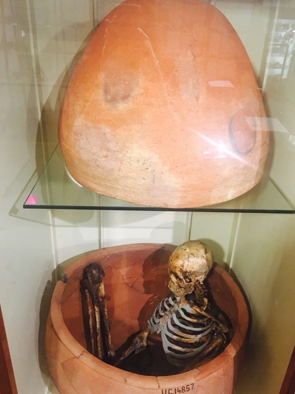
If you have ever visited the Petrie Museum you are unlikely to have missed the man buried in a pot. In the past the pragmatic scientist in me had just regarded this as a skeleton in a pot; spending most of my time studying it to see how many of the different human bones I could still name from my undergraduate anatomy class (not that many it turns out!). However, a group of visiting American college students made me think about it differently. They were discussing the use of human remains in museum collections, their purpose, and the importance of displaying them respectfully. This opened up an interesting debate: how should human remains be displayed and should they even be displayed at all?

Pot burial from Hemamieh, near the village of Badari UC14856-8

Pot burial from Hemamieh, near the village of Badari. UC14856-8

In the past human remains were regularly collected from excavation sites and displayed in museum cases with little thought put into the person that they once were. However, feelings towards the use of human remains in the UK have begun to change in recent years. In 2005 the Department for Culture, Media and Sport (DCMS) released a “Guidance for the Care of Human Remains in Museums” outlining a code of practice for the handling and displaying of human remains. Consequently, human remains have been given a unique status within collections and are no longer treated as objects.

Repatriation of human remains has also become more common with many indigenous communities requesting the return of their ancestors. Since 1996 both the UK and Australian governments have been committed to the return of indigenous remains to Australia and New Zealand. Over 1000 remains have been returned including 13 skulls and one limb, which were repatriated from UCL in 2007.

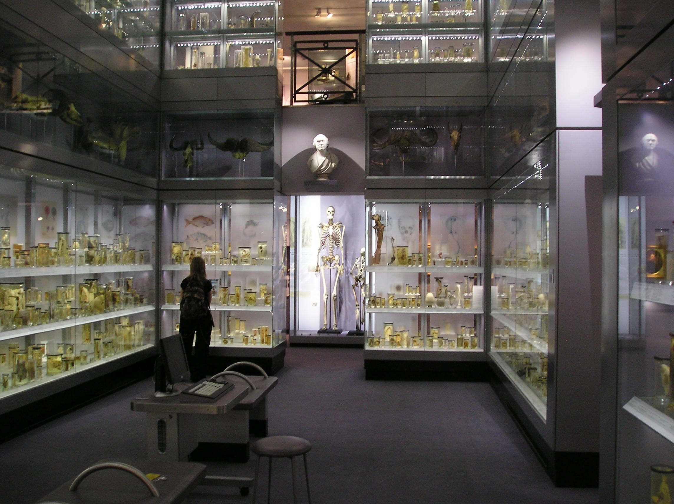
A display that has caused a large amount of controversy is the skeleton of “The Irish Giant” Charles Byrne. To avoid being dissected and used for medical research, Charles Byrne requested to be buried at sea. His request was ignored and his skeleton currently resides in a cabinet at the Huntarian Museum. Many have advocated for the return of his bones from the Huntarian allowing his burial wishes to be fulfilled. The Huntarian Museum, however, claim there is no direct evidence of his burial wishes and the educational benefits he provides to living and future generations of visitors is of greater value.

This brings up another important question: Does the educational benefit of human remains outweigh the rights of the dead? Human remains are an important teaching tool for anthropology and archaeology and are vital to the study of medical sciences. Use of human remains in exhibitions can also greatly stimulate a learning experience, allowing a much stronger connection to the culture that is being represented. A survey by English Heritage showed that only 9% of people opposed the display of human bones in museums suggesting there is still high demand for public display of human remains in museums. However, museums must be careful to utilise human remains in an appropriate context in order to educate rather than just to attract audiences.

Charles Byrne's skeleton on display in the Huntarian Museum. Photo credit: CC image courtesy of Paul Dean

Charles Byrne’s skeleton on display in the Huntarian Museum. (Photo credit: CC image courtesy of Paul Dean)

Many people do not consider the ethical issues of how the dead should be displayed in a collection until they are asked: what if that was your grandfather or great-great grandmother? Would you still consider this respectful? However, for many of the ancient human remains collected, including the man buried in the pot, their ancestry has been lost and we cannot know how their descendants, or they themselves, would feel about how their body is being used in the name of education. Although admittedly it is hard to argue that this is what the ancient Egyptians would have wanted.

In our current legal system we rely on our family and loved ones to carry out our burial wishes. But in their absence we too would have no control over this, much like the ancient Egyptians on display. I’m not sure I would be willing for my skeleton to be used to educate future generation about the irreversible impact mobile phones had on our postures and spines (I’m imagining my skeleton hunched over my iPhone whilst scrolling through Instagram). But others might feel differently.

As attitudes towards the displaying of human remains change, museum’s policies will have to adapt. Maybe in the future forms of consent will be required, similar to signing up for organ donation. But how many people will actually be willing to donate their bodies to museums? I guess only time will tell.


Sword Swallowing & Surgical Performance

By Gemma Angel, on 11 March 2013

Sarah Chaneyby Sarah Chaney


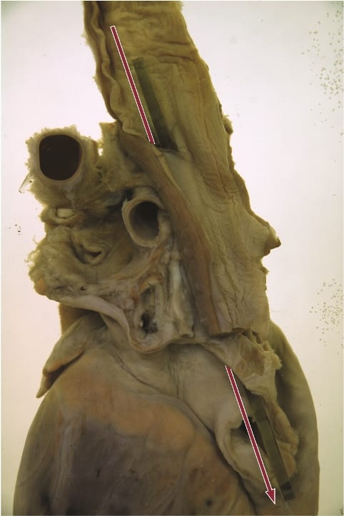
We know sadly little about the sword swallower’s sword that resides in the UCL Pathology Collection: not even how long it has been here. What we do know is that this performer was very unlucky. Perhaps he (or, indeed, she) didn’t tilt his head back far enough. Perhaps he moved during the process of insertion. Whatever the case, the sword pierced the flexible tube of the oesophagus, leading to the performer’s death. The heart and oesophagus were preserved – perhaps as a warning of the dangers of such feats – alongside the weapon that led to his demise.

Fatally ruptured oesophagus, caused by the sword swallower's sword. Photograph Gemma Angel, UCL Pathology Collections.

Fatally ruptured oesophagus, caused by the sword swallower’s sword. Photograph Gemma Angel, UCL Pathology Collections.

Sword swallowing seemingly originated in India some 4,000 years ago, but reached the western world of Ancient Greece and Rome in the first century AD. The performer tilts his or her head back, extending the neck, and learning to relax muscles that usually move involuntarily. A rigid weapon can then be passed down as far as the stomach, usually for just a few seconds, before removal. It is dangerous, certainly, but few performers suffer the fate of the individual preserved in the UCL collections. According to one recent article in the British Medical Journal, most serious incidents occur owing to distraction or attempts at exceedingly complex feats:

For example, one swallower lacerated his pharynx when trying to swallow a curved sabre, a second lacerated his oesophagus and developed pleurisy after being distracted by a misbehaving macaw on his shoulder, and a belly dancer suffered a major haemorrhage when a bystander pushed dollar bills into her belt causing three blades in her oesophagus to scissor. [1]

In many ways, sword swallowing is the opposite of the ingestion of other foreign bodies: rather than swallowing, the performer maintains absolute control over the process of consumption, taming the body’s reflexes and realigning the organs. As Mary Cappello notes in her fascinating literary biography of surgeon Chevalier Jackson (1865 – 1968), who was an expert in foreign body removal, sword swallowing was recognised by doctors as inspirational to their own techniques. Jackson took his lead from German professors Alfred Kirstein and Gustav Killian, who lectured that sword swallowing proved the possibility of passing a rigid tube into the oesophagus, in order to remove lodged objects. Jackson, who developed his own oesophagoscope in 1890, admitted that the abilities of circus performers had opened his eyes to the opportunity of removing foreign objects without dangerous surgery. He even taught his children how to “scope” themselves.[2]

In an intriguing parallel, the insertion of some foreign objects into the human body thus assisted with the removal of others. At the turn of the 20th century, the removal of foreign bodies lodged in the throat and airways frequently required an incision to be made into the trachea or oesophagus, an operation which could prove fatal. In the records of the Royal London Hospital, from 1890 to 1910, we find no mention of oesophagoscopy or bronchoscopy: instead, surgery or the probang or “coin-catcher” was the norm. This latter instrument was generally a simple hook, inserted without any kind of viewing device or illumination. The practitioner would feel blindly for the object, and either attempt to hook it out, or push it into the stomach. This might lead to numerous complications. In 1903, surgeons at the Royal London attempted to remove a halfpenny from the throat of a five-year-old boy by pushing it into the stomach. However, it was subsequently reported that the coin catcher broke off in the boy’s throat, necessitating a major operation from which the child did not survive.[3] Small wonder that, less than a decade later, Jackson declared such objects “rough, unjustifiable, brutal”.[4]

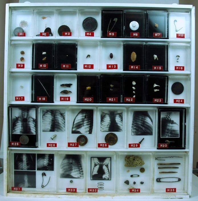
foreignbodies

UCL Pathology Collections contains many examples of foreign objects removed from the
human body: this purpose built display showcases many such objects, some with
small x-rays of the objects prior to removal.

X-ray imaging techniques aided the removal of foreign objects by instruments, and foreign body specimens are often accompanied by photographs showing the item’s location in the human body. The above set of items is found in the UCL Pathology Collection, the objects having been gathered by several surgeons in the 1920s – ‘50s. At some point, the individual boxes made for each specimen were mounted together, in a specially designed plastic surround. Fittings on the back indicate that the case was made to hang on a wall. But why? To decorate the office of a surgeon, showing off his achievements? To offer a warning to others to take care (particularly parents, for all these objects were removed from children and infants)?

Chevalier Jackson claimed that his collection of more than two thousand foreign bodies (now housed in Philadelphia’s Mutter Museum) was not a curiosity, but indicative of the everyday nature of foreign body ingestion and inspiration. Yet many of these specimens are not everyday. The two boxes of multiple objects in the bottom right, for example, were removed from the vaginas of young girls (six and eight years old respectively). The case notes do not indicate how these objects arrived in their location. Did the girls insert them themselves, or might it be a sign of sexual abuse? In her research into the medical histories of Jewish immigrants to the East End of London in the late nineteenth and early twentieth centuries, Carole Reeves came across a case of multiple foreign body insertion in a young woman, whose vagina was found to be tightly packed with pins. Reeves speculated that Leah G. might have inserted these items in an effort to ward off potential (and actual) abusers.[5]

In most instances, we can uncover little about the motivations of those in the late 19th and early 20th centuries whose foreign bodies are recorded in medical records: surgeons were often little interested in how the object came to be in its current location, but only in its removal. Yet this may often make such displays still more intriguing than otherwise. As Mary Cappello put it, in a video discussion of the UCL artefact pictured above for the Damaging the Body website: “What is the border or boundary between human flesh, between human life and the object world?”


References:

[1] Brian Witcombe and Dan Meyer, “Sword Swallowing and its Side Effects”, in British Medical Journal, 333 (2006), 1285-7, p. 1287.

[2] Mary Cappello, Swallow: Foreign Bodies, Their Ingestion, Inspiration and the Curious Doctor who Extracted Them, New York, London: The New Press (2011). Website: http://www.swallowthebook.com/

[3] Royal London Hospital Archives, Surgical Index 1903, LH/M/2/9, patient no. 4086.

[4] Chevalier Jackson, Lecture to the Kings County Medical Society, December 19 1911, quoted in Cappello, p. 208.

[5] Carole Anne Reeves, Insanity and Nervous Diseases Amongst Jewish Immigrants to the East End of London, 1880 – 1920 (Unpublished PhD thesis, University of London, 2001), p. 213.

A Dark Chapter in Tattoo History: Nazi Prisoner Tattoos

By Gemma Angel, on 28 January 2013

  by Gemma Angel


Edit note: This post was amended on 25 August 2023 in order to remove an image of the tattoo, remove broken links, provide further detail on the UCL Pathology Collections and update the pejorative use of ‘inmates’ to describe interned people.

Over the course of the past 3 years working on the history of preserved tattooed human skin, I have frequently met with difficult questions: Material and conservation concerns pose the question how were they preserved? Historical and anthropological approaches lead me to ask why were they collected? – which of course raises the inevitable consideration of who were these people? Who did the tattoos belong to in life, and who collected them postmortem? This has proven to be the most thorny question of all – identifying both the tattooees and the collectors of tattooed skin is challenging in itself – but quite often it is the relationship between these two groups that raises the most controversial issues.

Frequently, a relationship of power and domination emerges, in which one group has the authority to study, scrutinize, classify – and ultimately, to dissect and preserve – the bodies of others under their institutional control. This is certainly the case for tattoos collected during the late 19th century by physicians and criminologists, who studied the tattoos of criminals and military personnel in prisons, barracks and hospitals. The criminals, soldiers and common men in these institutions very likely did not give consent for their tattoos to be excised and preserved after death; a practice that was rarely questioned during the late 19th century.

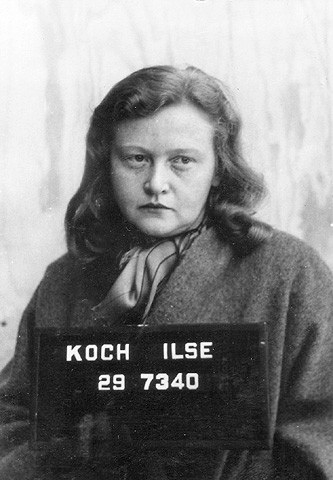
Ilse Koch was the wife of Karl Otto Koch,
Kommandant of the Buchenwald and Madjanek
concentration camps. She was convicted of war
crimes in 1947 and sentenced to life imprisonment.

The collecting of tattooed human skin – both for research purposes and as a kind of fetishized collector’s item – tailed off dramatically after the end of the second World War in the wake of revelations of Nazi concentration camp atrocities. Reports emerged from Buchenwald of the manufacture of everyday items such as gloves, knife sheaths, book-bindings and lampshades from the skins of murdered inmates.[1] In particular, stories of the collection of tattooed human skin, removed from the bodies of inmates at the behest of Ilse Koch, the wife of Kommandant Karl Otto Koch, caused a scandal in the Allied press. Although photographs documenting some of these objects were taken when the camps were liberated, no other material evidence of them was recovered to be entered into Koch’s trial at Dachau in 1947, or later at her second trial at Augsburg in 1950. Without material proof, Koch could not be convicted of the charges relating to the human skin objects.

The Nazis did not just collect the tattoos of prisoners as grotesque trophies of war, however – they also used tattooing as a weapon of dehumanization and control. From May 1940, prisoner numbers were introduced for all concentration camp prisoners deemed capable of work at the Auschwitz concentration camp complex – those sent directly to the gas chambers were not registered and did not receive numbers. These numbers were initially sewn onto prisoner uniforms. However, as the daily mortality rate increased and clothes were removed, this soon proved impractical as a way of identifying the dead. Tattooing of prisoner numbers was thus introduced at Auschwitz in the autumn of 1941. Tattoos were applied to either the inner or outer side of the left forearm on registration at the camp. More than 400,000 inmates were forcibly tattooed in this way at Auschwitz.

The SS introduced number sequences beginning with ‘A’ in mid-May 1944 – 20,000 men and 30,000 women were assigned numbers in this series. One of these women was Holocaust survivor Henia Bryer, who was liberated from Bergen-Belsen concentration camp in 1945. Henia still bears the number ‘A26188’ inscribed on her inner arm. She described her reasons for keeping her prisoner tattoo in a recent BBC programme:

I was offered by various surgeons to remove it, and they were very glad to do it, but I wanted to keep it on. So that when people say that it didn’t exist – these days, that the Holocaust didn’t exist, it’s a figment of your imagination – I wanted to show them. And many people don’t even know what this number means, still today. I wanted to keep it as a witness … as a sign that it really happened.

Tattoos carry with them a powerful capacity to evoke memory, and this quality is often a motivating factor for many people who choose to become tattooed with marks commemorating important life events or rites of passage. For those, like Henia, who have been forcibly tattooed, the mark may come to stand as testament to a personal and collective history of suffering and survival. For other Holocaust survivors, these marks became unwelcome reminders of trauma, and were removed by surgeons after the war. Some of these tattoos were retained in pathology collections, perhaps for reasons similar to those described by Henia above – ‘to stand witness’ to Nazi war crimes that should never be excised from the historical memory.

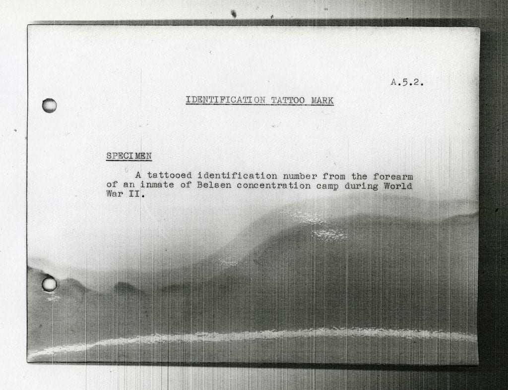
Original documentation record for object no. A.5.2. UCL Pathology Collections.


Whilst working with the UCL Pathology Collections, I came across such a tattoo: a small skin specimen in a perspex vitrine, catalogued as A.5.2 (pictured above). The original documentation associated with this object is the brief catalogue entry: “A tattooed identification number from the forearm of an inmate of Belsen concentration camp during World War II.”* No other information is known about this individual – since only people interned at Auschwitz were tattooed, it is very likely that this person, like Henia, endured the “death marches” from Auschwitz to Bergen-Belsen, where they were later liberated. Many Nazi records were destroyed at the end of the war, and the many thousands of files that do remain are scattered across Europe – even today, it is difficult to trace the identities of interned people based on their tattooed prisoner numbers.

Nor is it known exactly where this specimen was acquired, or who preserved it. Over the past 25 years, UCL Pathology Collections have absorbed a number of collections originating from other London medical institutions as a result of the creation of the Bloomsbury Health Authority in 1982 and subsequent UCLH NHS Trust established in 1994. A large number of the pathology specimens received by UCL arrived in a state of neglect, requiring intensive conservation and re-cataloguing – a task made all the more difficult for the lack of associated documentation. Specimens such as A.5.2 may make us uneasy, particularly when they are unprovenanced and inherited. Their histories are lost and fraught with ethical entanglements – but they cannot simply be discarded, and perhaps should not be hidden away. Henia reminds us of the need to remember past trauma, and of the role that material culture can play in this process. Nazi prisoner tattoos are a powerful reminder of the lived experience of war and genocide. As objects, their presence in pathology collections is undoubtedly troubling; yet they remain as an important testament to the horrors of the Holocaust – they are fragments of lives that should never be forgotten.


References:

[1] Flint Whitlock: The Beasts of Buchenwald, (2011) Cable Publishing, p.81. See also, Alexandra Przyrembel: ‘Transfixed by an Image: Ilse Koch, the ‘Komandeuse of Buchenwald’, in German History, Vol. 19 No. 3, (2001).

*The current catalogue entry for this specimen reflects recent research and has been updated to amend the pejorative term ‘inmate’. A.5.2

[analytics-counter]

Tattooing in Ancient Egypt Part 2: The Mummy of Amunet

By Gemma Angel, on 10 December 2012

  by Gemma Angel


In my previous post, I wrote about the possible connection between objects in the Petrie Museum, and ancient Egyptian tattooing practices. One of the greatest challenges in reconstructing the body modification practices of ancient peoples is in interpreting the fragmentary remains of material culture found at excavation sites. As archaeologist Geoffrey Tassie writes:

The use of many artefacts can only be inferred from their context and association, and tattoo needles are no different, although, if sufficiently well-preserved, scientific analysis of their tips may identify traces of blood or the pigment used to create the tattoo.[1]

In the absence of any such scientific testing, uncertainty remains as to whether the 7 prick points in the Petrie collection were used for tattooing. However, the decorative markings on a collection of blue faience figurines are less ambiguous. Although ancient Egyptian textual records make no mention of tattooing, there is nevertheless a considerable amount of iconographic evidence for the practice, which includes the engraved markings on faience figurines such as those on display in the Petrie Museum. Interestingly, these “tattooed” figures are invariably female, suggesting that tattooing was practiced exclusively by women.[2]
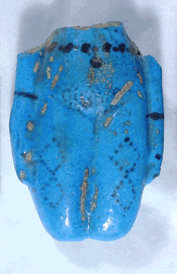
Blue faience figurine fragment,
showing tattoo markings on the
abdomen and thighs.
Image © UCL Museums & Collections

Faience figurines dating from the Middle Kingdom traditionally known as “Brides of the Dead”[3], frequently display a series of dotted geometric tattoo patterns, running in horizontal bands across the lower abdomen. Occasionally, the thighs are also decorated, as can be seen in the example shown (left). There are many examples of footless faience figurines such as these in museum collections around the world. According to Robert Bianchi, dependent upon their context, these figurines maybe interpreted ‘as guarantors of the deceased’s procreative abilities on analogy with those of the goddess Hathor’, who both represented fertility, childbirth and love, and welcomed the dead into the next life. Faience figurines are often found in tombs, interred with the dead in order to ensure resurrection.[4]

Tattooing practice in ancient Egypt is further supported by the discovery of a number of tattooed mummies in the late 19th and early 20th centuries. The most famous of these was discovered in Deir el-Bahari by French Egyptologist Eugène Grébaut in 1891. Dating from Dynasty XI (c.2134-1991 BC), a female mummy identified as Amunet, a Priestess of the goddess Hathor at Thebes, was found to have a number of tattooed markings on her body, which show striking correspondence with the patterns depicted on Middle Kingdom faience figurines. A design consisting of multiple diamond shapes composed of dots, are tattooed on the middle of her right thigh, similar to those engraved on the faience figure pictured above. As well as tattoos on her left shoulder and breast, and on her right arm below the elbow, Amunet also bore extensive tattooing over her abdomen: A series of dots and dashes forming an elliptical pattern of rows covers almost the entire abdominal wall in the suprapubic region (see sketch below right).
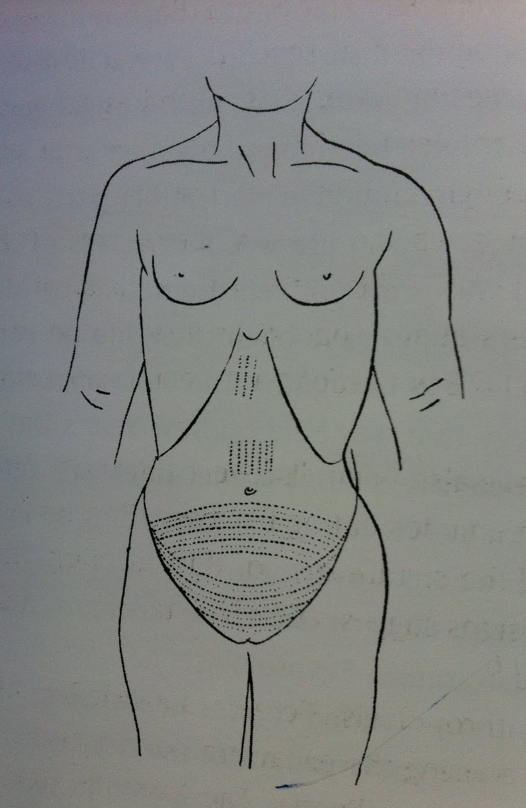
Drawing showing tattoo markings attributed
to the mummified remains of Amunet.
From Fouquet (1898), p.278

A further 2 female mummies, described as ‘Hathoric dancers in the court of King Mentuhotep,’ were excavated from pits located very near to the tomb of Amunet in 1923.[5] These women both bore similar body-markings to those of Amunet, in particular over the abdomen, which may suggest that these tattoos served fertility purposes:

Tattoos on the abdominal part of the female body would have become particularly notable when the woman became pregnant – the patterns would expand, forming an even more symbolically interesting pattern, like a web or netting design.[6]

The mummy of Amunet was unearthed at the height of the “Golden Age” of Egyptology, when the discovery of mass burials of mummified royalty and clergy became a source of popular fascination. As “Egyptomania” swept across Europe, some artists sought to commemorate the “great discoveries” of European explorers and scientists. For instance, the painting below, by French artist Paul Dominique Philippoteaux, depicts an historical event: The unwrapping of a mummy discovered at Deir el-Bahari, the same site where Amunet was buried. Although the mummy pictured dates from Dynasty XXI (c.970 BC) in the Third Intermediate Period, many of the men present in this scene were also involved in the excavation of Amunet. The eminent Dr. Daniel Fouquet takes centre stage, demonstrating to his learned audience of colleagues and lady spectators, as he unveils the mummified body of the “Priestess”, known as Ta-usa-ra. Mr. Grébaut, the leader of the expedition, also appears in the painting, second from left and wearing a fez.[7]

“Examination of a Mummy – The Priestess of Ammon” (1891)
Oil on canvas, by Paul Dominique Philippoteaux.
Photo credit: Peter Nahum at The Leicester Gallery, London.

In 1898, Fouquet wrote an article on “medical tattooing” practices in Ancient Egypt and the contemporary era, in which he describes the tattooed markings on the female mummies found at the Deir el-Bahari site. He speculated that the tattoos and other scarifications observed on the bodies may have served a medicinal or therapeutic purpose:

The examination of these scars, some white, others blue, leaves in no doubt that they are not, in essence, ornament, but an established treatment for a condition of the pelvis, very probably chronic pelvic peritonitis.[8]

Photograph showing the
tattooed abdomen of one of
female mummies found at
the Deir el-Bahari site,
possibly Amunet.

Whilst it is clear that the white scars Fouquet refers to are likely scarifications, the blue marks must be interpreted as tattoos – but whether or not they were primarily medicinal markings, or served a more ritual and symbolic function is uncertain. Based on the iconographic and material evidence of human remains, it certainly seems that some women in Ancient Egypt marked themselves as sexual beings; as Robert Bianchi writes:

The priestess Amunet and the figurines…have an undeniably carnal overtone. The eroticism that is undoubtedly associated with Egyptian tattoo of the Middle Kingdom correlates with the prevailing religious attitude that linked physical procreation with the loftier aspirations of resurrection in the Hereafter.[9]

Amunet’s mummified remains now lie in the Egyptian Museum of Antiquities, Cairo.


Did tattooing really have a medicinal purpose in the Ancient world? Check back for my next post on the history of tattooing as a therapeutic practice – and the health risks involved in becoming tattooed prior to modern antisepsis.


References:

[1] Geoffrey Tassie, ‘Identifying the Practice of Tattooing in Ancient Egypt and Nubia’, in Papers from the Institute of Archaeology, Vol.14 (2003), p86.

[2] According to Tassie, there is only one exception in the archaeological record – a Dynasty XII stele (a standing stone tablet) from Abydos:  ‘This depicts a figure, which is said to be male, with marks coming down over the chest. As the stele is extremely worn it is hard to distinguish whether the marks indeed represent tattoos.’ (Ibid, p.88).

[3] Robert Bianchi, ‘Tattooing and Skin Painting in the Ancient Nile Valley’, in Celenko, T. (ed.) Egypt in Africa, (1996), Indianapolis University Press, p.81.

[4] Ibid, pp.82-82.

[5] Tassie (2003), p.90.

[6] Ibid, p.91.

[7] Philippoteaux’s painting includes a plaque inscribed (in French) with the names of the sitters, as well as an historical description of the scene. From left to right: Marquis de Reverseaux (Ministre de France au Caire); Mr. Eugène Grébaut (Directeur Génerale du Service des Antiquities); Dr. Daniel Fouquet (Médecin au Caire); E. Brugshe Pacha (Conservateur du Musée); Mr. Georges Daressy (Conservateur adjoint du Musée) – pictured taking notes; Mr. H. Bazil (Secrétaire complable du Musée); Mr. J. Barois (Secrétaire Génerale du Ministére du Travaux Publies); Mr. U. Bouriant (Directeur de la Misien Archéologique française au Caire).

[8] Daniel Fouquet, ‘Le Tatouage Medicale en Egypte dans l’Antiquite et a l’Epoque Actuelle’, in Archives d’Anthropologie Criminelle, Tome 13 (1898), p.271.  Available online at Criminocorpus. Translated from the French: L’examen de ces cicatrices, les unes blanches, les autres bleues, ne laisse aucun doute dans l’espirit, il s’agit la non d’un ornement, mais bien d’un traitement institué pour une affection du petit bassin, très probablement une pelvi-péritonite chronique.

[9] Bianchi (1996), p.82.

[10] See also: Carolyn Graves-Brown, Dancing for Hathor. Women in Ancient Egypt, (2010), London  New York: Continuum Books.

[analytics-counter]

Tattooing in Ancient Egypt

By Gemma Angel, on 19 November 2012

  by Gemma Angel


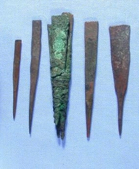
Of all the objects on display in the Petrie Museum of Egyptology, there are two particular groups of items that immediately jumped out at me when I first began to look closely at the collections. These objects are grouped closely together in one of the densely packed cabinets towards the back of the gallery, near the sarcophagi – but no connection is explicitly made between them in the displays. This is not surprising, given that the first collection of objects – a series of tapered and sharpened metal implements – are described in the museum catalogue as “prick points” of indeterminate use:

Tool set identified as a “stock of seven prick points” for removing thorns, bronze; three bound together with thread, a fourth with thread round; three separate; cleaned and treated. No information on findplace within Madinat al-Ghurab; purpose and date uncertain.[1]

Seven “prick points” on display in the Petrie Museum,
possibly used for tattooing.
Image © UCL Museums & Collections

The prick points were excavated by Petrie at Gurob in 1880, alongside cosmetic items and fine pottery ware, which is suggestive of a cosmetic use. [2] When I first saw these objects, they brought to mind an article that I had read by Charlotte Booth, titled Possible Tattooing Instruments in the Petrie Museum. Booth carried out a close material analysis of the prick points, and found that they were made by folding the corners of a flat rectangle of bronze inwards at one end, and then beaten into a smooth finish. [3] Each of the 7 prick points were manufactured in this fashion. Interestingly, 3 of them are bound together with thread, which has become permanently fused to the surface by corrosion (pictured left: 3rd from right). Petrie believed that these objects were in fact tweezer points used for removing thorns:

Slips of bronze were made with long sharp points, and kept sometimes in a bunch of half a dozen; but they were not fastened to the tweezers. [4]

However, as Booth points out, although the points are very sharp, they do not appear to be fine enough for removing thorns from the skin. If they had served such an everyday purpose as Petrie suggested, then surely similar instruments would be more commonly found amongst toiletry objects in ancient tomb excavations? Could they perhaps have served a different, more specialised purpose?

A modern tattoo needle bundle, showing
a large number of fine needles in a
round arrangement, used for shading.
Needles may also be arranged in ‘flat’
or ‘magum’ formations.

To the eye of a tattooist, it is easy to see a resemblance between these ancient bronze points and modern tattoo needles, as they share a number of similarities: Tattoo needles are not hollow like hypodermic needles, as some people assume, but are actually solid. Groupings of very fine needles are bound together in a bundle, much like the Egyptian prick points, except that modern needles are soldered in place onto a stainless steel needle bar, rather than bound together with thread. Modern tattoo needles may be grouped in bundles of anything between 3 and 7 individual needles for line work, and as many as 14, 16 or 18 for shading and colouring. The needles operate by drawing ink up into the narrow spaces between them via capillary action as they’re repeatedly dipped into a small inkwell. This remarkably simple yet effective technology hasn’t changed in hundreds – perhaps thousands – of years. Writing on tattooing in Upper Egypt in the 1920’s, Winifred Blackman describes tattoo instruments consisting of 7 needles fixed to the end of a stick used by the Fellahin; [5] if these needles were indeed similar to those in the Petrie collection, this would suggest that the practice dates back at least 4,000 years in Egypt.

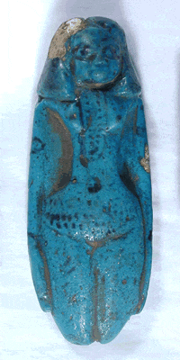
Blue faience figurine, decorated
to show hair, jewellery and tattoos.
Image © UCL Museums & Collections

Material evidence of ancient tattooing is extremely rare in the archaeological record, not least because of the difficulty in identifying artefacts and determining their original purpose. So what evidence is there that the ancient Egyptians practiced tattooing at all? The second group of objects which caught my attention at the Petrie museum provide some clues: two small blue faience figurines. Both of these objects depict nude female figures with black glazed decoration – one is broken at the waist, with only the lower portion of the torso preserved (object no. UC16724). The second figure is intact, with black detail indicating hair and what are assumed to be beads around her neck (object no. UC16725, pictured left).

Both figures are also decorated with a series of dotted lines across the abdomen, which are thought to represent tattoo markings. These markings bear striking similarity to tattoos found on ancient female mummies, which you can read more about in my next post on the Mummy of Amunet.


References:

[1] Petrie Museum online catalogue, object no. UC7790. See additional publications for more information on the origins of these objects.

[2] Charlotte Booth, ‘Possible Tattooing Instruments in the Petrie Museum’, in The Journal of Egyptian Archaeology, Vol. 87 (2001), p.172.

[3] Ibid, p.173.

[4] W.M.F. Petrie, Tools and Weapons, (London, 1917), pp.51-52.

[5] Winifred Blackman, The Fellahin of Upper Egypt, (London, 1927), pp.50-55.

[analytics-counter]

Justice for Magdalens

By Gemma Angel, on 12 November 2012

Lisa Plotkin by Lisa Plotkin


Many visitors to the Grant Museum of Zoology reacted very strongly to the recent exhibit Buried on Campus, which ran from April 23rd until July 13th. In fact, almost every single conversation I struck up with visitors during my time on the museum floor had something to do with the story the exhibit was telling – namely, the discovery of the remains of 180 individuals were discovered buried underneath the main quad at UCL in 2010. Visitors were surprised, inquisitive, and incredulous. Where did the bones come from? they wanted to know. When were they buried? Was UCL aware of this all along? Conspiracy theories abounded.

And it was not just the visitors who were asking questions. My fellow engagers did as well, with both Katie and Sarah writing blog posts on the topic. It seemed everyone was interested in reclaiming these human remains and reinserting them into an historical narrative. Buried on Campus also touched on a topic that is prominent in my own doctoral research, recalling the same questions that Sarah, Katie, and visitors alike asked: mainly, where in history do these bones belong and what are we going to do with them now?

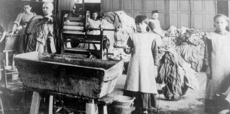
From the main quad of UCL I will detour to Dublin, where in 1993 an order of nuns put up a portion of their estate for sale to a real estate developer. Subsequently, the remains of 155 women were found buried in unmarked graves on the grounds. Their discovery was a catalyst for which revealed the hidden history of the Magdalen Asylums – or the Magdalen Laundries, as they were also known. If you haven’t already heard of Magdalen Asylums then perhaps you can guess their purpose from their namesake. These asylums were built to serve as penitentiaries for the housing and redemption of ‘fallen’ women. Their target demographic was prostitutes, but other so-called ‘deviant’ women were also admitted, for crimes ranging from unmarried motherhood to mental instability. While Magdalen Asylums were first created in the late 18th century, it wasn’t until post-famine Ireland that they were transformed from voluntary refuges for cast-out women to punitive institutions, which sometimes detained their inmates unwillingly for life.

Magdalen Asylums were supposed to offer a new vision for post-famine Irish society. They would solve the problems of poverty, family breakdown, and moral decline. As the Reverend Father Kerr explained:

The appalling demoralization of our times is evidenced mostly in the decline of that virtue amongst woman-kind which is her chief glory and title to our esteem. To-day in ever increasing numbers “Magdalen” has imitators in her sin. But few will share her penance.[1]

But that was no longer true due to these Asylums; now fallen women could share her penance. And repent they did. The women penitents were required to work long hours in laundries for no pay. They were to live under a regime of prayer, silence, and complete obedience and they were separated from their children. They could no longer leave voluntarily, as the late 19th century ushered in a preference for permanent inmates.

Magdalen Asylum.


This 1897 account sums up the situation succinctly:

At five o’clock I was at the Magdalen Home and was introduced by the Mother Superior of the Convent of Mercy to the Sister-in-Charge and six nuns who managed the laundry. We were all seated in the Sisters’ parlour where I put my questions. Most were answered by the Sister-in-charge.

         “How many girls have you?”

         “Seventy-three”          

         “How many are unmarried mothers?”

         “About 70 percent”

         “And the others?”

         “Some are sent here when they leave the Industrial School because they need special

         care.”

         “Are they mental defects”?

         “No.”

         “Backward”?

         “Yes.”

         “Are the girls paid?”

         “No, they earn their keep.”

         …

         “Are the girls free?”

         “Yes.”

         “Can a girl leave whenever she chooses?”

         MOTHER SUPERIOR: “No, we’re not as lenient as all that. The girl must have a suitable

         place to go…”

         “How long do they stay”?

         “Some stay for life” [2]

These asylums continued to operate up until the 1980s. In 1997 the documentary Sex in a Cold Climate premiered, featuring the testimonials of four former penitents. The women spoke of having to endure physical and sexual abuse while incarcerated against their will and one woman spoke of being forcibly separated from her child. Since then there have been numerous documentaries chronicling the physical and psychological abuse that the estimated 30,000 Magdalens suffered. Calls for justice for these forgotten women resound in Ireland today, and on June 6th, 2011 the United Nations Committee Against Torture put out a statement urging the Irish government to investigate the claim that thousands of girls and women were tortured in Catholic laundries.

As I reflect on the impact that Buried on Campus had on the UCL community, I can only imagine the impact that the 1993 discovery must have had in Ireland. Visitors to the Grant wondered at our complicity in the deaths of those 180 people buried underneath our lecture halls, and many were adamant that the narratives of these unknown people should be reasserted into UCL’s history. I am happy to say that over the past two decades Irish historians, and the Irish public, have given us a great model to follow in doing just that. And whilst justice was not granted to the unknown 155 buried in a mass grave within a convent’s walls, their story is being told and their memory is not forgotten.


References:

[1] Rev. Father Kerr, Galway. ‘The Good Shepherd Nuns at Home’, in The Fold of the Good Shepherd  (1931), p. 10.

[2] Halliday Sutherland: Irish Journey (1958), pp.81-83.

Buried on Campus: When Are Remains Human?

By Gemma Angel, on 10 September 2012

by Sarah Chaney


Katie’s recent post on the ethics of displaying human remains in museums, along with the recent Grant Museum exhibition on the topic, raised some important questions about collection and display. Unsurprisingly, this is a frequent topic of concern in medical museums – particularly in hospital museums, teaching collections tend to focus around anatomy and pathology, a large part of which consists of specimens of human remains. But what exactly constitutes “human remains”? This is, on occasion, a surprisingly difficult question to answer.

Many medical collections were created in the eighteenth and nineteenth centuries, as anatomy schools grew and increasingly began to offer practical training for medical students, alongside lectures. One such fascinating collection can be found at Benjamin Franklin House, in Craven Street near Charing Cross. An ordinary Georgian townhouse, which was home to American Founding Father Benjamin Franklin for nearly sixteen years, the building also housed a private anatomy school. The school was run by the landlady’s son-in-law, William Hewson; who had previously worked with the well known anatomist William Hunter. Hewson sadly died young, of septicaemia contracted during a dissection, but the remains of his school were uncovered during the restoration of the house in recent years, in a pit where the back yard would have been.

It is not unusual for bones to be unearthed when foundations are laid for new buildings in London, something explored not so long ago in an exhibition at the Wellcome Collection, London’s Dead. These skeletal remains are what first springs to mind when we think about human tissue. It is skeletons that we often expect to see in museums; the absence of flesh suggests age and the process of decay. How long, though, do we continue to regard these skeletons as the remains of our loved ones? Cemeteries, for example, tend to offer burial plots for periods of only around fifty or sixty years. By this time, the body will have decomposed, and the land can legally be re-used. In Sheffield, part of the former city cemetery has been turned into a park. In London, memorial grounds are built over all the time. Once people have faded from memory, it seems that their remains do too.

But what other human remains do we find in medical collections? Many of the specimens are dissections of various sizes and complexity. It seems obvious which of these are tissue and which not, but even here the recent Human Tissue Act has struggled to define humanity. We might assume that any part of our physical form constitutes human tissue but, legally speaking, this is not the case. Body parts that regularly grow and are removed, for example, are something of a grey area; for example, locks of hair, often kept as mementos of a loved one, can legally be kept or displayed by any museum. But what about blood? The status here is uncertain. On the other hand, a tumour which has been surgically removed is considered human tissue, despite the fact that the person operated on may well have considered it to be alien to their own person.

Stained Brain Specimens in the UCL Pathology Collection. Courtesy of Bethlam Heritage.

One of the things that intrigues me most, however, is the place of foreign bodies in medical collections. Foreign bodies are objects that have been swallowed, inhaled or otherwise inserted into the human body. Often, particularly when these items end up in the bladder, the body creates deposits around these objects, protecting organs from sharp edges or corrosive material. When removed, the foreign body may be invisible within layers of mineral coating. These objects are faintly mysterious: created by the human body, they are nonetheless not considered to be human at all. They lie beyond the regulations on human tissue, but could not have come into existence in the first place without having had a relationship with that tissue.

Dem Bones, Dem Bones, Dem Dry Bones … Excavating Memory, Digging up the Past

By Gemma Angel, on 16 July 2012

by Katie Donington


Above all, he must not be afraid to return again and again to the same matter; to scatter it as one scatters earth, to turn it over as one turns over soil. For the ‘matter itself’ is no more than the strata which yield their long-sought secrets only to the most meticulous investigation. That is to say, they yield those images that, severed from all earlier associations, reside as treasures in the sober rooms of our later insights – like torsos in a collector’s gallery.[1]

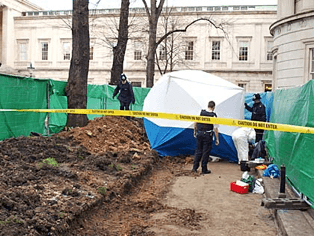
The Buried on Campus exhibition at the Grant Museum ran from April 23rd to July 13th 2012. Following the 2010 discovery of human remains beneath the Main Quad of UCL, research was undertaken to determine the reason for their presence. Forensic anatomist Wendy Birch and forensic anthropologist Christine King, members of the UCL Anatomy Lab, were able to date the bones which were over a hundred years old. The bones themselves also gave clues to the reason for their presence. Several items had numbers written on them and others displayed signs of medical incisions. This led the team to the conclusion that the bones represented a portion of the UCL Anatomy Collection which had been buried at some point after 1886.

The issue of displaying human remains in a museum of zoology was discussed by Jack Ashby, Grant Museum Manager in a recent blog post:

The whole topic of displaying human remains has to be considered carefully and handled sensitively… One of the questions we asked our visitors last term on a QRator iPad was “Should human and animal remains be treated any differently in museums like this?” and the majority of the responses were in favour of humans being displayed, with the sensible caveats of consent and sensitivity.[2]

The discovery and exhibition of human remains raises interesting questions about the relationship between archaeology, history, science, memory and identity. It also links into debates over the ethics of display in relation to human beings. Who were these people? Why did their bodies end up in an anatomy collection? Did they consent or were they compelled? Is it possible or desirable to attempt to retrieve or reconstruct the object as subject?

The case of the bones buried on campus reminds me of another example in which the physical act of excavation was transformed into an act of historical re-inscription. In 1991, workmen digging the foundations of a new federal building close to Wall Street uncovered the remains of 419 men, women and children. Archaeologists, historians and scientists were called in and they were able to identify the area as a 6.6 acre site used for the burial of free and enslaved Africans by examining maps from the seventeenth and eighteenth centuries.

The Maerschalck Map of 1754, showing the Negro Burial Grounds near the “Fresh Water” (the Collect Pond). Image © The African Burial Ground Project.


The bones offered specific information which helped to give a partial identity to the people interred. Using ‘skeletal biology’[3] it was possible in some cases to pin point where in Africa individuals had come from – Congo, Ghana, Ashanti and Benin, as well as revealing whether they had been transported via the Caribbean. Bone analysis spoke of the appalling conditions of slavery; fractured, broken, malformed and diseased bones articulated stories of unrelenting labour, nutritional deficiency and coercive violence.

Objects found inside some of the burials created a sense of the uniqueness of each person as well as the care taken by loved ones as they performed burial rituals. The lack of items found also indicated the social status of the majority of people buried on the site.

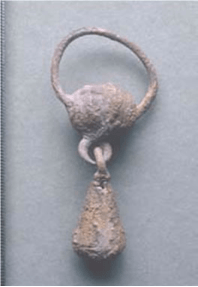
This pendant (image courtesy of the African Burial Ground Project) was recovered from burial 254, a child aged between 3 ½ and 5 ½ years old. It was found near the child’s jaw and may have been either an earring or part of a necklace. The objects and bones represented a visceral historic link to the African American community in New York. The sense of ownership they felt towards this history and the individuals who had emerged from the soil, led to active community engagement in the project. In line with the wishes of the African American community, all original items were facsimiled before being reinterred along with all 419 ancestral remains in a ceremony in 2003. A memorial and museum were also built on the site (see image below, courtesy of the African Burial Ground Project).

The emergence of the skeletons was interpreted by some as a literal rendering of the way in which America has been haunted by its relationship with slavery. As physical anthropologist Michael Blakely, who worked on the site explained; ‘with the African Burial Ground we found ourselves standing with a community that wanted to know things that had been hidden from view, buried, about who we are and what this society has been.’[4]

The context of the two sites is of course very different. However, a comparison of them does raise questions about the uses of human remains and their relationship to history, memory and identity. The bones at UCL formed part of an anatomical teaching collection; a composite of individuals whose bodies somehow became the property of medical institutions. Those people often consisted of those on the margins of society; the poor, the criminal and the exoticised ‘others’ of empire.[5] Debates over the repatriation of human remains in museum collections highlight their importance to people’s sense of identity and history. Without family or community groups to claim the individuals discovered at UCL, it seems that they are destined to remain object rather than subject – ‘severed from all earlier associations… torsos in a collector’s gallery’.


Have your say – what do you think should happen to the bones at UCL?


[1] Walter Benjamin, ‘Excavation and Memory’, in Selected Writings, Vol. 2, Part 2 (1931–1934),ed. by Marcus Paul Bullock, Michael William Jennings, Howard Eiland, and Gary Smith, (Massachusetts, Belknap Press of Harvard University Press, 2005), p. 576.

[2] http://blogs.ucl.ac.uk/museums/2012/04/24/buried-on-campus-has-opened/

[3] http://www.archaeology.org/online/interviews/blakey/

[4] http://www.archaeology.org/online/interviews/blakey/

[5] Sadiah Qureshi, ‘Displaying Sara Baartman, The Hottentot Venus’, History of Science, Volume 42 (2004), pp.233-257.

http://www.negri-froci-giudei.com/public/pdfs/qureshi-baartman.pdf