X Close

Researchers in Museums

Home

Engaging the public with research & collections

Menu

Archive for the 'Stacy Hackner' Category

Question of the Week: Do boys and girls enjoy different museum exhibits/items?

By Stacy Hackner, on 26 March 2014

Stacy Hackner_Thumbnail

By Stacy Hackner

This is actually a more complicated question than one would think, especially considering the recent controversies regarding “pink is for girls, blue is for boys” toys, the Independent’s refusal to review children’s books aimed at a particular gender, and Waterstones‘ refusal to sell such books. It’s also an interesting question to ask as most of us would consider museums fairly gender-neutral spaces. According to research, museum visitors are more likely to be female, educated, older, and white — but that’s a fairly narrow demographic. Clearly there are many visitors who are male or other genders, not in (or after) higher education, young, and of varying ethnicities. There are also two competing (but false) ideologies: that girls would prefer museums because they like quiet learning and being indoors, and that boys will prefer museums because they can interact with objects and tend to like “the gross stuff”. Studies from the 1990s showed that while boys and girls both visited all exhibits at a science museum, they interacted with the exhibits in different ways and for different amounts of time – i.e. boys preferred the water jets and girls preferred face paint. (What these activities have to do with science is unclear.) The researchers showed that children display “typical gender roles” when playing and advise museums to design displays accordingly. Another article encourages girls to visit science museums because they’re an informal and thus less intimidating environment than the classroom. However, it’s important to consider these articles in the context of the views of gender held at the time – I’d hope we’re less stereotypical these days.

In my experience in the Petrie and the Grant, I’ve found both of these stereotypes completely untrue. All kids who come to the Grant like “the gross stuff”, or as they’re properly termed, the wet specimens. I’ve had both boys and girls come up to ask me questions about dinosaurs and bones and worms and mummies and jewelry and the jar of moles. Both boys and girls want to dress up in the Petrie’s reproduction Egyptian clothing, especially the loincloth. Teenage boys, including a Scouts troop I engaged with, are particularly fascinated by the baculum — but then, so were a duo of thirty-year-old women. Above all, kids of all genders are natural scientists: curious, inquisitive, and unafraid to ask crazy questions. Children who visit museums are happier and, in a country where most museums are free, it’s always worthwhile for them to come and explore.


Sources

Falk, JH. 2009. Identity and the Museum Visitor Experience. Walnut Creek, CA: Left Coast Press.

Kremer, KB and GW Mullins. 1992. Children’s Gender Behavior at Science Museum Exhibits. Curator: The Museum Journal Volume 35, Issue 1, pages 39–48.

Ramey-Gassert, L, HJ Walberg III, and HJ Walberg. 1994. Reexamining connections: Museums as science learning environments. Science Education, Volume 78, Issue 4, pages 345–363.

Question of the week: Do other animals have belly buttons?

By Stacy Hackner, on 19 March 2014

Stacy Hackner_Thumbnail

by Stacy Hackner

This question was thrown at me at the end of a conversation about juvenile bone growth, and I was completely blindsided. I know my cat definitely has a bump in the place his navel should be, and I assumed all placental mammals have them.

Further research shows that indeed, all placental mammals start with a belly button (or navel, or umbilicus if you’re scientific). The navel is the remnant of the umbilical cord, which attaches a fetus to the mother’s placenta to deliver nutrients in utero. Thus animals that hatch from eggs don’t have them – this includes marsupials like kangaroos and wombats, which have not evolved a placental structure and instead incubate their young in a pouch. However, in most other mammals (and certain humans) they’re obscured by fur, and in some species they are a thin scar rather than a small bump, and fade over the course of the animal’s lifetime.

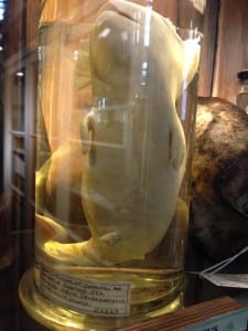
beluga

Umbilicus evident on a Grant Museum specimen of a fetal beluga whale.


The Biomechanics of Breasts

By Stacy Hackner, on 24 February 2014

Stacy Hackner_ThumbnailBy Stacy Hackner

Have you ever wondered what biomechanics has ever done for you? Well, if you’re a runner, it can tell you a lot about your gait and efficiency. It tells us why people with long legs are good at running and people with long arms are good at swimming, and the forces they use per stride or stroke. It can teach us proper runner techniques. If you’re a female runner, you may have encountered a problem biomechanical researchers are actively working to solve: bouncing breasts.

I’ve only been a runner since I started my PhD. As you may remember from my last post, I learned from my research that it’s very important for your bone strength to practice weight-bearing activity (sorry, astronauts), which includes running. As professional running goes, for some reason marathon organizers decided to exclude women from participating until the mid 1980s, when just a few women snuck into the Boston and London marathons and achieved quite good times (see Heminsley’s book for an exciting run-down of the sneaking). Since then, women have been participating in most major sports, including (very recently) American football; 36.5% of 2012 London Marathon finishers were female (Brown et al 2013). But sports equipment for women is still catching up, and biomechanics – long applied to gait and stride, torso and head movements – is now being brought in to design a better bra. Most biomechanical studies of breasts involve attaching markers to women on treadmills in clothed and unclothed conditions and filming them with an infrared camera – a slightly awkward study for the volunteers, but it’s worth it for the results.

The breast in three dimensions. Zhou et al 2012.

The breast in three dimensions. Zhou et al 2012.

First, let’s look at a breast from an anatomical perspective. Breasts are composed of milk glands and ducts, fat, connective tissue, and Cooper’s ligaments. The latter are fibers that attach the breast to the underlying fascia and pectoral muscles; throughout life, and in vigorous exercise, they can stretch or break and cause sagging and breast pain. Imagine a laundry line with wet clothes hanging on it – now shake it: that’s what happens during intense exercise. Of course, these forces have been measured, which can be difficult as unlike bones and muscles, they squish and deform, and each of the above types of tissue reacts to running forces differently. During the beginning of a running stride, the breasts are found to accelerate at up to 3 G (where G is the force of gravity – at that point in time, it’s like the breast weighs 3 times as much). This is considerably more than the rest of the trunk, and puts strain on Cooper’s ligaments. You can then imagine that each breast goes through a cycle of acceleration, stasis, and deceleration for each stride, like your head when stopping and starting in a car. For each stride, the breasts move forward into the air and backward into the ribcage, in what is called anterior displacement.

The figure-8 movement of breasts in a digital reconstruction. Image via ShockAbsorber website.

The figure-8 movement of breasts
in a digital reconstruction.
Image via ShockAbsorber website.

Now it gets more serious. In addition to being displaced anteriorly, breasts move in three dimensions. When running, the goal is generally to move forward. In order to do this, you need to move up as well. And with each step, you also sway side to side. Breasts respond to this combination of forces by actually moving in a figure-8, experiencing additional vertical and horizontal displacement. Studies show that it’s these three directions of displacement rather than acceleration that cause breast discomfort and pain; the worst seems to be vertical displacement, which peaks at mid-flight. At this point, the Cooper’s ligaments are basically floating upwards and then being tugged back down during deceleration (not to mention the fat and glands moving about internally). This is important to know for bra design, as many sports bras take the approach of “flattening everything is best” – however, as we’ve now learned, flattening will only reduce anterior displacement! Flattening bras can also cause breast pain, so it’s lose-lose situation.

Now let’s discuss what a bra actually does. The everyday padded bra is an attempt to compromise comfort, sexuality, and stabilization, often emphasizing one to the detriment of the other two. The goal is to hold the breasts in an uplifted position so they appear firm and don’t jiggle around too much while walking or climbing stairs. Sports bras, on the other hand, prioritize stabilization, as they’re to be worn in high-impact activities. Many sports bras take the approach that flatter is better, which as I’ve shown is not quite the case, but they do prevent one kind of displacement. Regular, everyday support bras lift the breasts up, reducing strain on the Cooper’s ligaments, but in tests of treadmill running do little to prevent any kind of displacement. Running bare-chested causes the most displacement, and – in the case of marathons – can lead to a breast injury experienced by men as well, where the nipples chafe against the fabric of the shirt. (This is actually very common, and there are marathoner web forums devoted to sharing prevention tips.) A newer kind of sports bra attempts to encapsulate rather than flatten, and researchers from biomechanics and textile manufacturing have been collaborating on new design. This bra holds each breast separately and matches the figure-8 to the overall movement of the torso, reducing displacement in all three directions.

As more women get into sports (which is particularly important for the prevention of osteoporosis), making us comfortable and keeping us engaged should be a high priority for sports equipment manufacturers. Most runners can find shoes that fit, as shoes have been tested and re-designed for the last thirty years, and have a high profile in the press. Despite the increase in women running, 75% of female London marathoners still reported a problem with their sports bra, with the prevalence higher among larger-breasted women. However, proper fitting technical sports bras receive significantly lower press coverage than proper running shoes. Clearly, there is more work to be done!


Sources

Brown, N., J. White, A. Brasher, and J. Scurr. 2013. An investigation into breast support and sports bra use in female runners of the 2012 London Marathon. Journal of Sports Sciences 2013:1-9.

Heminsley, A. 2013. Running Like a Girl. London: Huntchinson.

Scurr, J., J. White, and W. Hedger. 2010. The effect of breast support on the kinematics of the breast during the running gait cycle.  Journal of Sports Sciences 28(10): 1103-1109.

Zhou, J., W. Yu, and S.P. Ng. 2012. Studies of three-dimensional trajectories of breast movement for better bra design. Textile Research Journal 82(3): 242-254.

Update: The post originally stated that Cooper’s ligaments connect breast glands to the clavicle; this was incorrect. 

How are Ancient Nubians Like Astronauts?

By Stacy Hackner, on 6 January 2014

Stacy Hackner_Thumbnail By Stacy Hackner

Some respected individuals (supervisors, mentors, parents) have advised me to not get distracted by the primrose paths that crop up during a PhD. These primrose paths are always deliciously exciting, offering opportunities to study wonderful new topics that one can justify as marginally related to one’s thesis and therefore potentially of use. Of all the primrose paths I’ve followed, I never expected the most relevant one to be about astronauts.

Credit: Wikipedia Commons

Sudanese pyramids. (Wikimedia commons.)

My thesis explores ancient Nubia, the region that is now northern Sudan, from roughly 3000 years ago to medieval times. Unlike their contemporaries, the Egyptians, the Nubians didn’t have a system of writing until the Meroitic period (300 BCE-400 CE), a time of Egyptianizing influence. They built small pyramids and imported Egyptian goods, attesting to the influence of their famous northern neighbors. In the absence of writing (and even in the presence of texts, as humans tend to play with the truth), archaeologists try to build a picture of the ancient society using physical evidence, including human remains. Fortunately, the dry climate and sandy soil usually result in excellent bone preservation, allowing me to identify differences in bone shape. But wait – let me back up a little.

We aren’t entirely sure how bone works. There are two types of cell responsible for bone maintenance – osteoblasts and osteoclasts. Osteoblasts build bone, and osteoclasts take it away. The body is highly responsive to changes in activity, and bone is constantly updating itself accordingly. The general principle is that your body thinks what’s happening now will happen forever. Think about when you’re running a race: it’s hard to start because your body’s been used to standing still and needs some time to amp up your heart rate and muscle contractions. When you finish the race, your heart keeps pounding for a minute or two because it hasn’t quite got the signal to stop running yet. Bone works in a similar way. In response to physical stress, bone will accumulate more osteoblasts to strengthen itself. Each step makes tiny microfractures, which tells the bone “Come on, I’m breakin’ here! Give me more strength!” and the osteoblasts pile on. In the absence of activity – during periods of prolonged sitting or lying down – the osteoclasts come in to take away unnecessary bone. “You’re not using this one, right? Then we can send the calcium somewhere else.” The thing is, scientists don’t know all the signals involved in this process. We know what happens, but not the channels of communication. I like to imagine bone cells having little conversations with each other, but clearly it’s all on a neurochemical level we haven’t yet discovered.

The concept of bone building in necessary areas is keenly presented in studies of elite athletes. In a study by Haapsalo et al (1998) of young female tennis players, the players gained significant bone mineral content in the bones of their dominant (forehand and serving) arm. When the authors looked at a control sample (girls who did not play tennis), there was minimal or no difference between their arms; there was also minimal difference between the nondominant arms of the tennis players and the controls.

Another study, by Shaw & Stock (2009), examined differences between university athletes who competed in either hockey or long-distance running. They found significant differences in the actual shape of the tibia (shin bone) due to the physical stress of these activities. The tibias of the long-distance runners were more elongated front to back while the tibias of the hockey players were more even side-to-side, showing a distinct difference in the direction of activity in these sports. Clearly, osteoclasts were being sent to the bone locations these athletes needed them most: for runners, the front, and for hockey players, the sides. It is important to point out that many of the studies investigating activity and bone growth look at adolescents, since their bones develop until the end of puberty. After that, it seems to take a lot more effort to alter bone shape and density.

Credit: wikipedia

Her bones are losing mineral content by the minute! (Wikimedia commons.)

And what about the other side of the cycle? The osteoclasts? That’s where the astronauts (and cosmonauts) come in. The constant pounding of our feet against the floor keeps our bones as strong and dense as they need to be for everyday use. In zero gravity, though, there’s no pounding, just the occasional soft push off the wall of the space station. The osteoblasts don’t have any stress to react to, and the osteoclasts assume the extra bone is useless, so it starts to be resorbed. It helps that astronauts are some of the most-studied individuals on our planet (and definitely the most studied off the planet!). During spaceflight, urine calcium output is found to increase, indicating that bone is being sapped of minerals, and post-spaceflight bone scans reveal a condition called “spaceflight osteoporosis”, similar to the osteoporosis experienced on earth – but the bone density is only lost from the legs, feet, and hips, all weight-bearing regions, including an 8% loss in four months (compared to 1% loss per year for earth-bound sufferers of osteoporosis). The upper body and head generally remain unaffected (unless one of the astronauts was a tennis player, of course). One study found that after a “long-duration” spaceflight of 4-6 months, it took up to three years for astronauts to recover the bone density they’d lost in space (Sibonga et al, 2007). It makes one really appreciate gravity.

So how do I apply this to ancient populations? The data from astronauts indicates that most of the density lost is from trabecular bone, from the internal core, rather than from the outside. This means that even if ancient bones have lost density due to age, either before death or after burial, it’s likely to have happened from the inside out and thus the external shape should remain intact. This gives me more confidence in figuring out what kinds of activities they performed during adolescence, which in most cultures was when young people started to take up adult cultural roles. I hope to compare the shape of the bones of Nubians to those of athletes and to other populations whose activities are known in order to draw a better picture of their society.

Sources

For an amusing (but factual) look at the craziness that is astronaut and cosmonaut research, check out Mary Roach’s “Packing for Mars: The Curious Science of Life in the Void” (Norton & Co, 2010).

Haapasalo, H, P Kannus, H Sievänen, M Pasanen, K Uusi-Rasi, A Heinonen, P Oja, and I Vuori. 1998. Effect of long-term unilateral activity on bone mineral density of female junior tennis players. Journal of Bone and Mineral Research 13/2, 310-319.

Shaw, CN and JT Stock. 2009. Intensity, Repetitiveness, and Directionality of Habitual Adolescent Mobility Patterns Influence the Tibial Diaphysis Morphology of Athletes. AJPA 140, 149-159.

Sibonga, JD, HJ Evans, HG Sung, ER Spector, TF Lang, VS Oganov, AV Baulkin, LC Shackelford, and AD LeBlanc. 2007. Recovery of spaceflight-induced bone loss: bone mineral density after long-duration missions as fitted with an exponential function. Bone 41, 973-978.