X Close

Researchers in Museums

Home

Engaging the public with research & collections

Menu

Archive for the 'Foreign Bodies Exhibition' Category

Sword Swallowing & Surgical Performance

By Gemma Angel, on 11 March 2013

Sarah Chaneyby Sarah Chaney


We know sadly little about the sword swallower’s sword that resides in the UCL Pathology Collection: not even how long it has been here. What we do know is that this performer was very unlucky. Perhaps he (or, indeed, she) didn’t tilt his head back far enough. Perhaps he moved during the process of insertion. Whatever the case, the sword pierced the flexible tube of the oesophagus, leading to the performer’s death. The heart and oesophagus were preserved – perhaps as a warning of the dangers of such feats – alongside the weapon that led to his demise.

Fatally ruptured oesophagus, caused by the sword swallower's sword. Photograph Gemma Angel, UCL Pathology Collections.

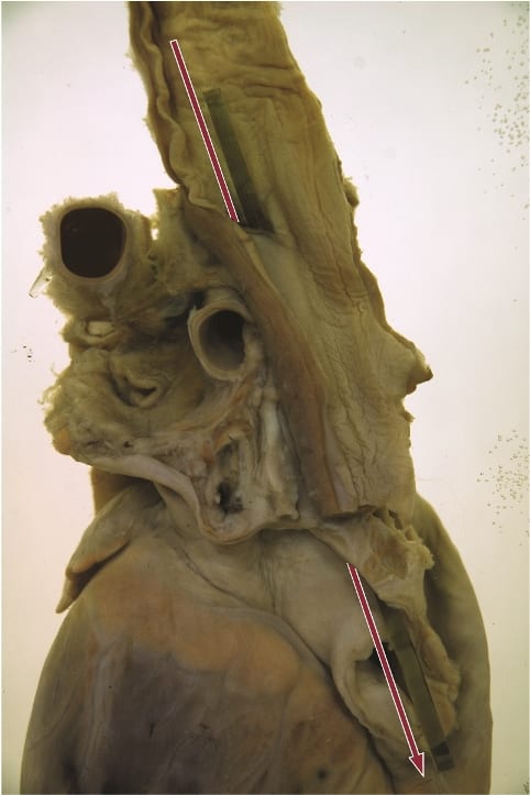
Fatally ruptured oesophagus, caused by the sword swallower’s sword. Photograph Gemma Angel, UCL Pathology Collections.

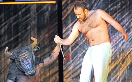
Sword swallowing seemingly originated in India some 4,000 years ago, but reached the western world of Ancient Greece and Rome in the first century AD. The performer tilts his or her head back, extending the neck, and learning to relax muscles that usually move involuntarily. A rigid weapon can then be passed down as far as the stomach, usually for just a few seconds, before removal. It is dangerous, certainly, but few performers suffer the fate of the individual preserved in the UCL collections. According to one recent article in the British Medical Journal, most serious incidents occur owing to distraction or attempts at exceedingly complex feats:

For example, one swallower lacerated his pharynx when trying to swallow a curved sabre, a second lacerated his oesophagus and developed pleurisy after being distracted by a misbehaving macaw on his shoulder, and a belly dancer suffered a major haemorrhage when a bystander pushed dollar bills into her belt causing three blades in her oesophagus to scissor. [1]

In many ways, sword swallowing is the opposite of the ingestion of other foreign bodies: rather than swallowing, the performer maintains absolute control over the process of consumption, taming the body’s reflexes and realigning the organs. As Mary Cappello notes in her fascinating literary biography of surgeon Chevalier Jackson (1865 – 1968), who was an expert in foreign body removal, sword swallowing was recognised by doctors as inspirational to their own techniques. Jackson took his lead from German professors Alfred Kirstein and Gustav Killian, who lectured that sword swallowing proved the possibility of passing a rigid tube into the oesophagus, in order to remove lodged objects. Jackson, who developed his own oesophagoscope in 1890, admitted that the abilities of circus performers had opened his eyes to the opportunity of removing foreign objects without dangerous surgery. He even taught his children how to “scope” themselves.[2]

In an intriguing parallel, the insertion of some foreign objects into the human body thus assisted with the removal of others. At the turn of the 20th century, the removal of foreign bodies lodged in the throat and airways frequently required an incision to be made into the trachea or oesophagus, an operation which could prove fatal. In the records of the Royal London Hospital, from 1890 to 1910, we find no mention of oesophagoscopy or bronchoscopy: instead, surgery or the probang or “coin-catcher” was the norm. This latter instrument was generally a simple hook, inserted without any kind of viewing device or illumination. The practitioner would feel blindly for the object, and either attempt to hook it out, or push it into the stomach. This might lead to numerous complications. In 1903, surgeons at the Royal London attempted to remove a halfpenny from the throat of a five-year-old boy by pushing it into the stomach. However, it was subsequently reported that the coin catcher broke off in the boy’s throat, necessitating a major operation from which the child did not survive.[3] Small wonder that, less than a decade later, Jackson declared such objects “rough, unjustifiable, brutal”.[4]

foreignbodies

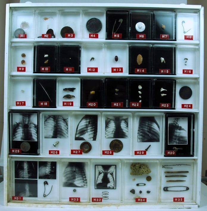
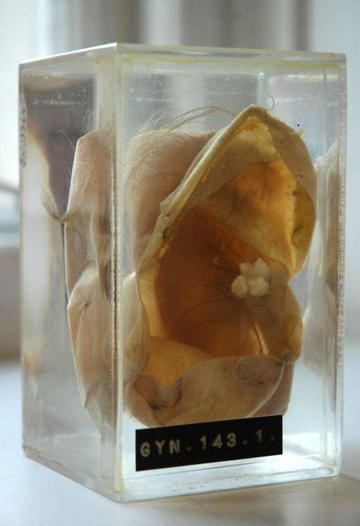
UCL Pathology Collections contains many examples of foreign objects removed from the
human body: this purpose built display showcases many such objects, some with
small x-rays of the objects prior to removal.

X-ray imaging techniques aided the removal of foreign objects by instruments, and foreign body specimens are often accompanied by photographs showing the item’s location in the human body. The above set of items is found in the UCL Pathology Collection, the objects having been gathered by several surgeons in the 1920s – ‘50s. At some point, the individual boxes made for each specimen were mounted together, in a specially designed plastic surround. Fittings on the back indicate that the case was made to hang on a wall. But why? To decorate the office of a surgeon, showing off his achievements? To offer a warning to others to take care (particularly parents, for all these objects were removed from children and infants)?

Chevalier Jackson claimed that his collection of more than two thousand foreign bodies (now housed in Philadelphia’s Mutter Museum) was not a curiosity, but indicative of the everyday nature of foreign body ingestion and inspiration. Yet many of these specimens are not everyday. The two boxes of multiple objects in the bottom right, for example, were removed from the vaginas of young girls (six and eight years old respectively). The case notes do not indicate how these objects arrived in their location. Did the girls insert them themselves, or might it be a sign of sexual abuse? In her research into the medical histories of Jewish immigrants to the East End of London in the late nineteenth and early twentieth centuries, Carole Reeves came across a case of multiple foreign body insertion in a young woman, whose vagina was found to be tightly packed with pins. Reeves speculated that Leah G. might have inserted these items in an effort to ward off potential (and actual) abusers.[5]

In most instances, we can uncover little about the motivations of those in the late 19th and early 20th centuries whose foreign bodies are recorded in medical records: surgeons were often little interested in how the object came to be in its current location, but only in its removal. Yet this may often make such displays still more intriguing than otherwise. As Mary Cappello put it, in a video discussion of the UCL artefact pictured above for the Damaging the Body website: “What is the border or boundary between human flesh, between human life and the object world?”


References:

[1] Brian Witcombe and Dan Meyer, “Sword Swallowing and its Side Effects”, in British Medical Journal, 333 (2006), 1285-7, p. 1287.

[2] Mary Cappello, Swallow: Foreign Bodies, Their Ingestion, Inspiration and the Curious Doctor who Extracted Them, New York, London: The New Press (2011). Website: http://www.swallowthebook.com/

[3] Royal London Hospital Archives, Surgical Index 1903, LH/M/2/9, patient no. 4086.

[4] Chevalier Jackson, Lecture to the Kings County Medical Society, December 19 1911, quoted in Cappello, p. 208.

[5] Carole Anne Reeves, Insanity and Nervous Diseases Amongst Jewish Immigrants to the East End of London, 1880 – 1920 (Unpublished PhD thesis, University of London, 2001), p. 213.

Pulling Teeth: Ovarian Teratomas & the Myth of Vagina Dentata

By Gemma Angel, on 4 March 2013

  by Gemma Angel


In preparation for our upcoming exhibition, Foreign Bodies, several members of the engagement team went to visit UCL Pathology Collections, to have a look at a collection of foreign objects removed from the human body. We soon encountered a number of other specimens which resonated with the exhibition theme in various ways: From a liver infected with syphilis, to a ruptured oesophagus and the sword swallower’s sword that caused the fatal injury; to a feotus inadvertently discovered during a hysterectomy, which was performed to extract a large tumour on the uterus.

The UCL Pathology Collections comprise over 6,000 specimens dating back to around 1850, many of which have been absorbed from other London medical institutions over the past 25 years, and these are currently in the process of being re-catalogued and conserved. It is a fascinating, not to mention an educationally invaluable collection – not least because it contains many specimens that demonstrate gross clinical manifestations of diseases which are now very rare in the Western world. Some of these diseases, such as syphilis, are unfortunately making a comeback, so it seems more important than ever that medical students are able to recognise the clinical signs of these infections. Pathology collections are a highly valuable medical teaching resource; particularly since these kinds of collections are now unlikely to be expanded in the wake of the 2004 Human Tissue Act.

As with many historical pathology collections, UCL possesses its share of medical anomalies or curiosities. Fragments of preserved skin belonging to a tattooed man certainly seem to fall into the category of the anatomically curious – there is certainly nothing pathological about this specimen. One of the biggest surprises I encountered during my visit to the collections, was the revelation that the female reproductive anatomy can, and occasionally does, grow teeth.

Teratoma with Tooth and Hair

Dermoid cyst (cystic teratoma) with fully developed
tooth and hair. UCL Pathology Collections.

The specimen shown here (right) is a dermoid cyst, or cystic teratoma, which has formed inside an ovary. When I first came across it, I experienced a strong visceral reaction: I didn’t have to be a medical student to recognise that this tooth, entwined in long hair drifting in the liquid-filled vitrine, was out of place – so much so, that the sight of it provoked an immediate and simultaneous sense of revulsion and fascination. The term teratoma is derived from the Greek, tera, meaning monster, and literally means “monstrous growth”; it was easy for me to see how such biological anomalies could become the stuff of nightmares. Despite the ominous name, however, ovarian teratomas are usually benign, and arise from totipotent stem cells which are capable of developing into any type of body cell. One 1941 pathology text describes these tumours as follows:

Dermoid cysts are usually globular in shape and dull white in color. They contain structures associated with epidermal tissues, such as hair, teeth, bone, sebaceous material resembling fat … The following is a partial list of tissues which have been found in dermoids: Skin and its derivatives, sebaceous glands, hair, sweat glands, and bone, especially the maxillae containing teeth. Up to 300 teeth have been found in one cyst … Long bones, digits, fingernails, and skull have been found. Brain tissue and its derivatives, intestinal loops, thyroid tissue, eyes, salivary glands, may occasionally be found. Even rudimentary fetuses have been described, such as a pelvis with hairy pubes and a vulva and clitoris. Brains with ventricles, spinal cords and a few complete extremities, have been observed. [1]

Although teratomas can develop in almost any part of the body – including the brain, neck, bladder, and the testes in men – being confronted with a toothy tumour in the female reproductive organs brought to mind mythic archetypes of the sexually devouring and deadly woman. I was immediately struck by the parallels between this specimen and the image of the vagina dentata. I am not the first to make such an observation,[2] and whilst I am not suggesting that there is any explanatory relationship to be found between the biological phenomena and the myths, it is certainly an intriguing association. The toothed vagina appears in the creation myths and folk stories of many cultures, from Native America, Russia and Japan (amongst the Ainu), to India, Samoa and New Zealand. [3] Funk and Wagnalls Standard Dictionary of Folklore, Mythology and Legend records this entry concerning vagina dentata:

The toothed vagina motif, so prominent in North American Indian mythology, is also found in the Chaco and the Guianas. The first men in the world were unable to have sexual relationships with their wives until the culture hero broke the teeth of the women’s vaginas (Chaco). According to the Waspishiana and Taruma Indians the first woman had a carnivorous fish inside her vagina. [4]

Many 19th and 20th century European interpretations linked the motif to Freudian concepts of castration anxiety, in which young males are said to experience an unconscious fear of castration upon seeing female genitalia. Whilst a Freudian analysis is undoubtedly culturally and historically specific, many vagina dentata legends explicitly articulate male fears of castration in the act of normal sexual intercourse, and warn of the necessity of removing the teeth from women’s vaginas, in order to transform her into a nonthreatening and marriageable sexual partner. A particularly telling collection of stories comes from India, in which the ferocious sexual appetites of beautiful young women are tamed and ‘made safe’ to men through the violent breaking of the teeth hidden inside their vaginas. [5]

Lloyd, Charles Augustus, d 1930. Lloyd, Charles A fl 1880s-1912 (Photographer) : Maori wood carving of the goddess Hine-nui-te-po, and Maui. Original photographic prints and postcards from file print collection, Box 14. Ref: PAColl-6585-10. Alexander Turnbull Library, Wellington, New Zealand. http://natlib.govt.nz/records/22708288

Māori wood carving of the goddess Hine-nui-te-pō and Māui.
Photograph by Charles Augustus Lloyd, c.1880s-1912.
Alexander Turnbull Library, Wellington, New Zealand.

The toothed vagina motif is not exclusively associated with male fears of the ‘castrating female’, however. In some traditions, the terrible power of the vagina dentata lies principally not in fears of the sexual act, but in its associations with death. The Māori legend of Māui and Hine-nui-te-pō is particularly interesting in this respect. Hine-nui-te-pō was the goddess of death and gatekeeper of the underworld, whom the trickster demigod Māui sought to kill in order to win immortality for humankind. When Māui asks his father what his ancestress Hine-nui-te-pō is like, he responds by pointing to the icy mountains beneath the fiery clouds of sunset. He explains:

What you see there is Hine-nui, flashing where the sky meets the earth. Her body is like a woman’s, but the pupils of her eyes are greenstone and her hair is kelp. Her mouth is that of a barracuda, and in the place where men enter her she has sharp teeth of obsidian and greenstone. [6]

Undeterred by his father’s grave warnings, Māui sets off on his quest with a gathering of bird companions. He proposes to kill Hine-nui-te-pō by entering her vagina and exiting through her mouth whilst she is sleeping, thus reversing the natural passage into life via birth. Māui finds the great goddess sleeping “with her legs apart” such that they can clearly see “those flints that were set between her thighs”, and he transforms himself into a caterpillar in order to crawl through her body. But his bird companions are so struck by the absurdity of his actions, that they laugh out loud and wake Hine-nui-te-pō from her slumber. Angry at Māui’s impiety, she crushes him with the obsidian teeth in her vagina; thus Māui becomes the first man to die and seals the fate of all humankind, who were ever after destined to die and be welcomed into the underworld by Hine-nui-te-pō. In this version of the myth, the vagina dentata appears as an inverse manifestation of the generative, life-giving powers of woman, which Māui attempts to subvert – he endeavours to overcome the forces of life and death, and therefore “by the way of rebirth he met his end.” [7]

Ovarian Dermoid Cyst

X ray of a dermoid cyst, showing a cluster of teeth in the pelvic cavity.

The mythical theme of the vagina-with-teeth can in most cases be read as an attempt to render the potentially dangerous sexuality of women nonthreatening to patriarchal power, through heroic acts of “pulling the teeth”. Some authors have even suggested a correspondence between this mythic construct and practices of clitoridectomy and ‘female circumcision’ in some cultures. [8] Whilst there can be little correlation between ancient stories and the observation of biological phenomena such as dermoid cysts, the removal of these peculiar tumours and their retention in pathology collections nevertheless reminds us of the remarkable complexity and diversity of human understandings of the body, and their wider cultural significance. For those readers interested in the practical removal of teratomas such as those discussed here, a demonstration of the surgical procedure can be viewed in this educational film (contains scenes of graphic live surgery).


References:

[1] Harry Sturgeon Cross and Robert James Crossen: Diseases of Women, St. Louis (1941), p.685.

[2] See, for example, Bruce Jackson: ‘Vagina Dentata and Cystic Teratoma’, in The Journal of American Folklore, Vol. 84 No. 333 (July-Sept 1971), pp.341-342. Available on JSTOR: http://www.jstor.org/stable/539812

[3] Verrier Elwin: ‘The Vagina Dentata Legend’, in British Journal of Medical Psychology, (1943) Vol. 19, pp. 439-453.

[4] Maria Leach (ed): Funk and Wagnalls Standard Dictionary of Folklore Mythology and Legend, Volume 2 J-Z (1950), p.1152.

[5]  Verrier Elwin: ‘The Vagina Dentata Legend’, in British Journal of Medical Psychology, (1943), Vol. 19, pp.439-453. A particularly illustrative example of one of these stories is recounted by Elwin on pp.439-440:

There was a Baiga girl who looked so fierce and angry, as if there was magic in her, that for all her beauty, no one dared to marry her. But she was full of passion and longed for men. She had many lovers, but – though she did not know it – she had three teeth in her vagina, and whenever she went to a man she cut his penis into three pieces. After a time she grew so beautiful that the landlord of the village determined to marry her on the condition that she allowed four of his servants to have intercourse with her first. To this she agreed, and the landlord first sent a Brahmin to her  – and he lost his penis. Then he sent a Gond, but the Gond said, “I am only a poor man and I am too shy to do this while you are looking at me.” He covered the girl’s face with a cloth. The two other servants, a Baiga and an Agaria, crept quietly into the room. The Gond held the girl down, and the Baiga thrust his flint into her vagina and knocked out one of the teeth. The Agaria inserted his tongs and pulled out the other two. The girl wept with the pain, but she was consoled when the landlord came in and said he would now marry her immediately.

[6] Antony Alpers: Maori Myths and Tribal Legends, Pearson Education, New Zealand (1964), p.67.

[7] Ibid, p.70.

[8] See for example, Jill Raitt: ‘The “Vagina Dentata” and the “Immaculatus Uterus Divini Fontis”‘, in Journal of the American Academy of Religion, Vol. 48 No. 3 (Sept. 1980), pp.415-431. Available on JSTOR: http://www.jstor.org/stable/1462869

[analytics-counter]

The Other Minotaur

By Gemma Angel, on 25 February 2013

Tzu-i Liaoby Tzu-i Liao


The much celebrated production of The Minotaur at the Royal Opera House [1] by Harrison Birtwistle and David Garsent drew the curtains in applause in January this year. The reworking of the ancient myth of the Minotaur – half-bull, half-man – presents new dimensions not only of theatre but also of the concept of “foreign bodies“. In classical Greek representations the Minotaur is a creature with the head of a bull and the body of a man [2], or as Ovid writes, “part man and part bull.”[3] Since his father Minos, King of Crete, provoked Poseidon the sea god, his mother Pasiphae is cursed to fall in love with a foreign white bull, and made Daedalus produce a cow costume in order to seduce the beautiful bull. The Minotaur is thus born of this species barrier-crossing union, and kept deep inside the labyrinth so that his strange shape and conduct would not “harm” others. Later Theseus, a foreign hero from Athens, came to Crete and won the heart of the princess Ariadne, with whose help he manages to kill the abominable monster and find his way out of the labyrinth.

minotaur theseus

While traditional depictions of the myth usually focus on the triumph of Theseus, the Birtwistle production invites the audience to reflect upon the boundary between the human and non-human animal. John Tomlinson, who plays the Minotaur, wears a bull-shaped mask which is semi-transparent and allows the audience not just to hear the Minotaur’s heartbreaking lows, but also to see the facial expressions of the human within the bull. The Minotaur is not presented as “half-bull, half-man,” but rather as a human being trapped within the terrifyingly foreign body of a beast. He feels the endless walls and the hostility around him; he sees and understands that people fear his strange looks; he desires the young and healthy bodies of virgins; he dreams of the being able to speak up for himself in human language – like all humans do. To what extent he comprehends the duality of his physical nature is not clear. Yet it is evident that when he dreams he is capable of and eager to express himself like all others, and what he talks about, again and again, is how his body stands like the walls of labyrinth between him and the others and thus keeps him from a normal human life. In many ways the mythic figure is presented as a person victimized due to his monstrous appearance and incapacity to of behave and communicate like everyone else. Of course, the myth gives no clue whether the Minotaur could have been a “normal” human being had he been treated like a normal child instead of being imprisoned in the maze. Birtwistle’s production urges us to look through the Minotaur’s intimidating physicality and feel his very human sorrow and hatred.

Gehörnter_Gott,_EnkomiAnother interesting interpretation of Birtwistle’s production is the  pronounced parallel between the Minotaur and Theseus. While both are foreigners and extraordinary in their different ways, one is hidden away and feared, and the other is loved and praised. Minotaur the monster is born from the union of the lustful Cretan Queen and a sacred bull from abroad [4]; Theseus the hero obtains the tool he needs to escape from the labyrinth after seducing the Cretan princess Ariadne, and is himself the son of Poseidon. The most obvious parallel occurs in the solos of Pasiphae and Ariadne, performed before and during their seduction of the sacred bull and Theseus respectively. The score was similar and the same motif is used in both scenes: Both of the Cretan women dance and sing near the cow costume Pasiphae wore when the Minotaur was conceived, caressing the body of the cow and yearning for the touch of the foreigner. The costume becomes a symbol not only of the (strange yet exotic) physical nature of the foreign male, but also the instrument which shapes the fate of the two foreign male bodies. Birtwistle’s production thus plays with conceptions of the foreign, not only dressing both men in almost an OLYMPUS DIGITAL CAMERAidentical way (both wear loose trousers only, with the exception of the Minotaur’s mask), but also amplifying the duality of their bodies and their fates.

The theme of foreign bodies runs through many ancient myths, as well as being a significant force in the socio-political environment from which these stories emerge (our upcoming exhibition demonstrates just how complex this realisation can be!). Birtwistle’s production elaborates on this concept, repeatedly asking his audience how one should face and understand a foreign body in the community. Keeping the Minotaur hidden away in the labyrinth is perhaps not the best  solution anymore. Perhaps the question we should be asking is this: is the Minotaur inherently “Minotaur the monster”? Or can we find a Theseus hidden within the Minotaur?


[2] As seen in the Minotaur myron in National Archaeological Museum in Athens; the Attic red-figure kylix from Etruscan Vulci; or the bronze Horned God from Enkomi, Cyprus.

[3] Ars Amatoria 2.24.

[4] Ovid seems to hint that the bull is actually a god in disguise: “[T]he bull’s form disguised the god, Pasiphae, my mother, a victim of the deluded bull, brought forth in travail her reproach and burden” (Heroides 4.53) – which extends the parallel further.


Foreign Bodies: Attack of the Clones

By Gemma Angel, on 18 February 2013

Profile  by Felicity Winkley


One of the most controversial specimens amongst the Grant Museum’s encyclopaedic collection is a preserved domestic cat; in fact, on one occasion, I was standing quite close to this object at the exact moment when a small child laid eyes upon it and promptly burst into tears. The fact that the sight of preserved animals, particularly domesticated or fluffy ones, provokes such a response would be ample topic for a debate in its own right, however in this instance I am more interested in the way the Grant have developed the subject in their museum signage. Beside the exhibit, they point out that in 2004 the first domesticated cat was cloned for $50,000 – a kitten called ‘Little Nicky’, commissioned by a Texan woman called Julie after the original cat ‘Nicky’ had died [1] – and ask whether or not this was a good thing to do? nickyAt the time of the cloning in 2004, the response from the scientific community was negative: it was thought a fatuous use of the technology to reproduce a domestic pet, as well as inhumane given the animal’s short life-expectancy (roughly a third of cloned cats did not survive beyond 60 days).[2] Today, expanding the subject beyond the cloning of domestic animals, as part of the successful QRator scheme (in which visitors are invited to record their responses to topical questions relating to the collections), the Grant Museum asks the public to contribute to a wider debate: Should we clone extinct animals?

The argument is a complex one. For one thing, extinct animals may have died out because of their own comparative weaknesses, and therefore any attempts to reintroduce them may prove futile. The journalist Chris Packham, for example, has famously lambasted attempts to conserve the Giant Panda, criticising the huge amounts of money spent on attempting to breed an animal which is so reluctant to reproduce itself. He suggests that the Giant Panda is “a species that of its own accord has gone down an evolutionary cul-de-sac” and therefore should be allowed to die out, not least because any attempts to reintroduce it into the wild will be limited by the increasingly diminishing area of its potential habitat anyway.[3] Where cloning animals and reintroducing them is concerned, habitat is also an issue in terms of preempting any potential environmental changes that might have occurred since the species was last present in the wild. The repercussions of reintroducing clones despite drastic ecosystem change are fairly clearly (if not necessarily realistically!) laid out for us to see in Jurassic Park. Although the author accepts this is an extreme example, it is nevertheless an effective visualisation of what can occur when we tamper with complicated systems of which we have limited understanding.

Jurassic Park III


But what of those species made extinct by human influence, and through no fault of their own? The quagga, hunted to extinction in 1883, and the thylacine, in 1936, are both on display in exhibition cases at the Grant Museum. If we accept, then, the fault of human oversight, perhaps these two could justifiably be cloned and reintroduced into the wild – but given the cost of the procedure and the potentially limited life-span of the animal subjects, wouldn’t the enormous investment be better applied to conserving those species still alive today but in dire need of assistance? The Amur leopard population, for example, is currently at a critical low, with just 7-12 thought to remain in the wild in China and 20-25 in Russia.[4]

Taking into account all of these conflicting arguments where cloning is concerned, it was with some interest, therefore, that I read a few weeks ago about a Harvard professor’s hopes for recruiting a female volunteer willing to surrogate a baby created with Neanderthal DNA.[5] Geneticist Professor George Church has recently completed enough Neanderthal bone-sample analysis to accurately isolate the genetic code that would enable him to create artificial Neanderthal DNA, according to his publication Regenesis: How Synthetic Biology Will Reinvent Nature and Ourselves.[6] Having previously been involved in the Human Genome Project, which successfully mapped human DNA, Professor Church would insert artificial Neanderthal DNA into stem cells and inject these into a human embryo in the earliest stages, allowing this to develop in the laboratory before implanting it into the womb of a potential surrogate mother. He believes that Neanderthals, whose population flourished in Europe and extended throughout the Middle East and into China between 70,000 and 30,000 years ago, were highly intelligent. This impression is certainly supported by archaeological evidence: the skulls of Neanderthals held large brains, “in the range of and exceeding the cranial capacity of modern humans” state Lewin and Foley.[7] As such, Professor Church proposes that a cloning and reintroduction of Neanderthals could be useful to increase diversity, and introduce an alternative way of thinking into society:

When the time comes to deal with an epidemic or getting off the planet or whatever, it’s conceivable that their way of thinking could be beneficial. They could maybe even create a new neo-Neanderthal culture and become a political force. The main goal is to increase diversity. The one thing that is bad for society is low diversity.[8]

Aside from the obvious concerns about the potential risks to the surrogate mother of a baby created via this method, critics have also challenged the ethics of the proposed experiment. Whilst the Charter of Fundamental Rights of the European Union prohibits reproductive human cloning in member states of the EU, and it is likewise illegal in the UK under the Human Fertilisation and Embryology Act 2008 – because the project proposes the cloning of a Neanderthal rather than a Homo Sapiens, there are fears that current legislation may not apply. In any case, there is no uniform guideline agreed for the United States of America on human cloning, whether reproductive or therapeutic. But were Professor Church to have his way, how would a new Neanderthal cope in modern-day society? Physically, could their immune system withstand it? Emotionally, would they successfully integrate, or be outcast as a monster? Whatever the answer – and luckily at the moment our concerns are purely speculative – there is no denying that a neo-Neanderthal person would be the ultimate foreign body.


Musical Apes: Can Baboons Play the Harp?

By Gemma Angel, on 11 February 2013

Suzanne Harvey #2  by Suzanne Harvey


Excuse me, is that baboon playing a harp?

This is a question that I long to be asked when I’m working at the Petrie Museum of Egyptology. Whilst there are many examples of baboon figurines in the collection, my favourites have always been the selection from Amarna, in which the animals are shown performing acrobatics, drinking beer and playing the harp. Baboons appear throughout Egyptian mythology, and the majority of the figurines at the Petrie and elsewhere depict baboons sitting in a realistic manner, rather than performing any elaborate tricks. These figurines are often found at burial sites. Hapi, an Egyptian God of the underworld, is depicted with the head of a Hamadryas baboon and is said to protect the lungs of the deceased.[1] For this reason, it is common to find the baboon head of Hapi as a lid of canopic jars containing lungs. So, if baboons are typically viewed as sacred animals used in funerary reliquary, why is that baboon playing a harp?

Monkeys of several species were kept as pets in Ancient Egypt, so it is possible that they were trained to perform tricks. Having studied olive baboon infant tantrums, I know from experience that they are athletic animals who are often keen to throw themselves around – but acrobatics seems a stretch, and whilst I’m sure they could be trained to pluck harp strings, I doubt it would be easy listening. Since my own biological and behavioural approach does little but rule out possibilities, there must be another explanation for the existence of these unusual figurines. The Petrie Museum attracts a lot of visitors who are either professional Egyptologists or well-read enthusiasts of the subject. So, in my first ever research engager micro crowd-sourcing exercise, I’ve compiled some of their theories here:


1. The figurines were crafted at a time when the Pharaoh Akhenaten brought in monotheism, demanding that his subjects worship only one God, the Sun God. Therefore, worship of the baboon God was forbidden, and these less serious depictions of baboons may have become fashionable – Suggested by a retired German doctor who researches the beginnings of monotheism in ancient cultures as a hobby.

2. They are part of a culture of fantastical animal stories used for entertainment, and would have been high status decorations in a wealthy household – Suggested by an American Professor of art, interested in representations of animals in Egypt.

3. As baboons, particularly alpha males, could be seen as the reincarnation of dead ancestors in the form of the baboon deity Babi (not to be confused with the baboon-headed deity, Hapi…) the statues may show baboons engaged in activities that dead relatives enjoyed – Suggested by a UCL masters student studying ancient writing.

Any or all of these theories may be relevant, but overall, it seems that perspectives from art, theology and graphology lead to more interesting interpretations of this object than my own biological anthropology approach. For anyone interested in cross-disciplinary or multiple interpretations of museum objects, our upcoming exhibition Foreign Bodies will be on display in UCL’s North Cloisters, with additional featured objects in all 3 of the UCL Museums from March 18th – featuring (amongst others) my favourite baboon harpist.


References:

[1] Hans Kummer: In Quest of the Sacred Baboon. (1995), Chichester: Princeton University Press.

Toxic Tattoos: Mercury Based Pigments in the 19th and 20th Centuries

By Gemma Angel, on 4 February 2013

  by Gemma Angel


In January this year, myself and fellow Research Engager Sarah Chaney went to visit the UCL Geology Collections, to see if there were any mineral or rock samples in the collection that would fit in with our upcoming cross-collections exhibition, Foreign Bodies. Neither of us being geologists, we didn’t have particularly high expectations – how interesting can rocks be, really? As it turned out, the answer to that question is – very! We spent a fascinating hour in the Rock Room, where we quickly realised that there were many specimens that could be interpreted as foreign bodies in one way or another: The fossilised forms of plants and animals in rock; a rusted nail fused into a lump of lava; and perhaps the ultimate foreign body, a beautifully patterned fragment of meteorite.

One particular sample drew my attention – a surprisingly heavy lump of purplish-red rock with pretty pink and bright red veins (pictured below). When I asked if I could have a closer look, I was told that I would have to wear gloves to handle this piece of rock, as it was in fact toxic. The rock sample was cinnabar, the common ore of mercury. I am well aware of the toxicity of mercury from my own research – gloves are also required when I’m handling preserved tattooed human skins as part of my work at the Science Museum archives. It is speculated that one of the substances used in the dry-preservation process of human skin is mercuric sulphide, and many of the specimens betray the typical orange-red staining that this chemical causes. But there is another unexpected connection between mercury and my research. Cinnabar has been used to make bold red pigments since antiquity – and this pigment was also historically used in European tattooing.

Cinnabar ore and powder (8.5% Hg) sample, in the UCL Rock Room.
UCL Geology Collections.


Red mercuric sulphide occurs naturally, and has been manufactured for use as a pigment since the early Middle Ages. The pigment was referred to interchangeably as vermilion or cinnabar, although vermilion became the more commonly used term by the 17th century. [1] Vermilion is now the standard English name given to red artists’ pigment based on artificially produced mercuric sulphide. [2] Since the toxic effects of mercury were historically well known, it might seem strange that cinnabar was used in tattooing at all. In fact, mercury has been used in medicine to treat a range of ailments throughout history, most notably syphilis. In European tattooing, red pigments were not commonly used pre-20th century, with red inks tending to be used sparingly for small areas of embellishment.

Most cinnabar was mined in China and by the mid 19th century, Chinese vermilion was generally considered to be the purest form, producing a superior hue to the European variety. The cinnabar ore on which vermillion production depended was costly; as a result, European vermilion was often mixed with inexpensive materials including brick, orpiment, iron oxide, Persian red, iodine scarlet, and minium (red lead). Whilst these additives also produced a bright red pigment, their relative impermanence made it an inferior choice for artists’ colours.

This may explain why there is marked variability amongst preserved tattoos containing red inks, in terms of both permanence and vibrancy of colour: The more commonly available and cheaper European variety of vermilion used by some 19th century tattooists likely contained additives which reduced colour saturation, and made the pigment more susceptible to light-degradation over time. The Wellcome Collection possesses only a handful of tattoos containing red dye, and most of these are very degraded, such that little colour is visible. In these cases, the red has often faded far more dramatically than the black ink used in the same tattoos. However, there are one or two preserved specimens containing exceptionally bright ink, which has lost none of its vivid red colour, an example of which can be seen below.

Tattooed human skin with bold red pigment, likely cinnabar.
Science Museum object no. A687. Photograph © Gemma Angel,
courtesy of the Science Museum London.


Since heavy mineral pigments do not generally lose saturation over time, it is possible to speculate that the bold red ink seen here very likely contains a high concentration of cinnabar, although it is impossible to know for certain without physical testing. There are, however, historical references to the use of mercury-based pigments in tattooing, most of which can be found in 20th century medical journals. As may be expected, these sources focus on the toxic effects of cinnabar-based tattoo pigments. In particular, mercury dermatitis in tattoos was sometimes reported during the early-mid 20th century, often many years after the tattoo was acquired by the patient.

In 1930, one such case appeared in the Archives of Dermatology and Syphilology, written by Dr. Paul Gerson Unna. His patient, a 63-year-old man who had been tattooed in his youth, suddenly developed itching, swelling and blistering in the red portions of the tattoo, following a mercury-based treatment for haemorrhoids. Three years later, Dr. D. B. Ballin reported a case in which a young male patient had developed itching, swelling and oozing in the red portions of a tattoo, 2 years after he had been tattooed. The patient was treated by the removal of the affected areas using a dermal punch, and the tattooed skin samples were sent for histological testing; however, the resultant scar tissue in the punched areas later developed the same reaction.

Photograph from Ballin’s 1933 report,
Cutaneous Hypersensitivity to Mercury from Tattooing
Caption reads: “Forearm of patient showing sensitivity
to mercury as a result of tattooing.”

Throughout the 1940s and 50s, cases of mercurial sensitivity and dermatitis in red tattoos appear sporadically in the medical literature, [4] though the apparent causes of the onset of symptoms vary. According to Keiller and Warin:

In some cases the use of mercurial applications elsewhere has led to the development of sensitivity and the red areas of the tattoo have subsequently become swollen. Other cases are reported in which the sensitivity has developed spontaneously. [5]

Interestingly, there were also reports of the apparent ‘positive’ effects of cinnabar tattoo pigments in cases of cutaneous syphilis during the early 20th century. It was observed that the red portions of a tattoo were seldom effected by syphilis sores – even in cases where adjacent areas of skin tattooed in black ink were engulfed by the infection.


References:

[1] R. D. Harley: Artists’ Pigments c.1600-1835: A Study in English Documentary Sources, (1982) Butterworth Scientific, p.125.

[2] Rutherford J. Gettens et. al. : ‘Vermilion and Cinnabar’, in Studies in Conservation, Vol. 17 No. 2. (May 1972), p.45. Available on JSTOR: http://www.jstor.org/stable/1505572

[3]  D. B. Ballin: ‘Cutaneous Hypersenistivity to Mercury From Tattooing’, in Archives of Dermatology and Syphilology, Vol. 27, No.2 (February 1933), pp.292-294.

[4] See, for example: Howard I. Goldberg: ‘Mercurial Reaction in a Tattoo’, in Canadian Medical Association Journal, Vol. 80 (Feb. 1 1959), pp.203-204. Available online: http://www.ncbi.nlm.nih.gov/pmc/articles/PMC1830587/ ; also R. A. G. Lane et. al.: ‘Mercurial Granuloma in a Tattoo’, in Canadian Medical Association Journal, Vol. 70 (May 1954), pp.546-548. Available online: http://www.ncbi.nlm.nih.gov/pmc/articles/PMC1825326/

[5] F. E. S. Keiller & R.P. Warin: ‘Mercury Dermatitis in a Tattoo: Treated With Dimercaprol’, in The British Medical Journal, Vol. 1, 5020 (Mar. 23, 1957), p.678. Available on JSTOR: http://www.jstor.org/stable/20361174

[6] For more on the history of tattooing and skin disease, see Gemma Angel: ‘Atavistic Marks and Risky Practices: the Tattoo in Medico-Legal Debate 1850~1950’, in J. Reinarz & K. Siena (eds.) A Medical History of Skin: Scratching The Surface, Pickering Chatto, (2013) pp.165-179.

[analytics-counter]

Tattooing in Ancient Egypt Part 2: The Mummy of Amunet

By Gemma Angel, on 10 December 2012

  by Gemma Angel


In my previous post, I wrote about the possible connection between objects in the Petrie Museum, and ancient Egyptian tattooing practices. One of the greatest challenges in reconstructing the body modification practices of ancient peoples is in interpreting the fragmentary remains of material culture found at excavation sites. As archaeologist Geoffrey Tassie writes:

The use of many artefacts can only be inferred from their context and association, and tattoo needles are no different, although, if sufficiently well-preserved, scientific analysis of their tips may identify traces of blood or the pigment used to create the tattoo.[1]

In the absence of any such scientific testing, uncertainty remains as to whether the 7 prick points in the Petrie collection were used for tattooing. However, the decorative markings on a collection of blue faience figurines are less ambiguous. Although ancient Egyptian textual records make no mention of tattooing, there is nevertheless a considerable amount of iconographic evidence for the practice, which includes the engraved markings on faience figurines such as those on display in the Petrie Museum. Interestingly, these “tattooed” figures are invariably female, suggesting that tattooing was practiced exclusively by women.[2]

Blue faience figurine fragment,
showing tattoo markings on the
abdomen and thighs.
Image © UCL Museums & Collections

Faience figurines dating from the Middle Kingdom traditionally known as “Brides of the Dead”[3], frequently display a series of dotted geometric tattoo patterns, running in horizontal bands across the lower abdomen. Occasionally, the thighs are also decorated, as can be seen in the example shown (left). There are many examples of footless faience figurines such as these in museum collections around the world. According to Robert Bianchi, dependent upon their context, these figurines maybe interpreted ‘as guarantors of the deceased’s procreative abilities on analogy with those of the goddess Hathor’, who both represented fertility, childbirth and love, and welcomed the dead into the next life. Faience figurines are often found in tombs, interred with the dead in order to ensure resurrection.[4]

Tattooing practice in ancient Egypt is further supported by the discovery of a number of tattooed mummies in the late 19th and early 20th centuries. The most famous of these was discovered in Deir el-Bahari by French Egyptologist Eugène Grébaut in 1891. Dating from Dynasty XI (c.2134-1991 BC), a female mummy identified as Amunet, a Priestess of the goddess Hathor at Thebes, was found to have a number of tattooed markings on her body, which show striking correspondence with the patterns depicted on Middle Kingdom faience figurines. A design consisting of multiple diamond shapes composed of dots, are tattooed on the middle of her right thigh, similar to those engraved on the faience figure pictured above. As well as tattoos on her left shoulder and breast, and on her right arm below the elbow, Amunet also bore extensive tattooing over her abdomen: A series of dots and dashes forming an elliptical pattern of rows covers almost the entire abdominal wall in the suprapubic region (see sketch below right).

Drawing showing tattoo markings attributed
to the mummified remains of Amunet.
From Fouquet (1898), p.278

A further 2 female mummies, described as ‘Hathoric dancers in the court of King Mentuhotep,’ were excavated from pits located very near to the tomb of Amunet in 1923.[5] These women both bore similar body-markings to those of Amunet, in particular over the abdomen, which may suggest that these tattoos served fertility purposes:

Tattoos on the abdominal part of the female body would have become particularly notable when the woman became pregnant – the patterns would expand, forming an even more symbolically interesting pattern, like a web or netting design.[6]

The mummy of Amunet was unearthed at the height of the “Golden Age” of Egyptology, when the discovery of mass burials of mummified royalty and clergy became a source of popular fascination. As “Egyptomania” swept across Europe, some artists sought to commemorate the “great discoveries” of European explorers and scientists. For instance, the painting below, by French artist Paul Dominique Philippoteaux, depicts an historical event: The unwrapping of a mummy discovered at Deir el-Bahari, the same site where Amunet was buried. Although the mummy pictured dates from Dynasty XXI (c.970 BC) in the Third Intermediate Period, many of the men present in this scene were also involved in the excavation of Amunet. The eminent Dr. Daniel Fouquet takes centre stage, demonstrating to his learned audience of colleagues and lady spectators, as he unveils the mummified body of the “Priestess”, known as Ta-usa-ra. Mr. Grébaut, the leader of the expedition, also appears in the painting, second from left and wearing a fez.[7]

“Examination of a Mummy – The Priestess of Ammon” (1891)
Oil on canvas, by Paul Dominique Philippoteaux.
Photo credit: Peter Nahum at The Leicester Gallery, London.

In 1898, Fouquet wrote an article on “medical tattooing” practices in Ancient Egypt and the contemporary era, in which he describes the tattooed markings on the female mummies found at the Deir el-Bahari site. He speculated that the tattoos and other scarifications observed on the bodies may have served a medicinal or therapeutic purpose:

The examination of these scars, some white, others blue, leaves in no doubt that they are not, in essence, ornament, but an established treatment for a condition of the pelvis, very probably chronic pelvic peritonitis.[8]

Photograph showing the
tattooed abdomen of one of
female mummies found at
the Deir el-Bahari site,
possibly Amunet.

Whilst it is clear that the white scars Fouquet refers to are likely scarifications, the blue marks must be interpreted as tattoos – but whether or not they were primarily medicinal markings, or served a more ritual and symbolic function is uncertain. Based on the iconographic and material evidence of human remains, it certainly seems that some women in Ancient Egypt marked themselves as sexual beings; as Robert Bianchi writes:

The priestess Amunet and the figurines…have an undeniably carnal overtone. The eroticism that is undoubtedly associated with Egyptian tattoo of the Middle Kingdom correlates with the prevailing religious attitude that linked physical procreation with the loftier aspirations of resurrection in the Hereafter.[9]

Amunet’s mummified remains now lie in the Egyptian Museum of Antiquities, Cairo.


Did tattooing really have a medicinal purpose in the Ancient world? Check back for my next post on the history of tattooing as a therapeutic practice – and the health risks involved in becoming tattooed prior to modern antisepsis.


References:

[1] Geoffrey Tassie, ‘Identifying the Practice of Tattooing in Ancient Egypt and Nubia’, in Papers from the Institute of Archaeology, Vol.14 (2003), p86.

[2] According to Tassie, there is only one exception in the archaeological record – a Dynasty XII stele (a standing stone tablet) from Abydos:  ‘This depicts a figure, which is said to be male, with marks coming down over the chest. As the stele is extremely worn it is hard to distinguish whether the marks indeed represent tattoos.’ (Ibid, p.88).

[3] Robert Bianchi, ‘Tattooing and Skin Painting in the Ancient Nile Valley’, in Celenko, T. (ed.) Egypt in Africa, (1996), Indianapolis University Press, p.81.

[4] Ibid, pp.82-82.

[5] Tassie (2003), p.90.

[6] Ibid, p.91.

[7] Philippoteaux’s painting includes a plaque inscribed (in French) with the names of the sitters, as well as an historical description of the scene. From left to right: Marquis de Reverseaux (Ministre de France au Caire); Mr. Eugène Grébaut (Directeur Génerale du Service des Antiquities); Dr. Daniel Fouquet (Médecin au Caire); E. Brugshe Pacha (Conservateur du Musée); Mr. Georges Daressy (Conservateur adjoint du Musée) – pictured taking notes; Mr. H. Bazil (Secrétaire complable du Musée); Mr. J. Barois (Secrétaire Génerale du Ministére du Travaux Publies); Mr. U. Bouriant (Directeur de la Misien Archéologique française au Caire).

[8] Daniel Fouquet, ‘Le Tatouage Medicale en Egypte dans l’Antiquite et a l’Epoque Actuelle’, in Archives d’Anthropologie Criminelle, Tome 13 (1898), p.271.  Available online at Criminocorpus. Translated from the French: L’examen de ces cicatrices, les unes blanches, les autres bleues, ne laisse aucun doute dans l’espirit, il s’agit la non d’un ornement, mais bien d’un traitement institué pour une affection du petit bassin, très probablement une pelvi-péritonite chronique.

[9] Bianchi (1996), p.82.

[10] See also: Carolyn Graves-Brown, Dancing for Hathor. Women in Ancient Egypt, (2010), London  New York: Continuum Books.

[analytics-counter]

Tattooing in Ancient Egypt

By Gemma Angel, on 19 November 2012

  by Gemma Angel


Of all the objects on display in the Petrie Museum of Egyptology, there are two particular groups of items that immediately jumped out at me when I first began to look closely at the collections. These objects are grouped closely together in one of the densely packed cabinets towards the back of the gallery, near the sarcophagi – but no connection is explicitly made between them in the displays. This is not surprising, given that the first collection of objects – a series of tapered and sharpened metal implements – are described in the museum catalogue as “prick points” of indeterminate use:

Tool set identified as a “stock of seven prick points” for removing thorns, bronze; three bound together with thread, a fourth with thread round; three separate; cleaned and treated. No information on findplace within Madinat al-Ghurab; purpose and date uncertain.[1]

Seven “prick points” on display in the Petrie Museum,
possibly used for tattooing.
Image © UCL Museums & Collections

The prick points were excavated by Petrie at Gurob in 1880, alongside cosmetic items and fine pottery ware, which is suggestive of a cosmetic use. [2] When I first saw these objects, they brought to mind an article that I had read by Charlotte Booth, titled Possible Tattooing Instruments in the Petrie Museum. Booth carried out a close material analysis of the prick points, and found that they were made by folding the corners of a flat rectangle of bronze inwards at one end, and then beaten into a smooth finish. [3] Each of the 7 prick points were manufactured in this fashion. Interestingly, 3 of them are bound together with thread, which has become permanently fused to the surface by corrosion (pictured left: 3rd from right). Petrie believed that these objects were in fact tweezer points used for removing thorns:

Slips of bronze were made with long sharp points, and kept sometimes in a bunch of half a dozen; but they were not fastened to the tweezers. [4]

However, as Booth points out, although the points are very sharp, they do not appear to be fine enough for removing thorns from the skin. If they had served such an everyday purpose as Petrie suggested, then surely similar instruments would be more commonly found amongst toiletry objects in ancient tomb excavations? Could they perhaps have served a different, more specialised purpose?

A modern tattoo needle bundle, showing
a large number of fine needles in a
round arrangement, used for shading.
Needles may also be arranged in ‘flat’
or ‘magum’ formations.

To the eye of a tattooist, it is easy to see a resemblance between these ancient bronze points and modern tattoo needles, as they share a number of similarities: Tattoo needles are not hollow like hypodermic needles, as some people assume, but are actually solid. Groupings of very fine needles are bound together in a bundle, much like the Egyptian prick points, except that modern needles are soldered in place onto a stainless steel needle bar, rather than bound together with thread. Modern tattoo needles may be grouped in bundles of anything between 3 and 7 individual needles for line work, and as many as 14, 16 or 18 for shading and colouring. The needles operate by drawing ink up into the narrow spaces between them via capillary action as they’re repeatedly dipped into a small inkwell. This remarkably simple yet effective technology hasn’t changed in hundreds – perhaps thousands – of years. Writing on tattooing in Upper Egypt in the 1920’s, Winifred Blackman describes tattoo instruments consisting of 7 needles fixed to the end of a stick used by the Fellahin; [5] if these needles were indeed similar to those in the Petrie collection, this would suggest that the practice dates back at least 4,000 years in Egypt.

Blue faience figurine, decorated
to show hair, jewellery and tattoos.
Image © UCL Museums & Collections

Material evidence of ancient tattooing is extremely rare in the archaeological record, not least because of the difficulty in identifying artefacts and determining their original purpose. So what evidence is there that the ancient Egyptians practiced tattooing at all? The second group of objects which caught my attention at the Petrie museum provide some clues: two small blue faience figurines. Both of these objects depict nude female figures with black glazed decoration – one is broken at the waist, with only the lower portion of the torso preserved (object no. UC16724). The second figure is intact, with black detail indicating hair and what are assumed to be beads around her neck (object no. UC16725, pictured left).

Both figures are also decorated with a series of dotted lines across the abdomen, which are thought to represent tattoo markings. These markings bear striking similarity to tattoos found on ancient female mummies, which you can read more about in my next post on the Mummy of Amunet.


References:

[1] Petrie Museum online catalogue, object no. UC7790. See additional publications for more information on the origins of these objects.

[2] Charlotte Booth, ‘Possible Tattooing Instruments in the Petrie Museum’, in The Journal of Egyptian Archaeology, Vol. 87 (2001), p.172.

[3] Ibid, p.173.

[4] W.M.F. Petrie, Tools and Weapons, (London, 1917), pp.51-52.

[5] Winifred Blackman, The Fellahin of Upper Egypt, (London, 1927), pp.50-55.

[analytics-counter]