X Close

Researchers in Museums

Home

Engaging the public with research & collections

Menu

Archive for the 'Current Engagers' Category

I spy with my little eye… Micrarium Top 5

By tcrnkl0, on 9 January 2018

Want a tour through the Grant Museum’s iconic display of the tiny creatures that populate our world? Well unfortunately, it’s much too small for that! However, here I’ll tell you about five of my favourite slides to be on the lookout for when you visit.

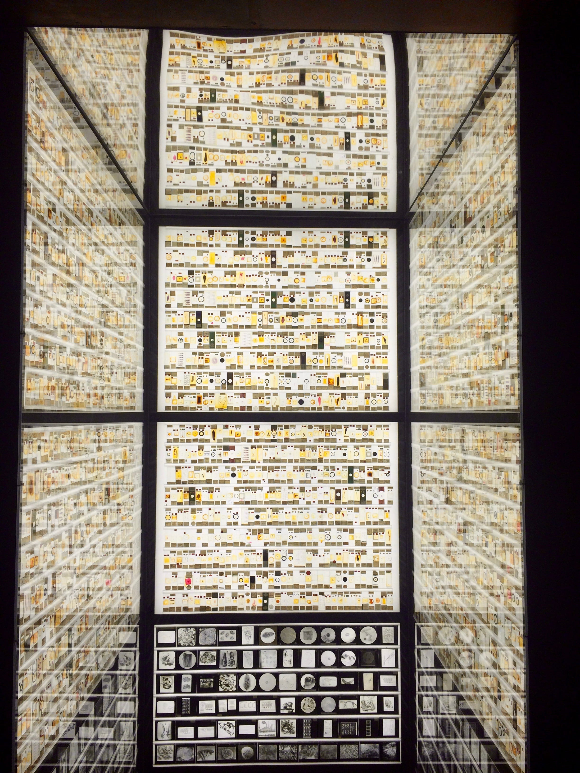
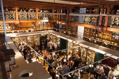
The Micrarium. Photo by author.

The Micrarium’s floor-to-ceiling lightboxes illuminate 2323 microscope slides featuring insects, sea creatures, and more, with another 252 lantern slides underneath. While this sounds like a lot of slides, it’s only around 10% of what the museum holds. Natural history museums often find it difficult to display their slide collections, but the diminutive creatures often featured on them make up most of our planet’s biodiversity.

I start most of my conversations with visitors during Student Engager shifts here – the Micrarium provides a clear illustration of my PhD research about how challenging aspects of diversity (of all kinds) are integrated into existing collections. It’s also an ideal place within the museum to try to pause people in the flow of their visit – it’s hard to resist stopping to snap a selfie or two.

Selfie by author.

The soft glow of the Micrarium’s backlit walls often draws people into the space without realising the enormity (or tininess!) of what they’re looking at. Over time, I’ve cultivated a number of favourites that I point out  in order to share the variety, strangeness, and poetry of the individual slides.

Small and mighty

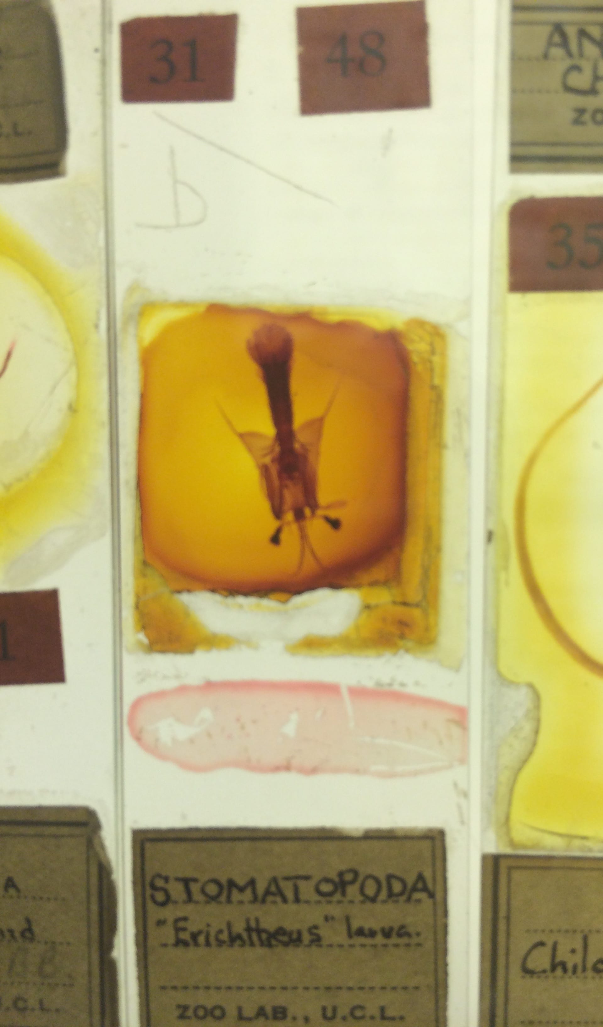
‘Stomatopoda “Erichtheus” larva’. Photo by author.

I was attracted to this slide because at first I thought it looked like a little flying squirrel. In actuality, it’s the larvae of a mantis shrimp.

The mantis shrimp is an incredible animal. To start, they have the most complex eyes of any animal, seeing a spectrum of colour ten times richer than our own. Its two ‘raptorial’ appendages can strike prey with an amount of force and speed, causing the water around them to boil and producing shockwaves and light that stun, smash and generally decimate their prey.

For more, check out this comic by The Oatmeal that illustrates just how impressive mantis shrimp are.

‘and toe of frog, wool of bat, and tongue of dog’

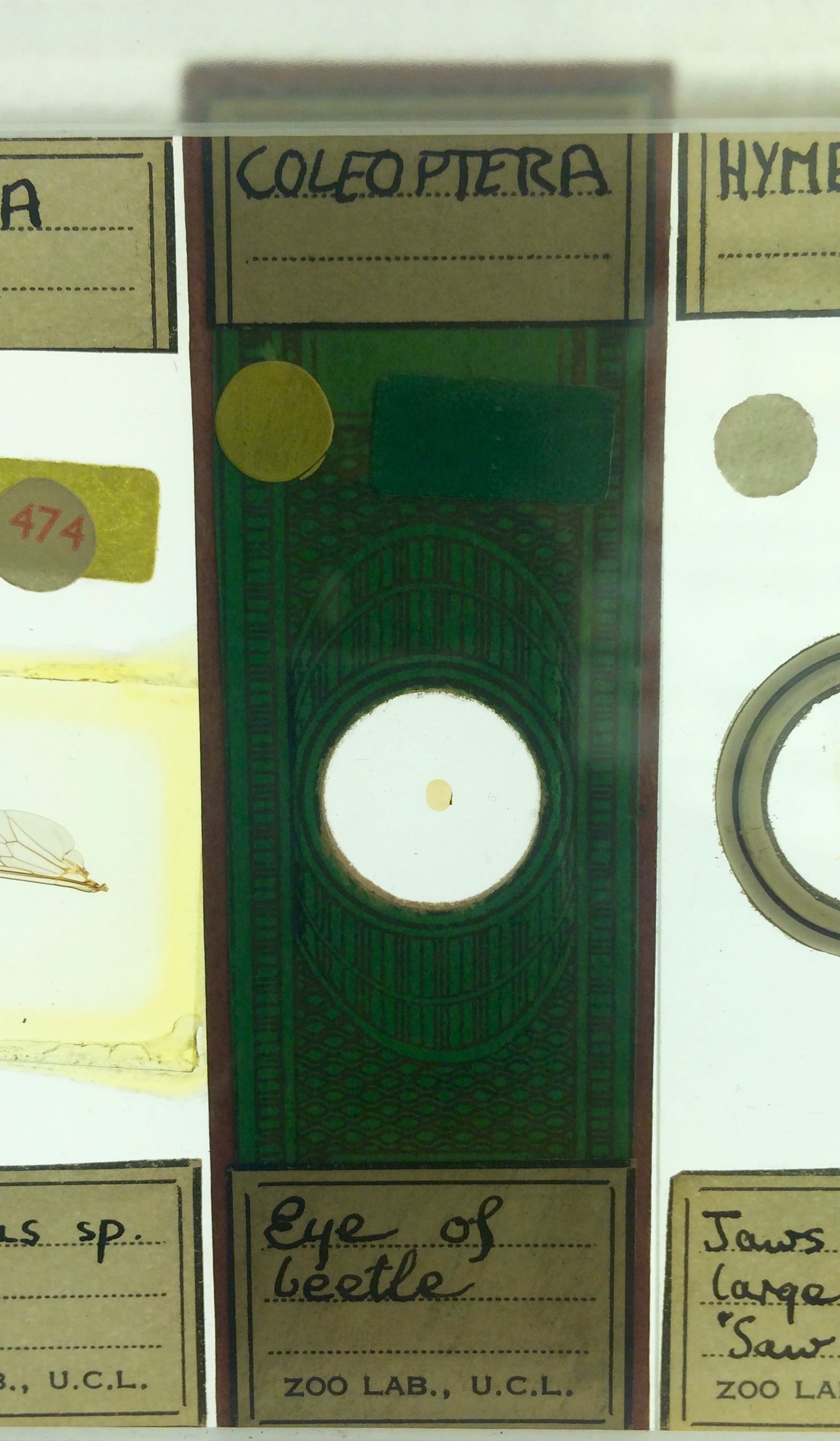
‘Eye of beetle’. Photo by author.

This is one of my favourite labels in the collection – was a zoologist also dabbling in witchcraft ingredients?  Probably not. But, I’d love to know what the slide was originally used for.

The slide itself also looks unusual due to its decorative paper wrapping. These wrappings were common to slides from the mid-19th century, which were produced and sold by slide preparers for others to study.

Many of the slides in the Micrarium were for teaching students who could check out slides like library books. So, perhaps it illustrated some general principles about beetle eyes rather than being used for specialist research.

Cat and Mouse

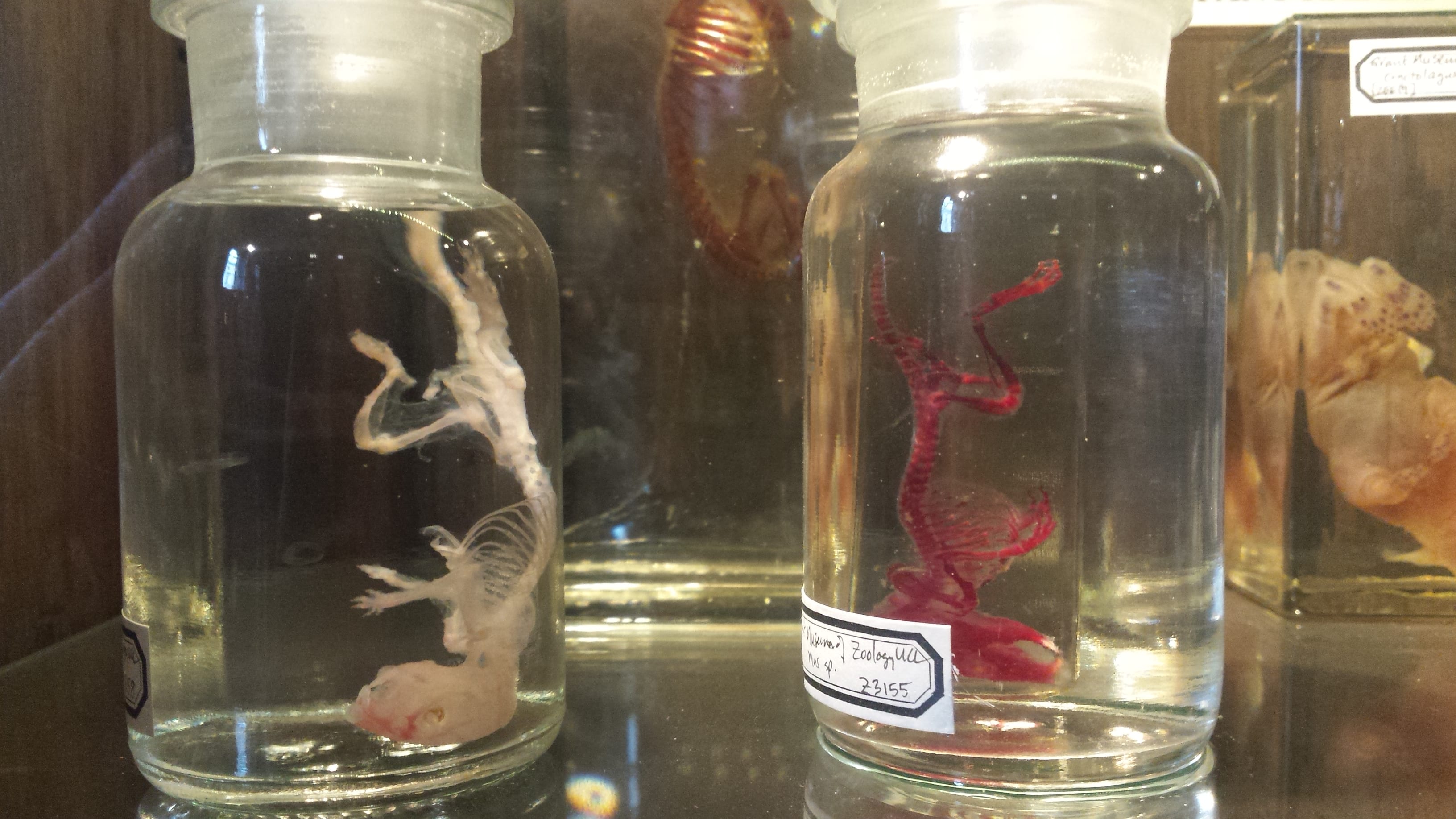
Fetal cat head (L). Embryonic mouse head (R). Photo by author.

One of the secrets of the Micrarium is that there are bits of larger animals hidden among all of the tiny ones. I like how the mice look surprisingly cheerful, all things considered. Bonus: see if you can also find the fetal cat paws!

Seeing stars

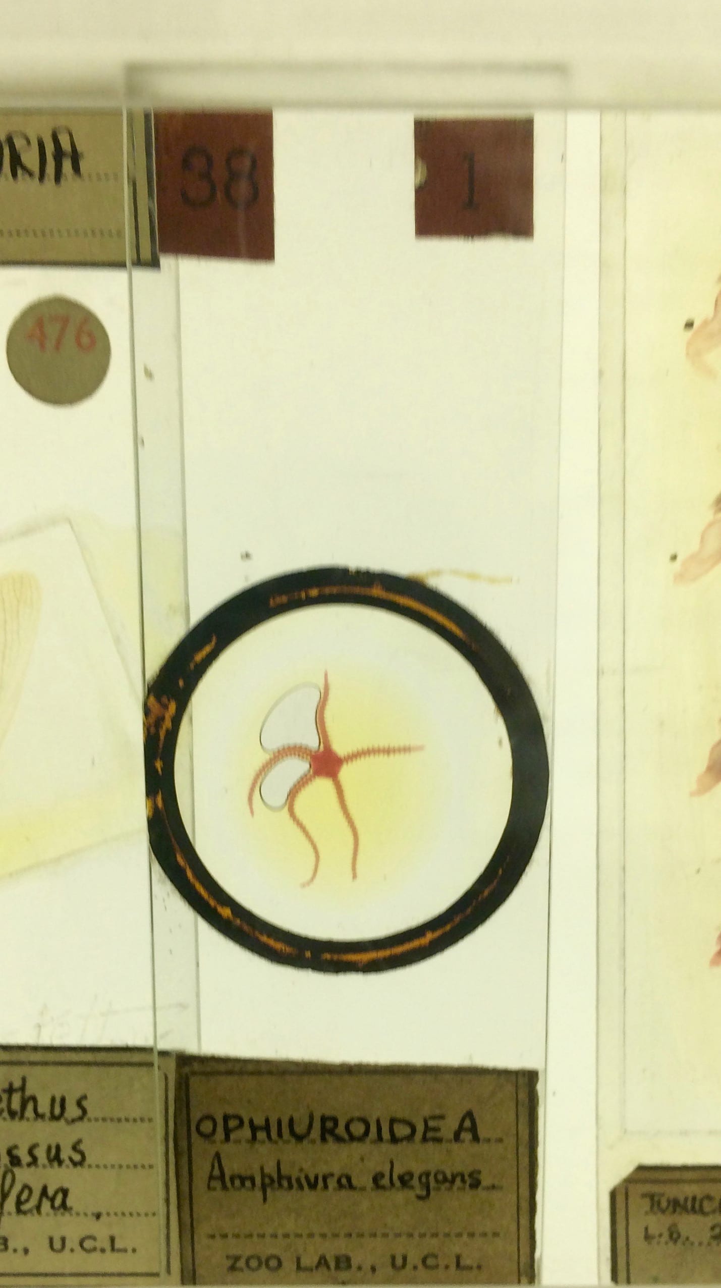
‘OPHIUROIDEA Amphiura elegens’. Photo by author.

This is a young brittle star, which in the largest species can have arms extending out to 60cm. Brittle stars are a distinct group from starfish; most tend to live in much deeper depths than starfish venture. They also move much faster than starfish, and their scientific name ‘Ophiuroidea’, refers to the slithery, snake-like way their arms move.

This slide can be found at child height, and it’s nice to show kids something they’re likely to recognise.

And finally:

Have you seen the bees’ tongue?

‘Apis (Latin for bee) tongue’ Photo by author.

Showing visitors this slide of the bee’s tongue almost always elicits surprise and fascination. Surprise at the seemingly strange choice to look at just the tongue of something so small and fascination at how complex it is.

We don’t normally think of insects having something so animal-sounding as a tongue (more like stabby spear bits to sting or bite us with!). But, bee tongues are sensitive and impressive tools: scientists have observed bee tongues rapidly evolving alongside climate change.

Good luck finding these…or your own Top 5! Share any of your favourites in the comments.

The Grant Museum blog did a similar post five years ago when the Micrarium opened. These don’t overalp with my Top 5 (which is easy to avoid when there are 2323 slides), so you should also check that out.

Season’s (Philosophical) Feastings

By Hannah L Wills, on 13 December 2017

Christmas is a time for overindulgence, so let’s have some tales of eighteenth-century feasting, with a twist from the history of science.

In my research, I examine the diary of Charles Blagden (1748-1820), physician, natural philosopher, and secretary to London’s Royal Society. One of the things I’ve been most struck by in my work on Blagden’s diary is the ever-presence of food and feasting within the social and scientific worlds of the late eighteenth century. Blagden’s diary reveals a near-daily itinerary of dining engagements where politicians, fellows of the Royal Society, and members of London’s well-to-do gathered to discuss news, politics, and the latest developments in natural knowledge over a range of lavish and often exotic meals. 

Scientific gatherings and feasts

A typical day for Blagden in the year 1795 began with a trip to the London home of Sir Joseph Banks, president of the Royal Society, for breakfast. Though the diary gives little indication of the food on offer, it does reveal that at these gatherings participants discussed news, politics, and natural philosophy, all over breakfast. On some occasions, Blagden and Banks conducted experiments, as revealed in Blagden’s diary entry for 19 February 1795: ‘Breakfasted at Sir Joseph Banks’s. all civil: made some experiments on crystallisation of nitre’.[i] This experiment was one that investigated the properties of a key ingredient in the manufacture of saltpetre (potassium nitrate) used in the manufacture of explosives.

On Thursdays, before the weekly meetings of the Royal Society, Blagden attended the Royal Society Club, a dining club for fellows of the Society held at the Crown and Anchor Inn on the Strand. While meetings of the club were intended to be social, scientific matters were inevitably discussed while members feasted on a variety of foods.[ii] The Royal Society archives contain some of the menus from these meetings, which at a dinner held on 23 October 1783 included ‘A Turtle’, which had for several days before the dinner been allowed to roam at Banks’s London home, ‘Scate’ (the fish skate), ‘Harricot of Mutton’ (a mutton stew), ‘a Hare’, ‘another dish of Turtle’, ‘Potatoes’, ‘Cold Ribs of Lamb’, ‘Breast of Veal’, ‘Haddock’ and finally ‘more of the Turtle’.[iii]

Feasting as research

As well as being a convivial aid to the discussion of natural philosophical topics, eating was also a central part of investigating nature. At gatherings hosted by Banks, visitors indulged in the consumption of various plants and animals, many sourced from exotic locations. One entry in Blagden’s diary reveals a particular gathering during which guests enjoyed several nuts brought by the botanist Richard Molesworth, named in Blagden’s diary as ‘Buticosa’ and ‘Sawena’. Blagden described them as ‘both pleasant to eat; one a sort of buttery nut, the other larger & more like walnut’.[iv]

Such behaviour might seem eccentric and even dangerous to us depending on the kinds of exotic fare on offer. Banks was frequently targeted by contemporary satire with his ‘philosophical’ feasting caricatured in a sketch by the artist Thomas Rowlandson. In ‘The Fish Supper’ (below) we see Banks’s guests, possibly including Blagden, eagerly preparing to devour an alligator specimen, while Banks, on the right-hand side of the image, greedily gnaws on a snake.

Thomas Rowlandson, Sir Joseph Banks about to Eat an Alligator (‘The Fish Supper’), 1788, ink and watercolour on paper (Image credit: © Tate (2014), CC-BY-NC-ND 3.0 Unported)


Festive feasting, with a bang

Experiments combined with dining did on occasion produce dangerous results. For a final festive example, we turn to an anecdote of the earlier eighteenth century. On Christmas Day 1750, Blagden’s contemporary Benjamin Franklin conducted an ill-fated experiment in cooking a turkey. Though today perhaps best known as one of the founding fathers of America, Franklin was also a renowned natural philosopher, famed for his electrical experiments. In April 1749, Franklin wrote a letter detailing an experiment he intended to make where ‘A turkey is to be killed for our dinner by the electrical shock, and roasted by the electrical jack’.[v] Franklin repeated this experiment on Christmas Day the following year with disastrous results, describing it as:

an Experiment in Electricity that I desire never to repeat… I inadvertently took the whole [shock] thro’ my own Arms and Body… the flash was very great and the crack as loud as a Pistol; yet my Senses being instantly gone, I neither Saw the one nor heard the other’.[vi]

Franklin’s turkey cooking is definitely a dining experiment not to be tried at home!


References:

[i] Royal Society Library, Charles Blagden’s Diary Vol 3, entry dated 19 Feb 1795, f. 47r.

[ii] For more information on the dining clubs of the Royal Society, including its membership, see T. E. Allibone, The Royal Society and Its Dining Clubs (Oxford: Pergamon Press, 1976).

[iii] Ibid., 121.

[iv] Royal Society Library, Charles Blagden’s Diary Vol 3, entry dated 17 Oct 1795, f. 70v.

[v] Meredith Man, ‘Ben Franklin on Cooking Turkey… with Electricity’, blog post for the New York Public Library website, published on 24 Nov 2014.

[vi] Ronald Clark, Benjamin Franklin: A Biography (London: Weidenfeld and Nicholson, 1983), 76.

This Little Piggy went to Market…

By uctzcbr, on 6 December 2017

One of the objects on display as part of the Grant’s Ordinary Animals exhibition is a jar of piglets. These particular specimens often receive a lot of attention, probably because the piglets are so small and visitors are surprised to discover what they are. I was happy to see them included in the exhibition because piglets are used by other researchers in my department (the Department of Crime and Security Science) and so they provide a useful launching pad to engage visitors in my research.

The specimen in question, courtesy of the Grant Museum.

This tactic has had varying success. The way pigs are used in Crime Research is fascinating but can also be a bit gruesome — perhaps too gruesome for some visitors. Pig skin, and flesh, is similar to that of humans and so can be used to conduct experiments in the absence of a human cadaver. For example, a colleague, Sian Smith, whose PhD research focuses on 3-D digitisation methods for sharp-force traumas, studies stab wounds she has made in pigs. A number of her experiments have required her to transport pig parts to Mile End cemetery where they are buried and left to decompose before being photographed so that the images can be used to create 3-D models. Her work has potential applications in crime scene forensics, as well as for providing evidence in court, or even archaeological research on burial sites and other human remains.

I think this story is fascinating and can start many different conversations about how crime research is conducted and used. But, I have learned very quickly not all visitors feel the same way. For example, I made the mistake of telling this exact tale to a pair of visitors who were vegan. Concerned by the use of living things in order to meet the needs of humans, they were not very impressed by this particular research project. Perhaps, I should have guessed that they were not my target audience. Whilst I have met many visitors in the museum who have backgrounds in forensics or who like to preserve animal remains as a hobby, many more visitors haven’t ever seen anything like the Grant Museum’s collection outside the museum itself. I now make sure I check with visitors whether or not they want all the gory details before launching into my stories.

It is worth pointing out, however, that a reason that piglets are often used in research is because it is not uncommon for mothers to kill their young accidentally (by rolling over on them) leaving farmers or other pig owners with piglets that they cannot raise but can sell to labs instead. The vegan visitors that I spoke to felt that — as the pigs were not killed for the purpose of research — it seemed reasonable to use their bodies in this manner. Incidentally, both visitors were organ donors and intended to leave their own bodies to science. We spent a great deal more time discussing the use of animals in scientific, but non-medical, research which made for very interesting chat, if not exactly where I saw the conversation from the start.

Why are animals 3D?

By ucbtch1, on 23 November 2017


Have you heard that our body is mainly composed of 70% water? Although true, the percentage varies from 55% of water in adult women, all the way up to 78% in babies, with the percentage for adult men somewhere in between. This is also true for animals, where some — like the jellyfish — have even 90% of their body composed of water. With this in mind, why don’t animals, including us, look like a soup? How can animals have a defined 3D structure?

Aurelia aurita, moon jellyfish, 5 preserved specimens (C193)

Aurelia aurita, moon jellyfish, 5 preserved specimens (C193)


Animals are made out of cells, the building blocks of our organs and tissues. But cells are basically a bag of water and chemicals; so again, why don’t animals look like giant bags of chemicals? The most obvious reason is that animals have bones that give structure to the rest of the body. But even bones are 31% water, and organs with no bones, such as hearts, still have a unique 3D form. Hearts have defined chambers (see the elephant heart below); they’re not just a mush of cells. The answer lies not in the cells themselves but in what surrounds them.

Elephant dried heart (Z639)

Elephant dried heart (Z639)

Cells are engulfed by the extracellular matrix (ECM) which is mainly composed of proteins. This matrix encompasses the space in-between cells, gives them structural support and acts like a scaffold. It can also act as a pathway for cells to migrate along and it gives out chemical and physical cues that cells respond to. The ECM varies from organ to organ. The brain, for example, is mainly composed of cells with an ECM of only 20% of the total mass. In contrast, cartilage has fewer cells and around 70% of its mass is ECM. Every cell type is surrounded by a specific matrix that will affect its function. Studying this extracellular environment is important to understand how cells develop, how they interact with each other, and how they react to disease.

At the same time, by studying the ECM, researchers can get an idea of how an organ or tissue is structured and how to replicate its intricate architecture. Scientists that work in tissue engineering use a technique which consists of washing away the cells of an organ, literally. By using detergents, the cells are washed away in cycles until just the extracellular matrix is left. In this manner, they can analyse its composition and experiment with the matrix with the end goal of growing an organ in the lab. Therefore, one day we could replace diseased or aged organs with new ones without the need for transplantation. The unique composition of the ECM provides cells with the support they need to survive, and at the same time, gives animals and their organs a defined 3D structure.


Sources:

https://water.usgs.gov/edu/propertyyou.html

Make a Museum about Pokémon!

By Josie Mills, on 21 November 2017

Post-it Note found on the visitor feedback board in the Petrie Museum of Egyptian Archaeology (if this was left by you - do get in touch!)

Post-it Note found on the visitor feedback board in the Petrie Museum of Egyptian Archaeology (if this was left by you – get in touch!)


While working in the Petrie Museum last week I glanced at the board reserved for visitor feedback, noticed this post-it and couldn’t resist taking a photo… As a child of the nineties I have a soft spot for Pokémon and have wholeheartedly embraced revival of the plucky little critters. I’ve particularly enjoyed the nostalgia of sharing the new games with my younger brother, Daniel; if only there were a degree in poké-studies!

Last year the app Pokémon Go made headlines world-wide and the presence of the virtual creatures in museums and archives was widely discussed within the heritage industry. Perhaps unsurprisingly, I’m team positive for Pokémon Go in museums, and the staff at UCL Museums have written some great blog posts on the subject. See this post by Grant Museum Manager Jack Ashby and Research Engager Arendse’s blog.


A wild Pigeotto appears as I am doing my PXRF analysis (Image: J Mills 2016)

A wild Pigeotto appears as I am doing my PXRF analysis (Image: Author’s own photo)


In fact, museums, archaeological ruins, and science labs also feature in the Pokémon games, serving as venues to transform fossils found in the wild into rare Pokémon. These Pokémon, like those mentioned in Arendse’s post, were inspired by real fossils, for example Omanyte from ammonites, and Aerodactyl from pterodactyl. My personal favourite is Relicanth, a fish bearing a close resemblance to the Lazarus species the coeleocanth!


Ammonite

Left: Omanyte (Image: Bulbapedia; Right: Ammonite (Image: British Geological Survey)


Omanyte and Ammonite

Here you can see the similarities between the shell of the Pokémon and the preserved ammonite Mantelliceras, which lived during the Late Cretaceous/Cenomanian period around 100 million years ago. This was a time when much of the Chalk in England formed and Cretaceous ammonite fossils are common in chalky areas of the South Coast. Interestingly fossil ammonites are often displayed ‘upside down’ (effectively with their head in the air!) whereas the Pokémon are orientated to move with their tentacles at the base, more like the original creatures.


Top: Aerodactyl (Image: https://bulbapedia.bulbagarden.net/wiki/Aerodactyl_(Pok%C3%A9mon ); Bottom: Pterodactyl (Image: http://dinosaurpictures.org/Pterodactyl-pictures)

Top: Aerodactyl (Image: Bulbapedia); Bottom: Pterodactyl (Image: Dinosaur Pictures)


Aerodactyl and Pterodactyl

Perhaps the most literal translation of the three are the obvious similarities between the Aerodactyl Pokémon and the extinct Pterodactyl, which lived during the Jurassic period 200-150 million years ago. The fossils on the game are rare and can only be found in certain areas; similarly pterodactyl fossils are also localised in real life, with most excavated from the Solnhofen limestone in Germany.

Top: Relicanth (Image: https://vignette3.wikia.nocookie.net/pokemon/images/a/ad/369Relicanth_AG_anime.png/revision/latest?cb=20141006041759); Bottom: Coelacanth (Image: http://vertebrates.si.edu/fishes/coelacanth/coelacanth_wider.html)

Top: Relicanth (Image: Bulbapedia); Bottom: Coelacanth (Image: Smithsonian)


Relicanth and Coelacanth

Even Relicanth’s name nods to the antiquity and mysterious-ness of the real-life fish the Coelacanth. The Coelacanth is a ‘Lazarus’ species, a term that refers to a taxon that was thought to be extinct but reappears again in the wild. The cryptic nature of the Coelacanth is reflected by the camouflage cartoon pattern of the Pokémon, a graphic allegory of the fish’s complex history!

Although the Pokémon hype has gradually dwindled, perhaps this message from our younger audience (or any nineties kids out there), highlights what we can take from Pokémon: the appeal of learning about animals and artefacts, the surprise of finding new things where you didn’t expect them, and the lure of encountering the rare and interesting.

More practically, specimens that have inspired both fossils and non-fossil Pokémon are on display across the Grant Museum. However, if anybody is interested in funding a museum solely about Pokémon, please get in touch: I know someone who might be suitable for curator *cough*…

Are emojis the hieroglyphics of the 21st century?

By Julia R Deathridge, on 7 November 2017

Emojis are everywhere. Whether they’re all over your social media, on advertisements on the tube, or adorning t-shirts and bags in Primark, you just can’t escape them! A recent survey by TalkTalk revealed that 72% of 18-25 year olds find it easier when texting to express themselves with emojis rather than using the written word. And it’s not just millenials who have been affected – even my Mum can’t send me a text without including one. But this is not the first time pictorial images have been used as a form of written communication.

During a recent shift in the Petrie museum, I realised that many of the hieroglyphic carvings on display held a strong resemblance to an emoji-filled text I had sent earlier that day; this left me wondering what the similarities are between the two languages. Are emojis a step forwards in how we communicate or are we reverting back to the language of the ancient Egyptians?

Hieroglyphic inscription from an anti-chamber wall (UC45400)

Hieroglyphic inscription (UC45400)

The History of Hieroglyphics

 Hieroglyphics are considered to be one of the oldest forms of written language, with the earliest known form dating back to 3300-3200BC. The term hieroglyphics was coined by the ancient Greeks to describe the ‘sacred carvings’ they observed on Egyptian monuments. In ancient Egyptian the word for hieroglyphics translates to mean ‘the word of the gods’, highlighting its importance in Egyptian culture.

Unlike emojis, which are used by more than 90% of the world’s online population, only a small percentage of Ancient Egyptians were taught how to write hieroglyphics, such as priests, royals and civil officials. Consequently, hieroglyphics were predominantly confined to religious texts, royal documents and the recording of historical events.

Divorce document inscribed in hieratic (UC19614)

Divorce document inscribed in hieratic (UC19614)

Over time, the use of hieroglyphics became more widespread in Egyptian civilization; this resulted in a simplified cursive form of the script, known as hieratic being developed. Despite hieroglyphics being the language most commonly associated with Ancient Egypt, hieratic was actually used for the bulk of written texts. Hieratic was simplified even further into demotic scripture in 7th century BC. Thereafter, hieroglyphics were primarily used for inscriptions on buildings, and as a form of decorative writing on furniture and jewellery.

Deciphering Hieroglyphics

Use of hieroglyphics declined rapidly in Egypt under Roman rule and their meaning was lost for almost 2,000 years until the rediscovery of the Rosetta Stone in 1799.

CC BY-SA 4.0. Attribution: © Hans Hillewaert

Rosetta Stone  CC BY-SA 4.0. © Hans Hillewaert

The Rosetta stone was the missing key to deciphering hieroglyphics, as it was engraved with a text written in three different ways: hieroglyphics, ancient Greek and demotic script. The French scholar Jean-Francoise Champillion used the Rosetta stone, alongside the work of other European scholars, to decipher the hieroglyphs and unlocked the language of the Ancient Egyptians once more.

Work deciphering the different written texts of Ancient Egypt is still ongoing. UCL’s Papyrus for the People Project aims to improve our understanding of the collection of written texts at the Petrie Museum and make them more accessible to the general public. You can read more about the project here.

Emojis vs Hieroglyphics

The term emoji originates from the Japanese for pictograph: e “picture” + moji “character”. Emojis are classified as a pictographic and ideographic writing system that uses symbols to represent an object or an idea rather than specific words. Although at first glance hieroglyphics may also appear to function in a similar way, the language is actually far more layered and complex.

Hieroglyphics are comprised of phonograms which represent sounds, logograms which represent words or phrases, and determinatives which are used at the end to clarify meaning of the word.

Hieroglyphic characters can also have multiple meanings depending on how they are used. For example the symbol for ‘house’, which was pronounced as pr, can also be used phonetically to represent the sound ‘pr’ in other words. Combinations of hieroglyphics characters could therefore be used to spell out larger words and composite phrases.

According to a journalist at the Guardian, emojis are an evolutionary step back, a return to the ‘static culture’ of ancient Egypt that was limited by its use of hieroglyphs. However, the hieroglyphics language was far more than ‘picture writing’. It allowed ancient Egyptians to compose a huge variety of texts from medical documents to poetry – texts that are significantly more advanced than what is possible to convey with emojis. Let’s just say if my doctor tried to write my medical report purely in emojis I would be concerned!

Emojis are a great form of communication and can add a creative flair to how we message one another. However, they will never be a replacement for the written word and I doubt they would have the capacity to help build and maintain an entire civilisation. If I change my mind and decide to write my thesis in emoji, I’ll let you know!

emoji wink


How to visualize the insides of an animal?

By ucbtch1, on 26 October 2017

When studying animals, sometimes we need to study them from the inside out —literally. One way to do this is to cut them open and looking at their internal structures, such as with the bisected heads or the microscope slides in the Grant Museum of Zoology. Another way to visualize the inside of an animal is to stain a particular body part while making everything else clear; researchers can do this by using chemicals and colour stains. For example, in the Grant Museum of Zoology, we can find specimens like the tarsier, with its skeleton stained in red, or the zebrafish with red and blue parts.

Adult tarsier stained with Alizarin Red to show calcium (Z2718)

Adult tarsier stained with Alizarin Red to show calcium (Z2718)

Adult zebrafish stained with Alcian Blue and Alizarin Red (V1550)

Adult zebrafish stained with Alcian Blue and Alizarin Red (V1550)


The process of staining these animals begins with the removal of the skin, viscera and fat tissue.  Then, soft tissues like muscle are cleared using a variety of different methods which mostly involve exposing the specimen to different baths of chemicals. Next, the bones are stained with Alizarin Red and the cartilage with Alcian Blue. It’s a long process that can take a couple of days because the stain needs to properly penetrate the tissues, but the results are amazing.

Initially, both Alizarin Red and Alcian Blue were used as textile dyes, but now they also have numerous biological applications. Alizarin Red staining is a method to visualize mineralized tissue because it stains calcium and Alcian Blue stains specific structures mainly found in cartilage. These stains constitute an important part of research because they allow researchers to visualize the intricate structure of tissues and thus understand how they form throughout development.

ADSCs

Image credit: Eleonora Zucchelli

In the lab where I study, researchers work with adipose (fat) derived stem cells which have the capacity to become different kinds of mature cells. These stem cells are grown under specific conditions and by changing these conditions scientists can direct them into becoming mature cells like fat, bone or cartilage — a process called differentiation. But this process can take anywhere from a couple of weeks up to a couple of months! In order to determine if the differentiation is working, researchers stain the stem cells with Alizarin Red and Alcian Blue to identify if they are in fact turning into bone or cartilage. In the images depicted, undifferentiated adipose derived stem cells (ADSCs) on the top appear clear but their differentiated counterparts are stained in blue or red. This means the differentiation is working.

There are many other stains used on animals or cells. The process of clearing and staining can be very complicated depending on the specimen and what one wishes to stain, but the results can be quite fascinating. What animal would you like to see stained from the inside.


Mouse stained with alizarin red (Z3155)

Mouse stained with alizarin red (Z3155)


References:

PUCHTLER, H., Meloan, S. N., & TERRY, M. S. (1969). On the history and mechanism of alizarin and alizarin red S stains for calcium. Journal of Histochemistry & Cytochemistry17(2), 110-124.

McLeod, M. J. (1980). Differential staining of cartilage and bone in whole mouse fetuses by alcian blue and alizarin red S. Teratology22(3), 299-301.


Question of the Week: What’s this Museum For?

By Hannah L Wills, on 19 October 2017

By Hannah Wills


A couple of weeks ago, whilst engaging in the Grant Museum, I started talking to some secondary school students on a group visit to the museum. During their visit, the students had been asked to think about a number of questions, one of which was “what is the purpose of this museum?” When asked by some of the students, I started by telling them a little about the history of the museum, why the collection had been assembled, and how visitors and members of UCL use the museum today. As we continued chatting, I started to think about the question in more detail. How did visitors experience the role of museums in the past? How do museums themselves understand their role in today’s world? What could museums be in the future? It was only during our discussion that I realised quite how big this question was, and it is one I have continued to think about since.

What are UCL museums for?

The Grant Museum, in a similar way to both the Petrie and Art Museums, was founded in 1828 as a teaching collection. Named after Robert Grant, the first professor of zoology and comparative anatomy at UCL, the collection was originally assembled in order to teach students. Today, the museum is the last surviving university zoological museum in London, and is still used as a teaching resource, alongside being a public museum. As well as finding classes of biology and zoology students in the museum, you’re also likely to encounter artists, historians and students from a variety of other disciplines, using the museum as a place to get inspiration and to encounter new ideas. Alongside their roles as spaces for teaching and learning, UCL museums are also places for conversation, comedy, film screenings and interactive workshops — a whole host of activities that might not have taken place when these museums were first created. As student engagers, we are part of this process, bringing our own research, from a variety of disciplines not all naturally associated with the content of each of the museums, into the museum space.


A Murder-Mystery Night at the Grant Museum (Image credit: Grant Museum / Matt Clayton)

A Murder-Mystery Night at the Grant Museum (Image credit: Grant Museum / Matt Clayton)


What was the role of museums in the past?

Taking a look at the seventeenth and eighteenth-century roots of the Ashmolean Museum in Oxford and the British Museum in London, it is possible to see how markedly the role and function of the museum has changed over time. These museums were originally only open to elite visitors. The 1697 statues of the Ashmolean Museum required that ‘Every Person’ wishing to see the museum pay ‘Six Pence… for the Space of One Hour’.[i] In its early days, the British Museum was only open to the public on weekdays at restricted times, effectively excluding anyone except the leisured upper classes from attending.[ii]

Another feature of these early museums was the ubiquity of the sense of touch within the visitor experience, as revealed in contemporary visitor accounts. The role of these early museums was to serve as a place for learning about objects and the world through sensory experience, something that, although present in museum activities including handling workshops, tactile displays, and projects such as ‘Heritage in Hospitals’, is not typically associated with the modern visitor experience. Zacharias Conrad von Uffenbach (1683-1784), a distinguished German collector, recorded his visit to Oxford in 1710, and his handling of a range of museum specimens. Of his interactions with a Turkish goat specimen, Uffenbach wrote, ‘it is very large, yellowish-white, with… crinkled hair… as soft as silk’.[iii] As Constance Classen has argued, the early museum experience resembled that of the private ‘house tour’, where the museum keeper, assuming the role of the ‘gracious host’, was expected to offer objects up to be touched, with the elite visitor showing polite and learned interest by handling the proffered objects.[iv]

Aristocratic visitors handle objects and books in a Dutch cabinet of curiosities, Levinus Vincent, Illustration from the book, Wondertooneel der Nature - a Cabinet of Curiosities or Wunderkammern in Holland. c. 1706-1715 (Image credit: Universities of Strasbourg)

Aristocratic visitors handle objects and books in a Dutch cabinet of curiosities, Levinus Vincent, Illustration from the book, Wondertooneel der Nature – a Cabinet of Curiosities or Wunderkammern in Holland. c. 1706-1715 (Image credit: Universities of Strasbourg)


How do museums think about their function today?

In understanding how museums think about their role in the present, it can be useful to examine the kind of language museums employ when describing visitor experiences. The British Museum regularly publishes exhibition evaluation reports on its website, detailing visitor attendance, identity, motivation and experience. These reports are fascinating, particularly in the way they classify different visitor types and motivations for visiting a museum. Visitor motivations are broken down into four categories: ‘Spiritual’, ‘Emotional’, ‘Intellectual’ and ‘Social’, with each connected to a different type of museum function.[v]

Those who are driven by spiritual motivations are described as seeing the museum as a Church — a place ‘to escape and recharge, food for the soul’. Those motivated by emotion are understood as searching for ‘Ambience, deep sensory and intellectual experience’, the role of the museum being described as akin to that of a spa. For the intellectually motivated, the museum’s role is conceptualised as that of an archive, a place to develop knowledge and conduct a ‘journey of discovery’. For social visitors, the museum is an attraction, an ‘enjoyable place to spend time’ where facilitates, services and welcoming staff improve the experience. Visitors are by no means homogenous, their unique needs and expectations varying between every visit they make, as the Museum’s surveys point out. Nevertheless, the language of these motivations reveals how museum professionals and evaluation experts envisage the role of the modern museum, a place which serves multiple functions in line with what a visitor might expect to gain from the time they spend there.

What will the museum of the future be like?

In an article published in Frieze magazine a couple of years ago, Sam Thorne, director of Nottingham Contemporary, invited a group of curators to share their visions on the future of museums. Responses ranged from the notion of the museum as a ‘necessary sanctuary for the freedom of ideas’, to more dystopian fears of increased corporate funding and the museum as a ‘business’.[vi] These ways of approaching the role of the museum are by no means exclusive; there are countless other ways that museums have been used, can be used, and may be used in the future. My thinking after the conversation I had in the Grant Museum focussed on my own research and experience with museums, but this is a discussion that can and should be had by everyone — those who work in museums, those who go to museums, and those who might never have visited a museum before.


What do you think a museum is for? Tweet us @ResearchEngager or come and find us in the UCL museums and carry on the discussion!


References:

[i] R. F. Ovenell, The Ashmolean Museum 1683-1894 (Oxford: Clarendon Press, 1986), 87.

[ii] Fiona Candlin has written on the class politics of early museums, in “Museums, Modernity and the Class Politics of Touching Objects,” in Touch in Museums: Policy and Practice in Object Handling, ed. Helen Chatterjee, et al. (Oxford: Berg, 2008).

[iii] Zacharias Konrad von Uffenbach, Oxford in 1710: From the Travels of Zacharias Conrad von Uffenbach, trans. W. H. Quarrell and W. J. C. Quarrell (Oxford: Blackwell, 1928), 28.

[iv] Constance Classen, “Touch in the Museum,” in The Book of Touch, ed. Constance Classen (Oxford Berg, 2005), 275.

[v] For this post I took a look at ‘More than mummies A summative report of Egypt: faith after the pharaohs at the British Museum May 2016’, Appendix A: Understanding motivations, 27.

[vi] Sam Thorne, “What is the Future of the Museum?” Frieze 175, (2015), accessed online.

Label Detective: Are Bacteria ‘Ordinary Animals?’

By tcrnkl0, on 17 October 2017

A few weeks ago, the Grant Museum opened a new exhibit, The Museum of Ordinary Animals: boring beasts that changed the world. As a detective of the mundane myself, I am a huge fan. But I’m particularly curious about the ordinary animals we can’t see.

Rather than focusing on a specific artefact label, I answer the title question by visiting two places in the Museum of Ordinary Animals exhibition that help raise questions about how things are organised and labeled in zoology more broadly.

Case notes: Bacteria are everywhere. As I mentioned in my previous post, we have 160 major species of bacteria in our bodies alone, living and working together with our organ systems to do things like digest nutrients. This is also happens with other animals — consider the ordinary cow, eating grass. Scientist Scott F. Gilbert tells us that in reality, cows cannot eat grass. The cow’s genome doesn’t have the right proteins to digest grass. Instead, the cow chews grass and the bacteria living in its cut digest it. In that way, the bacteria ‘make the cow possible’.

IMG_1102

The Ordinary Cow, brought to you to by bacteria. Credit: Photo by author

Scientifically speaking, bacteria aren’t actually ‘animals’; they form their own domain of unicellular life. But, as with the cow, bacteria and animals are highly connected. Increasingly, scientists say that the study of bacteria is ‘fundamentally altering our understanding of animal biology’ and theories about the origin and evolution of animals.

But, before we get into that, let’s go back to Charles Darwin (1809-1882). Darwin studied how different species of animals, like the pigeon, are related to each other, and how mapping their sexual reproduction shows how these species diversify and increase in complexity over time. This gets depicted as a tree, with the ancestors at the trunk and species diversifying over time into branches.

Picture1

Darwin’s Ordinary Tree of Pigeons. Photos by author

When scientists began to use electron microscopes in the mid-20th century, our ideas about what made up the ‘tree of life’ expanded. We could not only observe plants, animals, and fungi, but also protists (complex small things) and monera (not-so-complex small things). This was called the five kingdom model. Although many people still vaguely recollect this model from school, improved techniques in genetic research starting in the 1970s has transformed our picture of the ‘tree of life’.

It turns out we had given way too much importance to all the ordinary things we could see, when in fact most of the tree of life is microbes. The newer tree looks like this:

Credit: Wikipedia Commons

Credit: Wikipedia Commons

Now there are just three overarching domains of life: Bacteria, Eucarya (plants, animals, and fungi are just tiny twigs on this branch), and Archaea (another domain of unicellular life, but we’ll leave those for another day).

There’s a third transformation of the ‘tree of life’, and this one is my favourite. Since the 1990s, DNA technology and genomics have given us an even greater ability to ‘see’ the diversity of microbial life and how it relates to each other. The newest models of the tree look more like this:

Credit: Wikipedia Commons

Credit: Wikipedia Commons

This is a lot messier. Why? Unlike the very tiny branches of life (plants and animals) that we focused a lot of attention on early on in the study of evolution, most of life on earth doesn’t reproduce sexually. Instead, most microbes transfer genes ‘horizontally’ (non-sexually) across organisms, rather than ‘down’ a (sexual) genetic line. This creates links between the ‘branches’ of the tree, starting to make it look like….not a tree at all. As scientist Margaret McFall-Ngai puts it: ‘we now know that genetic material from bacteria sometimes ends up in the bodies of beetles, that of fungi in aphids, and that of humans in malaria protozoa. For bacteria, at least, such transfers are not the stuff of science fiction but of everyday evolution’.

Status: Are bacteria Ordinary Animals? We can conclusively say that bacteria are not animals. But, they are extremely ordinary, even if we can’t see them with the naked eye. In truth, they’re way more ordinary than we are.


Notes

As with the previous Label Detective entry, this post was deeply inspired by the book Arts of Living on a Damaged Planet, an anthology of essays by zoologists, anthropologists, and other scholars who explore how environmental crisis has highlights the complex and surprising ways that life on earth is tied together. Scott F. Gilbert and Margaret McFall-Ngai, both cited above, contribute chapters.

Add Like an (Ancient) Egyptian

By uctzcbr, on 12 October 2017

As student engagers, we work in each of the museums no matter how far from our own disciplines they are. I study cybercrime which is not clearly related zoology, art, or Egyptology; as a result, I have received many looks of surprise from visitors when they discover someone working in the museum is not an expert in the subject matter. To be a better student engager, I have learned a lot about the history of each museum and researched many objects so that I can answer questions and provide useful information to visitors, but I also like to talk about subjects related to my discipline. For the Grant Museum, this means talking about a study which looked at the trade (or lack thereof) of endangered animal souvenirs on the Dark Net; for the Art Museum, I talk about an art exhibition displaying objects purchased at random from Dark Net Markets. However, I have always struggled to link my research to Archaeology and the objects at the Petrie.

Instead, I like to talk about my undergraduate degree: Mathematics. There is evidence that the Ancient Egyptians had not only a counting system but prolifically and pragmatically used Mathematics. Records show that they used maths for accounting, architecture, and astronomy, amongst other things. Their techniques enabled a complex tax system and were even adopted by Greek mathematicians such as Pythagoras.

Papyrus showing mathematical calculations in Hieratic script.

Papyrus showing mathematical calculations in Hieratic script.

However, Egyptian mathematics was very different to that which we use today. Whilst they also used a base 10 system, at first they only had symbols for the numbers 1, 10, 100, 1,000, 10,000 and 100,000. This made writing numbers sometimes laborious – to write the number 7, you would need to write the hieroglyph for the number 1 seven times. The numbers 2-9 were added later after they began writing on papyrus using the Hieratic script instead of Hieroglyphs.  Fractions were denoted using a specific symbol and could only be of the form 1 , with a 1 as the numerator. This system made addition and subtraction simple but other tasks, such as multiplication, much more complex.

To do these more complex computations, the Ancient Egyptians would combine addition and subtraction in brute force methods that would provide approximations of the answer. For example, to multiply two numbers together, they would add the first number to itself the second number of times in a process of doubling not unlike the way computers are now programmed to do. As an illustration, to calculate 3 × 4 they would double 3 (that is 3 × 2) and then double 3 again (that is 3 ×(2+2)=3 ×4.

They would also rely on pre-calculated times tables to increase the speed of their work and prevent them from having to repeat the same problems again and again. This is believed to be the case because some of these tables have survived to today. For example, object UC32159 is a section of papyrus that displays division tables containing the answers to 2 being divided by the odd numbers from 3 to 31.

Remains of papyrus showing the division of 2, written in Hieratic script.

Remains of papyrus showing the division of 2, written in Hieratic script.

The collections in each of UCL’s museums are so large and varied that there will always be something relevant and of interest to anyone who visits.