 Close
Close

How to visualize the insides of an animal?
By ucbtch1, on 26 October 2017
When studying animals, sometimes we need to study them from the inside out —literally. One way to do this is to cut them open and looking at their internal structures, such as with the bisected heads or the microscope slides in the Grant Museum of Zoology. Another way to visualize the inside of an animal is to stain a particular body part while making everything else clear; researchers can do this by using chemicals and colour stains. For example, in the Grant Museum of Zoology, we can find specimens like the tarsier, with its skeleton stained in red, or the zebrafish with red and blue parts.

Adult tarsier stained with Alizarin Red to show calcium (Z2718)

Adult zebrafish stained with Alcian Blue and Alizarin Red (V1550)
The process of staining these animals begins with the removal of the skin, viscera and fat tissue. Then, soft tissues like muscle are cleared using a variety of different methods which mostly involve exposing the specimen to different baths of chemicals. Next, the bones are stained with Alizarin Red and the cartilage with Alcian Blue. It’s a long process that can take a couple of days because the stain needs to properly penetrate the tissues, but the results are amazing.
Initially, both Alizarin Red and Alcian Blue were used as textile dyes, but now they also have numerous biological applications. Alizarin Red staining is a method to visualize mineralized tissue because it stains calcium and Alcian Blue stains specific structures mainly found in cartilage. These stains constitute an important part of research because they allow researchers to visualize the intricate structure of tissues and thus understand how they form throughout development.
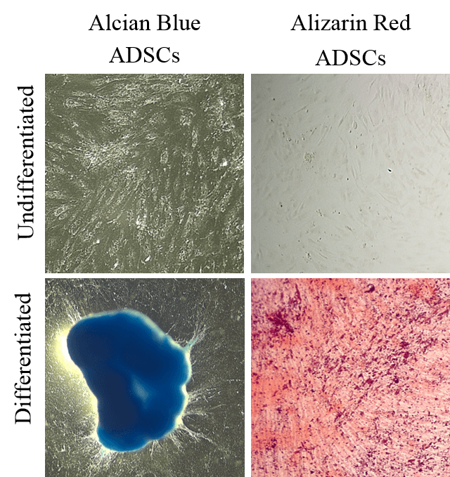
Image credit: Eleonora Zucchelli
In the lab where I study, researchers work with adipose (fat) derived stem cells which have the capacity to become different kinds of mature cells. These stem cells are grown under specific conditions and by changing these conditions scientists can direct them into becoming mature cells like fat, bone or cartilage — a process called differentiation. But this process can take anywhere from a couple of weeks up to a couple of months! In order to determine if the differentiation is working, researchers stain the stem cells with Alizarin Red and Alcian Blue to identify if they are in fact turning into bone or cartilage. In the images depicted, undifferentiated adipose derived stem cells (ADSCs) on the top appear clear but their differentiated counterparts are stained in blue or red. This means the differentiation is working.
There are many other stains used on animals or cells. The process of clearing and staining can be very complicated depending on the specimen and what one wishes to stain, but the results can be quite fascinating. What animal would you like to see stained from the inside.
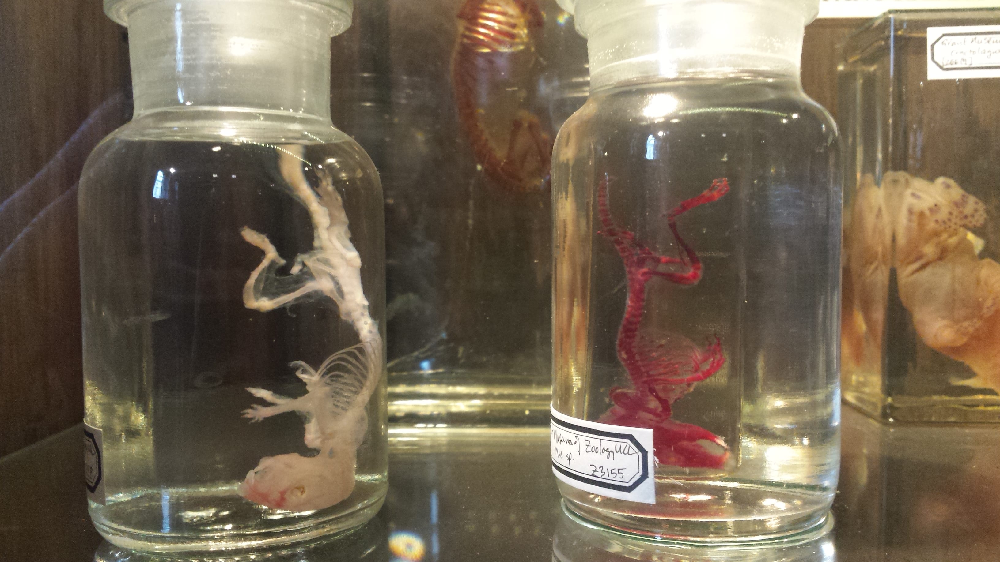
Mouse stained with alizarin red (Z3155)
References:
PUCHTLER, H., Meloan, S. N., & TERRY, M. S. (1969). On the history and mechanism of alizarin and alizarin red S stains for calcium. Journal of Histochemistry & Cytochemistry, 17(2), 110-124.
McLeod, M. J. (1980). Differential staining of cartilage and bone in whole mouse fetuses by alcian blue and alizarin red S. Teratology, 22(3), 299-301.