X Close

Researchers in Museums

Home

Engaging the public with research & collections

Menu

Archive for the 'Public Engagement' Category

Question of the Week: How do snakes poop?

By Arendse I Lund, on 9 November 2016

Sometimes kids ask the darndest things, and this week in the Grant Museum one kid asked me how do snakes poop? I didn’t know, and none of the books we consulted seemed to get to the bottom of this.
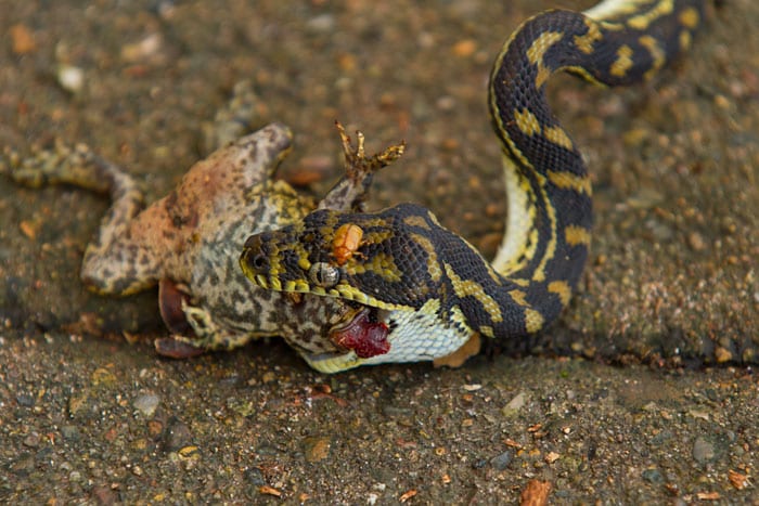
carpet snake eating toad

Juvenile carpet snake eating a cane toad (Photo: Andrew Mercer)

It turns out that a snake’s excretion process is highly variable from species to species. When a snake eats something—be it a mouse, deer, or hippo—it’s digested, and the gut extracts the nutrients. Poop consists of everything that couldn’t, for whatever reason, be extracted. Rat snakes defecate approximately every two days; bush vipers defecate every 3-7 days. A good rule of thumb is that if a snake eats frequently, it will defecate frequently. If a snake eats infrequently, it will defecate infrequently. Simple in theory, this means that a snake may defecate only a few times a year. (One snake was recorded holding it for 420 days!)

Because of this, up to 5-20% of a snake’s body weight at any given time may be fecal matter. In a human of 130 lbs, that would be 6.5-26 lbs of feces. If you think that number is incredible, it turns out that a snake eating a particularly large meal will potentially experience its body mass more than double!

Alazarin stained snake

Grass snake stained with Alazarin Red to show the skeletal structure (Grant Museum, X50)

How big a snake is and where it lives matters too. Scientists have found there to be a positive correlation between the ingestion to defecation period and the relative body mass of snake species. An arboreal snake will defecate soon after eating to maximize mobility; a terrestrial snake such as the Gaboon Viper, which lies still for days on end, doesn’t require the same speediness and doesn’t defecate as frequently. One theory suggests that holding onto its feces may help a snake as the increased size and weight could anchor it in attacks on larger and heavier animals. If the added weight becomes cumbersome, then the snake can simply dispel it. (Please don’t try this at home.)
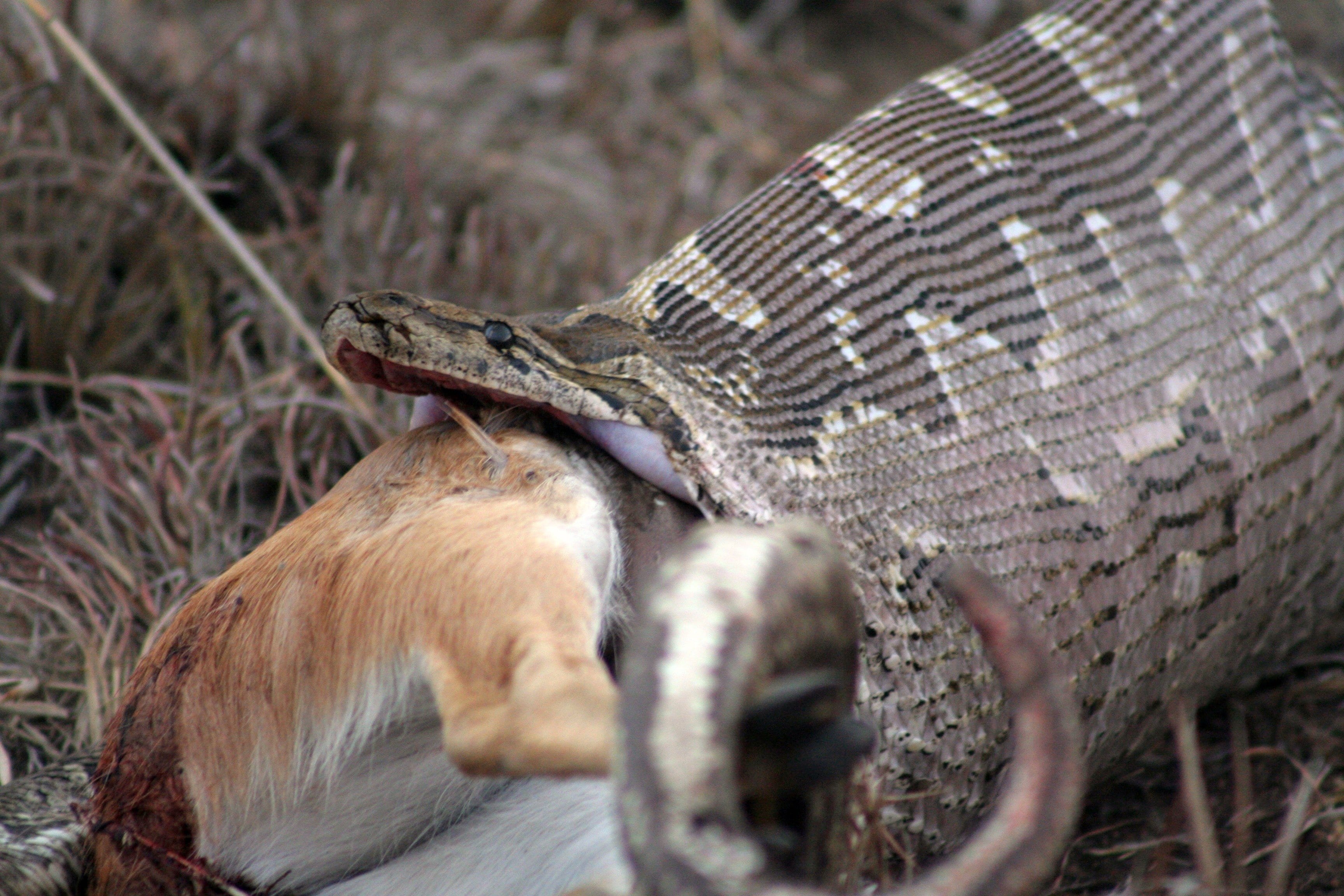
rock python eating antelope

An African Rock Python eating an antelope (Photo: Alex Griffiths)

So in the end, where does it all go? Once the meal is reduced to poop, the snake can get rid of it through an anal opening, or cloaca, which is Latin for ‘sewer.’ This opening can be found at the end of a snake’s belly and beginning of its tail; unsurprisingly, the feces are the same width as the snake’s body. A snake will use the same opening to defecate, urinate, mate, and lay eggs—now that’s multi-purpose!

Follow Arendse Lund on Twitter

The Student Engager Project featured on the LSE Impact Blog

By Kevin Guyan, on 12 August 2016

The Student Engager project featured on the LSE Impact Blog, an online hub for those researching and working at universities who wish to maximise the impact of academic work.  In the article, the project coordinator Kevin Guyan discusses the potential benefits of the public engagement project for training the next generation of researchers in ways to communicate research with non-academic audiences.


LSE Impact Blog

To read the full article visit the LSE Impact Blog.


On the Origin of Pokémon Species

By Arendse I Lund, on 27 July 2016

Arendseby Arendse Lund

Last week, I was in the Grant Museum of Zoology when a cry came that a Pokémon had been spotted! Mobiles out, the visitors advanced on the creature and succeeded in capturing it. Surrounded by zoological specimens meticulously collected over centuries, here were people amassing their own digital collection of creatures. From a mouse that shoots lightning bolts to a shellfish that poisons with its lick, these creatures, collectively called Pokémon, take a variety of forms and all have different abilities. Many of these Pokémon are based on real animals.

TapirFor example, take Drowzee: These psychic Pokémon eat dreams and are based on the chiefly nocturnal tapirs, animals indigenous to tropical America and Southeast Asia. Tapirs have short legs and the Malayan tapirs are two-toned just like their virtual counterparts; they also have long, flexible snouts which allow them to grab foliage beyond their reach and even act as a snorkel when swimming. The Japanese word for tapir, baku, refers to both the zoological animal and a spirit in folklore which consumes dreams—just like its Pokémon counterpart.

PangolinAnother example are Sandslash, which have long claws for burrowing, feature brown quills covering their bodies, and will roll into a ball in order to defend themselves from attack. These Pokémon take their inspiration from the pangolin, which are sometimes referred to as scaly anteaters. However, they are more closely related to giant pandas. The name derives from the Malay word pengguling, meaning “something that rolls up.” They have overlapping keratin scales armoring their backs, with tails strong enough to hang from trees, and a tongue that, when extended, is longer than its body. These incredible creatures are found in Asia and Africa but are sadly the most trafficked animals in the world.

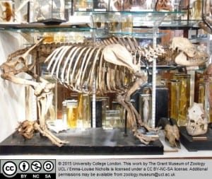
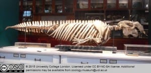
DugongThen there’s Seel, which evolves into Dewgong. In these two Pokémon, everything’s in the names: The former is based off of a seal and the latter a dugong. In appearance, dugongs are similar to manatees but both male and female dugongs grow two tusks—this is reflected in its adorable Pokémon counterpart. The semi-nomadic dugongs’ habitat extends throughout the Indo-West Pacific but they mostly stay in the bays around Australia; their conservation status is listed as vulnerable due to overdevelopment of the coastal areas and excessive fishing. Dugongs derive their name from the Malay word duyung, meaning “lady of the sea” and the species is possibly the origin of the mermaid myth.

There are many more examples of the real-life basis for Pokémon: axolotl for Mudkip, tadpoles for Poliwag, and racoons for Zigzagoon, to name just a few. The creator of Pokémon, Satoshi Tajiri, was heavily influenced by the type of creatures he found while insect collecting as a child. He used to sneak out into rice paddies and look under rocks for beetles. In a 1999 interview, he bemoaned the disappearance of those paddies and said of his love for the outdoors, “As a child, I wanted to be an entomologist. Insects fascinated me. Every new insect was a wonderful mystery. And as I searched for more, I would find more. If I put my hand in a river, I would get a crayfish. Put a stick underwater and make a hole, look for bubbles and there were more creatures.” These findings influenced many of the creatures that would make up the Pokémon world.

As both the Grant and Petrie Museums are PokéStops, it’s great to see people encouraged to check out the museum collections as they pursue their Pokémon. Perhaps most fascinating of all, these virtual Pokémon collections now spark conversations with strangers over techniques and the best places to acquire rarer species in a strikingly similar way to amassing physical collections of any sort.

And any avid collector, be it of stamps or insects, can understand the lure of the Pokémon slogan: “Gotta catch ‘em all!”

Question of the Week:

What is an axolotl and why is it so unique?

By ucbtch1, on 4 May 2016

By Citlali Helenes Gonzalez

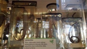
From the many specimens that are on exhibit at the Grant Museum of Zoology, it is hard to choose one that stands out. For me, it has to be the axolotl, a small little creature that passes by unnoticeable. It is not the biggest or the strangest but it has a unique feature that we humans desire to achieve: tissue regeneration. Although newts, salamanders and starfish can also regenerate tissue, the axolotl is probably the most interesting of these animals because of the extent of its capacity.

20160210_134444When we humans have an injury, our body starts forming scar tissue over the injury. With just a few exceptions, like the skin that is regenerating constantly and the liver that does something similar to regeneration, the rest of our body has very limited regeneration capacity.

The axolotl, on the other hand, after an injury, the tissue at the wounding site starts to regrow new healthy tissue instead of scar tissue. It does so to the extent that it can regrow a whole limb, jaw, tail, spinal cord and even some parts of its brain. Scientists have even transplanted organs from one axolotl to another with no rejection issues. And if all of this is not enough for you to be amazed, the axolotl is over 1000 times more resistant to cancer than mammals. This is why it has been used as a model animal for the study of regeneration and development.

Studying the axolotl has huge implications for medical research because if we can learn how the mechanisms of regeneration in this little animal work, then maybe we can simulate it in humans. And in the long run, if we learn how to regenerate and repair tissue, this could mean no more need for transplants, no more prosthesis of arms or legs, helping burn victims just to name a few benefits.

Unfortunately, this little animal native to Mexico has been listed as an endangered species due to the destruction of its natural habitat in the lakes and canals of Mexico City. But scientists across the globe are still studying them and looking closer into their genome to try to unlock the secrets of tissue regeneration. So next time you stop by the Grant Museum take a closer look at this fascinating animal that even though its small size has a lot to offer to human medical research.


Further reading:

Roy, S. and Lévesque, M., 2006. Limb regeneration in axolotl: is it superhealing? The Scientific World Journal6, pp.12-25.

http://blogs.scientificamerican.com/guest-blog/regeneration-the-axolotl-story/


Question of the Week:

Was Using Human Remains for Science Taboo?

By ucramew, on 20 January 2016

Misha Ewen

By Misha Ewen


During a shift in the Grant Museum of Zoology recently, an American high school student asked me about the history of the collection and how it has been (and still is) used to teach students about anatomy. We got on to talking about museum collections that have specimens of human remains, like the Hunterian Museum in London. His next question was, when did we stop feeling that studying human remains through dissection, for the purposes of science, was taboo?

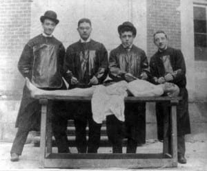
Nowadays, it’s commonplace for students studying anatomy to encounter human remains as part of their university degree, but this wasn’t always the case. In the early nineteenth century, there was a dire shortage in Britain of bodies for the purpose of medical research. For instance, the Edinburgh Medical College received fewer than five cadavers a year [1]. This was because only the remains of executed criminals could legally be used. The limitations put on scientific research because of this policy gave oxygen to the criminal business of ‘body-snatching’. When it began, the ‘snatchers’ invented a method to remove bodies from graves without detection: they used to dig holes, some distance away, and tunnel down into the graves before pulling bodies out by rope or hooks. Those who could afford it soon began to invest in mausoleums, vaults and table tombstones to ensure the safekeeping of their eternal resting places [2].

Medical students? Body-snatchers? Or both?

Medical students? Body-snatchers? Or both?

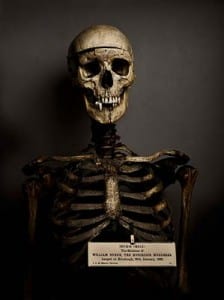
The business of bodysnatching, that fuelled medical research, soon turned even more sinister… In 1831 three men were arrested in London for the murder of vagrants, individuals whose deaths they thought would go unnoticed. On the day they were arrested, they had tried to sell the body of a fourteen year old boy to the lecturers of King’s College for twelve guineas [3]. There was also the famous case of William Burke and William Hare in Edinburgh, who murdered seventeen victims between 1827 and 1829, before selling the corpses to Dr Robert Knox at the Edinburgh Medical College. Unfortunately, this grisly business was inherently tied up in the advancement of medical knowledge.

The dissection of bodies was problematic, in both religious and moral terms, for contemporaries. In the first instance, many believed that their bodies had to remain intact for the afterlife, and dissection was also widely considered to be a punishment for the worst type of criminal. Take the fate of the Edinburgh bodysnatcher William Burke, for instance: he was executed by hanging in 1829 and his body was then publicly dissected at the Edinburgh Medical College [4]. And yet, in this period, recognition of the need for medical students to learn from human subjects was growing.

Courtesy of the Edinburgh City of Literature

Courtesy of the Edinburgh City of Literature

Public outcry, because of the black-market that had developed around medical research, helped the passing of a new bill: the 1832 Anatomy Act, which recognised that more bodies were needed for research and teaching. University College London’s Jeremy Bentham, who donated his own body to science (his auto-icon remains in the UCL South Cloisters), helped prepare the bill before his death in 1832. The act significantly extended access to cadavers, by allowing anatomists to dissect ‘unclaimed bodies’, individuals who died without anyone coming forward to pay for their burial. This was mostly people who died destitute in hospitals, workhouses and prisons. Dissection was no longer solely associated with individuals who were executed for murder, it was now also associated with the shame of dying in poverty [5].

It was really only in the mid-twentieth century that the donation of bodies to science became commonplace. Yet even now, we often feel squeamish about donating our bodies to science after we die. Attitudes certainly have changed, however, since 1832. From December 2015, individuals living in Wales will now have to opt-out if they don’t want their organs donated when they die, and legislation will certainly change soon in the rest of the United Kingdom.


[1] http://www.edinburgh-history.co.uk/burke-hare.html

[2] http://www.history.co.uk/study-topics/history-of-death/the-rise-of-the-body-snatchers

[3] http://www.exclassics.com/newgate/ng609.htm

[4] http://www.edinburgh-history.co.uk/burke-hare.html

[5] http://www.kingscollections.org/exhibitions/specialcollections/charles-dickens-2/italian-boy/anatomy-act

Further reading:

Colin Blakemore & Sheila Jennett, ‘body snatchers’, The Oxford Companion to the Body (2001). Encyclopedia.com. <http://www.encyclopedia.com>.

Stress: Using Oral History Interviews

By tcrnfmw, on 11 November 2015

Profileby Felicity Winkley 

This post is associated with our exhibit Stress: Approaches to the First World War, open October 12-November 20.


A visitor to Stress last week commented that the objects on display weren’t what she expected, that she had anticipated they would show much more directly the obvious effects, or stresses, of the First World War on men, women and children.

I wasn’t surprised by this response. We knew when we curated Stress that the interpretation of the objects was more convoluted than most traditional museum displays – the object labels are longer than best practice advises, the visual links between the cases difficult for the visitor to immediately grasp.

Part of this is owed to the fact that the objects have been chosen from the UCL collections – geology, pathology and science specimens among others – rather than from a military history assemblage. In equal measure, it is also because the objects have been chosen not only because of their relevance to the exhibition, but also according to the individual research interests of the curators.

One element of the exhibition breaks this mould however – the audio installation which plays two oral history interviews from the archives of the National Army Museum, recollections of two individuals who served in the First World War. For me, this part of the exhibition provides the visitor with the most direct link to the conflict – an immediate and very powerful ‘place-setting’ via the experiences being narrated, quite apart from the objects on display.

Although the audio is on a permanent loop, no matter at what stage you join in the story you are transported: with Adelaide Marian Davies, who served with the Women’s Army Aux Corps in France, you can picture the scene as she describes the dances held for troops at the Front, where it was forbidden for her rank to dance with the officers; with L/Cpl Billy Meade, you might join him at the Dardanelles, Ypres or later at the prisoner of war camp where he tasted Schnapps for the first time.

The Oral History Listening Post at Stress

The Oral History Listening Post at Stress

The resonance of these anecdotes illustrates just why oral history interviews are important, and why they are such a useful element to incorporate in exhibitions, or indeed many kinds of research. As opposed to much of the written historical record, oral histories are collected directly from the source and feel so much more authentic for it. For the purposes of my PhD, I used a ‘go-along’ interview technique, which involved talking to respondents whilst walking, in order to glean accurate insight to their experience of being in that environment. More recently, I volunteered as an oral history interviewer for the London Bubble’s After Hiroshima project which explores the responses of Londoners to the dropping of the first atomic bomb on 6 August 1945, both in the immediate aftermath and throughout the peace movement of the 1950s and 60s.

Oral histories lend themselves to many situations, not only to provide a means for gathering unique, illuminating and personal records and reflections, but also – in the process of their collection – to involve a wider community in research and offer an opportunity for participation in history and heritage in practice. We were thrilled to be joined on the Stress opening night by the family of Billy Meade, including his daughter (now 84), who had never before heard his recording.

Stress: Selecting and Engaging with Pathology Specimens in the Exhibition Space

By Sarah Savage Hanney, on 28 October 2015

By Sarah Savage Hanney

Over the past two and a half weeks, I have had the pleasure of engaging with visitors in the Stress exhibition at the North Lodge. When visitors first come into the space, many ask questions about the concept behind the exhibition and the selection of objects. In the planning process, each exhibition curator chose objects from the UCL Collections that related to his/her approach to stress in the First World War and significance within individual PhD research.

When the Student Engager group first discussed curating an independent exhibition using UCL Collections objects and specimens in summer 2014, I already knew exactly what collection I wanted to use: the Pathology Collection. Unlike the three UCL Museums on campus, the Pathology Collection was less accessible to the public due to human tissue licensing restrictions. For the previous Engager event Movement in May 2014, I used photographs of Pathology specimens to enhance visitors’ understandings of the effects of disease on the human body. Visitors were especially interested in the photographs of a coal miner’s lung, diseased human heart, and a haemorrhaged brain. I kept these interesting specimens in mind and hoped to use them in our future exhibition.

Luckily, the Pathology Collection received its license to display human tissue early in 2015 and it would be possible to display Pathology specimens in the Stress exhibition. The curator of the Pathology Collection, Subhadra Das, was incredibly helpful in suggesting specimens and organising the conservation work for the two final selections: the coal miner’s lung and the diseased human heart.

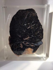
Visitors can now see the coal miner’s lung and diseased human heart suspended in a clear, preservative liquid on either end of the long wall display in the exhibition space. As a historian of medicine specialising in the early twentieth century, I wanted my contribution of specimens to highlight little known medical conditions that affected people in the First World War period.

Although we do not know specific dates for these specimens, the organs came from patients at University College Hospital in the first half of the twentieth century.

At first glance, the specimens can be a bit off putting. The coal miner’s lung barely looks like a lung apart from the general shape. The lung is nearly completely black and exposes the harsh reality of the health of British coal miners. For those men who remained in Britain mining coal for the war effort, their efforts would eventually cost them their healthy lungs.

Coal Miner's Lung- UCL Pathology Collection

Coal Miner’s Lung- UCL Pathology Collection


The diseased human heart also holds special significance for health in the First World War period. Over the course of the war, the British military medical officers discovered that many young men who enlisted to fight had pre-existing heart conditions that would affect their ability as fit, healthy soldiers. After speaking with visitors about this specimen, many visitors commented on how they never associated heart disease with the early twentieth century.

Diseased Human Heart- UCL Pathology Collection

Diseased Human Heart- UCL Pathology Collection


By having these specimens on public display, I hope that visitors contemplate the stress that the First World War placed on the physical bodies of those who fought and contributed to the war effort. The exhibition presents a rare opportunity for visitors to examine these remarkable specimens in person and engage in discussion with curators about their different approaches to stress in the First World War.

If you are interested in speaking with Engager Sarah Savage Hanney in the exhibition space, she will be at Stress each Friday from 1pm-5pm for the next three weeks.

Stress is open Monday through Friday from 1pm-5pm and on alternating Saturdays.

For more information about visiting the UCL Pathology Collection at the Royal Free Hospital Campus of the UCL Medical School.

Upcoming Events:

Commemoration Event, November 11, 2015  1pm-3pm UCL Art Museum

Bloomsbury Walking Tour, November 20, 2015  1pm-2pm UCL Quad

Question of the Week:

Why can’t I touch museum objects?

By Stacy Hackner, on 19 August 2015

DSC_0745By Stacy Hackner

For humans, touch is an important way to gain information about an object. We can tell if something is soft or hard, heavy or light, smooth or rough or fluffy, pliable, sharp, irregular. During my masters class on human dentition, I learned to identify teeth by touch to get around visual biases. We spent a significant amount of time touching objects in our environment, so we tend to get angry when museums tell us not to touch the objects.

I understand the desire to touch a piece of history. There’s a feeling of authenticity you get from holding something made by ancient people, and a sense of disappointment if you’re told the artifact is actually a replica. A British Museum visitor commented that “It was just lovely to know that you could pick something up that was authentic. It was just lovely to put your hands on something.” Another said “You do think sometimes when you’re looking in the cases, sometimes I’d like to pick that up and really look closely.”[i]

Even with “no touching” signs, museum visitors continue to touch things. Sometimes it’s by accident and sometimes they get a sneaky look on their faces, knowing they’re ignoring the signs; most often, they don’t realize what they’re doing is damaging the object.

Passive conservation of an object involves creating a stable environment so that the object can continue its “life” undisturbed. Sudden changes in humidity, temperature, and light can degrade the object. Touching it introduces dirt and oils from your skin onto its surface – the same way you’d leave fingerprints at a crime scene. Additionally, the oils can then attract dirt to linger, and acidic oils can also degrade metallic surfaces.

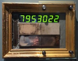
Yes, museum professionals handle objects for research purposes. However, we attempt to handle them as little as possible with clean hands and wear gloves when appropriate. This difference between museum staff and the public is also one of quantity: it’s ok if one person does it occasionally, but if everyone touches it on every visit, the grime adds up. In 2009, the Ashmolean Museum in Oxford introduced a “touchometer” that counts how many people have touched an object made of various materials. As you can see in the image below, after nearly 8 million touches, the left half of the object is severely degraded. The stone (centre) has developed a patina, the metal (bottom) has become shiny, and the cloth (left) has entirely worn away. (Also, people have scratched the frame.)

The Ashmolean's Touchometer. Thanks to Mark Norman.

The Ashmolean’s Touchometer. Thanks to Mark Norman, the Ashmolean’s Head of Conservation.

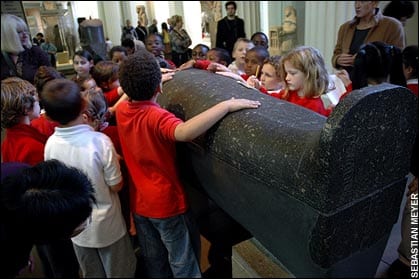
If you walk through the British Museum’s gallery of Egyptian statuary, you can clearly see the areas on artifacts that people like to touch – the corners and public-facing edges of sarcophaguses are darker than the wall-facing edges, and anything round and protruding tends to have a sheen that takes years of painstaking work to remove (hands, feet, and breasts of statues at human height are particularly vulnerable).

Schoolchildren touch a sarcophagus. Credit: Sebastian Meyer for The Telegraph.

The Grant Museum has specific objects that can be handled, and UCL Museums have object-based learning programs to introduce students and specific groups to handling museum objects. [ii] Many other museums have touch tables or touch sessions where you can feel the weight of hand axes or porcupine quills. Don’t despair if you’re asked not to touch something in a museum – we’re not angry, we just want to make sure they’re preserved for future museum visitors to enjoy.


Sources

[i] Touching History: An evaluation of Hands On desks at The British Museum. 2008. Morris Hargreaves Mcintyre.

[ii] UCL Museums Touch & Wellbeing; Object-Based Learning

Conservation Advice – Handling Museum Objects. 2015. Southeast Museums.

Question of the week:

Are there any objects depicting Queen Nefertiti at Petrie museum?

By rmjllil, on 22 July 2015

By Ann Liljas

Earlier this month a new statue of Queen Nefertiti was unveiled in Egypt. In case you haven’t heard about Nefertiti, she lived around 1370-1330 BC and was married to Pharaoh Akhenaten. Nefertiti means “the beautiful has come” and to many Egyptians she is a symbol of the country’s beauty and rich cultural heritage. The new sculpture was intended to be a replica of a famous 3,300-year old bust of the queen (currently housed in Neues Museum in Berlin) at the entrance of the city of Samalut. But immediately when the new statue was unveiled it was criticised for being an incredibly ugly copy. Within just a few days Egyptians had forced the authorities to remove the statue. A visitor to Petrie museum had heard about this controversial statue and asked me if there are any artefacts at Petrie museum depicting Nefertiti.

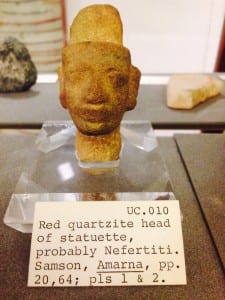
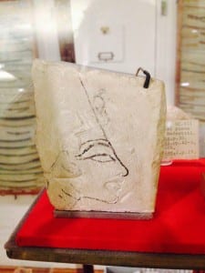
In 1891 Flinders Petrie excavated two artefacts that are likely to be representing Nefertiti at Amarna. One is a sketch of the queen wearing her distinctive tall crown on limestone. The other piece is a small head modelled from red quartzite which is believed to depict Nefertiti. The artefacts made an enormous impact on the contemporary art world in London when exhibited in the 1890s. Petrie also found earrings from the same province (Amarna) and time (1550-1300 BC) which may have belonged to Nefertiti. All these objects are on display at the Petrie museum.

Click here to see pictures of the new statue of Nefertiti published by the BBC.

image1 image2 image3

Stress: Building an Exhibition

By Kevin Guyan, on 20 July 2015


Kevin GuyanBy Kevin Guyan


With the announcement of the Student Engagers’ autumn exhibition, here is the first in a series of blog posts that share personal insights into the curatorial process.


Stress offers the student engagement team an opportunity to curate an exhibition that counters the traditional view of museums and galleries as fixed spaces that display objects that convey a message. Instead – I see this as a chance for us to experiment with bold and exciting ways to share knowledge and create a space on campus for three-way conversations between curators, objects and the public.

The inception of our exhibition first found life in the summer of 2014 during a conversation between the student engagement team and the UCL Art Museum. Over one year later and, as design ideas and draft event listings are shared via email, the exhibition at last feels like it is coming together.

As our more experienced colleagues in UCL Museums and Public and Cultural Engagement warned, ‘exhibition time’ runs significantly slower than ‘normal time’ and we now appreciate starting this project with 16 months to spare. I remember looking at action plans with distant dates and the feeling that our plans were a lifetime away. Those dates have now come and gone as we hurtle through 2015 and towards our opening night on Friday 9 October.

The meat and bones of the student engagement project is the presence of researchers in UCL’s three public museums. Therefore, one of the key hopes for Stress was to import this practice and create an exhibition where a researcher was always present, waiting and ready for conversation.

The researcher’s presence will also create a way to feed-back information from visitors into the planning of events, pre-empting many questions and queries fielded and offering a more tailored visitor experience. It further gives us opportunities to adapt the exhibition during its run. For example, conversations between engagers and visitors will inform the writing of blog articles that will then shape how future visitors perceive the objects on display.

Like our previous events and exhibitions, Movement, LandSCAPE and Foreign Bodies, the theme of Stress brings together the research interests of a diverse group of PhD students under one overarching theme. This means that the visitor experience will differ according to the researcher in the exhibition space and their interpretation of the objects on display.

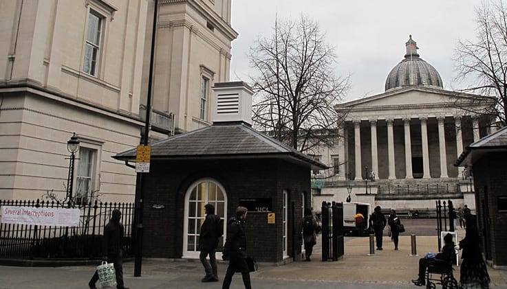
North Lodge

UCL’s North Lodge will house a team of postgraduate researchers throughout the exhibition.

I am excited to see how this works in reality – the continual presence of a researcher in the North Lodge exhibition space may prove overbearing and turn-off visitors looking for a space of solitude in busy Bloomsbury. More optimistically, the space will become a talking shop at the entrance to UCL’s campus and create a different, yet equally enriching, experience for visitors.

For me, public engagement is about more than sharing research ideas with other people. The benefits should reach far further than dissemination alone and empower researchers to enter into dialogues with people from different backgrounds. The process of sharing ideas with people unfamiliar with our own field will foster new and unexpected connections and force us to change the way we share our work, ultimately resulting in a deeper understanding for everyone involved.

We are attempting to build an exhibition with public engagement as a foundational building block and create a space that gives researchers and visitors opportunities to follow pathways unaware where they might lead. This is very much the ethos of the student engagement project – let’s see how our ideas work in reality.