X Close

Researchers in Museums

Home

Engaging the public with research & collections

Menu

Archive for the 'Museum Trail' Category

Mythical Hybrids and Fantastic Beasts

By Gemma Angel, on 13 May 2013

Gemma Angelby Gemma Angel


I’m going to describe a creature, and you have to try and guess what it is, based on the following three clues: 1) it lays eggs; 2) it has venomous claws; and 3) it uses electroreception to assist it in catching prey under water. You probably guessed some sort of reptile, right? Wrong. Ok, so those questions were a bit tricky. I’ll give you another three clues: 4) it’s semi-aquatic; 5) it has thick fur; and 6) despite laying eggs, it suckles its young on milk. Some of you will probably have worked out what this mysterious animal is by now. I am, of course, describing Ornithorhynchus anatinus, or as it is more commonly known, the platypus.

Growing up in Australia, I was fascinated by the native wildlife. As a curious 7-year-old recently emigrated from England, I tried to assimilate the unfamiliar Antipodean fauna into my limited understanding of the animal kingdom, largely through approximations: To me, the wombat was like a kind of stout, snub-nosed badger; sugar gliders were reminiscent of squirrels; and the echidna was a larger and longer-nosed version of the hedgehog. Kangaroos were a more difficult species to accommodate, with a face similar to a deer, and the hind legs of some sort of giant Alice-in-Wonderlandesque rabbit. But my system completely fell down when it came to the platypus. This creature was truly weird, a kind of animal cut-and-paste that defied all of the categories that I tried to fit it into. As it turned out, I wasn’t alone in my estimations of this remarkable and unique creature.

Platypus-sketch

Ornithornhynchus anatinus, John Gould (1863).

As an Australian native, the platypus has been known in Aboriginal culture for millennia – but it was not until 1797 that Europeans first encountered them. Captain John Hunter of the Royal Navy sent a pelt and a sketch back to Britain in 1798, [1] but the bizarre appearance of the creature baffled European naturalists. Some considered it to be an elaborate hoax, and Scottish zoologist Robert Knox believed the creature to be the work of an inventive Asian taxidermist. Even George Shaw, the first man to scientifically describe the platypus, admitted that “a degree of scepticism is not only pardonable, but laudable … I almost doubt the testimony of my own eyes.” [2]

Whilst it makes perfect sense that European observers would find the platypus strange, having never encountered anything like it in the Northern hemisphere outside of the bizarre chimerical creatures of mythology, it is perhaps more surprising that Aboriginal Dreamtime legends also describe the platypus as a peculiar exception within the animal realm. Known as the ‘mallangong’, tambreet’ or ‘duliawarung’ to local indigenous peoples, Aboriginal story-telling traditions use myth to explain the unique appearance and behavioural characteristics of the platypus. The platypus was believed to be the offspring of a mother duck and a father water rat, accounting for its unusual characteristics – inheriting the duck-bill, webbed feet and egg-laying abilities of their mother, and the thick fur, claws and four legs of their father. In an origin story of the platypus from Northern New South Wales, their poor mother Gaygar is ostracized by the other ducks because of her bizarre-looking hatchlings, and is forced to leave her home on Narran Lake. She takes her babies up into the Warrumbungle mountains, thereby accounting for why platypus are only found in particular regions. In another story from the New South Wales Central Coast, the animals argue amongst themselves about who is the most important creature. They form three exclusive groups, all convinced of their superiority: The animals with fur who can run across land, the birds who lay eggs, and the water creatures who can swim. All of the groups want the platypus to join them, since he shares characteristics with all of them, and each faction invites him to be part of their group. After thinking about this for some days, the platypus gathers all the animals to tell them his decision:

I don’t have to join anyone’s group to be special because I am special in my own way. Because I have fur and love to run across the land, I have a little bit of animal in me. I also have a little bit of bird in me because of my bill and the fact that my wife lays eggs. As well, I also have a bit of water creature in me because I love to swim and explore the underwater world. […] I don’t know why the ancestors have made us all different, but we must learn to accept these differences and live with each other. [3]

All of the animals listening, including people, agreed that the platypus was very wise; and the people decided that they would not hunt the platypus because he was so special. Non-human animal hybrids of Eurasian mythology have also often been considered special, such as the Griffin, which combined features of the lion and eagle, both of which were regarded as especially regal animals.

Animal-hybrids from diverse mythological traditions demonstrate the significance of animals within human culture, playing an important role in origin stories and cosmology, as well as in defining what it is to be human. In the Aboriginal story above for instance, the strange ‘hybrid’ character of the platypus reminds us to accept and learn from our differences. To early European observers, the platypus must have seemed like the ultimate foreign creature, an almost perfect embodiment of mythical animal-assemblages such as the Chimera, a fire-breathing, androgynous, composite creature of ancient Greek legend that had the head and body of a lion, a snake for a tail and the head of a goat emerging from its back. But the platypus does not merely look like an odd melding of different species; recent scientific research has revealed that the platypus also has a very complex genetic lineage. Studies on platypus venom, which is secreted from a gland in the male’s hind legs and delivered by a ‘spur’, or hollow claw-like structure, have shown that their venom contains 80 different toxins, which share genetic similarities to poisons produced by snakes, lizards, spiders, starfish and sea anenomes, as well as containing 3 proteins that are unique to the platypus. [4] Despite these genetic similarities, this research suggests that platypus venom is an example of convergent evolution, whereby similar traits in different genetic lineages can arise independently due to similar environmental pressures. The eye, wings and fins are all examples of convergent evolution. Thus it seems that whilst the platypus appears to closely resemble a range of other species – both on the surface and genetically – it is nevertheless a uniquely adapted and very special creature indeed.

Platypus taxidermy specimen at the Grant Museum of Zoology. © The Grant Museum, UCL.

Platypus taxidermy specimen at the Grant Museum of Zoology.
Photograph © The Grant Museum, UCL.


References:

[1] Brian K. Hall, The Paradoxical Platypus in BioScience, Vol. 49 No. 3 (March 1999), p. 211. 

[2] George Shaw, The naturalist’s miscellany – Platypus Anatinus, June 1799, Vol. 10, published by Frederick P. Nodder, (London 1813/14). Available online from the Library of NSW.

[3] Helen F. McKay, Pauline E. Jones, F. Francis & June E. Barber: Gadi Mirrabooka: Australian Aboriginal Tales from the Dreaming. Libraries Unlimited (2001), pp. 57-60 & 83-85.

[4] Ewen Callaway, Poisonous Platypuses Confirm Convergent Evolution in Nature, (October 12th 2012).

[analytics-counter]

Was Helen of Troy a Natural Blonde?

By Gemma Angel, on 6 May 2013

Tzu-i Liaoby Tzu-i Liao


Petrie Museum holds an extraordinary sample of pale yellow human hair, which is attached to a patch of scalp and entangled with darker curls that are most likely hair from a wig. It is very tempting to assume that the wig was used to conceal – even in the afterlife – the natural blonde hair colour of the wearer. On discovering this artefact in a tomb at Gurob, Petrie himself wrote that “the person was light-haired and wore a wig of black, hiding the foreign token.” Petrie based his argument on traditional studies of the ancient Mediterranean world. It is very likely that the vast majority of ancient Egyptians probably did have darker, coarser hair, and blonde hair in this context would have been very unusual. The use of wigs was not an uncommon practice amongst the ancient Egyptians, as many Egyptologists of Petrie’s era recognised. Indeed, bodily features were definitely a strong point of reference in recognising foreigners, and having different hair colour was surely one of them. Consider, for example, the common epithets (nick-names) for different ethnic groups used in ancient Greek literature: the hairy-headed Achaeans (kare komoontes), Abantes (Thracians), known for their long hair (opithen komoontes), and the bright-haired (likely golden, or blonde) Menelaus (xanthos) – including Helen of Troy (described as having bostrychous xanthes komes).

The example of Helen is particularly interesting for our discussion, not only because she might have been blonde, like the owner of the black wig in Petrie Museum, but also because she may have been to ancient Egypt as well. In Euripides’ tragedy Helen, the beautiful wife of Menelaus was not abducted to Troy; instead, she was sent by a god to Egypt to avoid this terrible fate. If Petrie’s theory about the purpose of the black wig was correct, Helen, as a refugee with conspicuous blonde hair, would probably have tried to ‘hide her foreign token’ with a similar black wig.

British Museum.

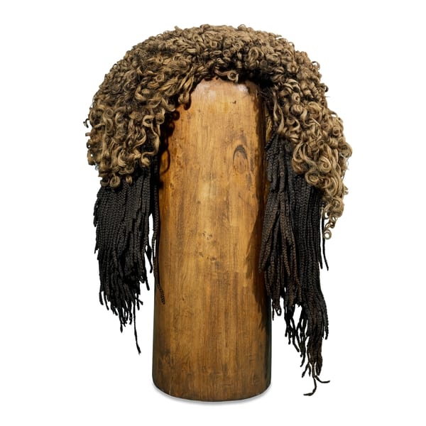
Ancient Egyptian mutli-tonal wig. From the British Museum collections.


While this all sounds very intriguing to a classicist like me, there is no textual or archaeological evidence which can confirm the theory that wigs were used to conceal particular hair types denoting unfavourable ethnic features. On the contrary, as the hairstyles represented in Egyptian art are often described by Egyptologists as wigs, it appears that wigs of this kind were more likely worn more commonly for other aesthetic reasons. Another wig from ancient Egypt (Thebes) in the British Museum is a good example of this counterargument. This piece consists of hair of two colours; a lighter shade arranged into looser curls on the top, and darker long braids underneath. The two colours are presented in different styles in one hairpiece, the lighter colour appearing more prominently over the darker hair. Having lighter hair colour does not seem to be too much of a concern here. The important thing is to have the style on display – or even, to distinguish the special status of the wearer. It is less likely that our blonde woman wore the black wig to make herself look just like everyone else. This wig was probably worn on special occasions, or to denote her high social status. J. Stevens Cox even suggests that the prevalent use of wigs as fashion and status markers in the Roman empire was in fact a result of contact with Egypt, where it was already a popular practice.[1]

The reason that Petrie so quickly assumed that the black wig was worn for purposes of concealment of ethnic identity, probably owes more to his interest in eugenics. Since the time of Aristotle, many people have made often spurious connections between appearance and character. In the era of eugenics, bodily features supposedly denoting psychological or moral character were not only categorised but also ranked in a hierarchy. Petrie’s interest in these theories are apparent in his archaeological work as well as in his writing; he extensively documented Egyptian monuments that displayed “racial types”; he collected glass and terracotta figurines or heads that depict ethnic phenotypes and labeled them somewhat arbitrarily (a selection of these are presented in our recent Foreign Bodies exhibition). As late as 1934, Petrie sent skulls of “racial types” to Francis Galton, the father of eugenics, for further research.[2] In the preface of his Janus in Modern Life [3], Petrie clarifies the purpose of such studies, describing them as “physiological research of the obscure causes of [present] troubles” (to use a medical metaphor), since “the present time seems to most people so infinitely more important…than the past or future,” and “[t]hey forget that it is only a fiction to speak of the present…and every such present has been entirely conditioned by its past…” Looking at his work retrospectively, it is clear that much of his own reading of the past was actually largely conditioned by his present. He made some of the very mistakes he set out to help the world to avoid.


References:

[1] J. Stevens Cox, “The construction of an Ancient Egyptian wig (c. 1400 B.C.) in the British Museum”, in Journal of Egyptian Archaeology Vol. 5 No. 63 (1977).

[2] For further information, please see the website of Petrie Museum exhibition Typecast.

[3] W. M. F. Petrie, Janus of Modern Life. G.P. Putnams’ Sons (1907).

Diagnosing Foreign Bodies

By Gemma Angel, on 25 March 2013

Sarah Chaneyby Sarah Chaney


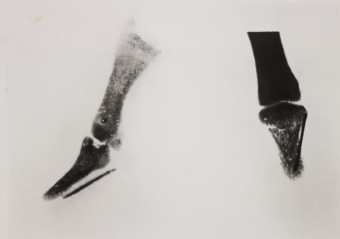
One of the most important diagnostic tools to assist in foreign body removal was the development of the x-ray. In 1895, Wilhelm Röntgen, a German physics professor, developed the x-ray photograph, which enabled the interior of the body to be made visible using electromagnetic radiation.[1]  In the late 1890s and early 1900s, medical reports on foreign bodies frequently focused on the use of the “Röntgen Rays” or “skiagraphs” (as x-rays were then widely called) to locate such objects. A few weeks after Röntgen published the first X-ray photograph, Norman Collie at UCL made his own x-ray tube in order to locate a broken needle in the thumb of a female patient.

World’s first diagnostic x-ray, by Norman Collie at UCL. UCL Special Collections, on display in the Octagon Gallery until 30 April: http://www.ucl.ac.uk/museums/whats-on

World’s first diagnostic x-ray, by Norman Collie at UCL.
UCL Special Collections, on display in the Octagon Gallery
until 30 April: http://www.ucl.ac.uk/museums/whats-on


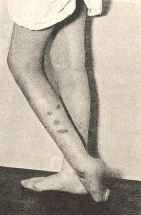
Many of the foreign bodies case histories in hospital records of the period focus on the use of this new diagnostic technique. However, x-rays could also lead to tension between patients and clinicians, when the photographs contradicted stories told by patients. One German publication of 1899 reported a soldier’s claim to have been bitten by a horse as “malingering” (a serious military crime) when broken needles were found in the wound, suggesting that the injury was self-inflicted.[2]

Image showing self-inflicted burns in a “hysterical” patient, from John Collie’s Malingering and Feigned Sickness (1913)

Image showing self-inflicted burns in a
“hysterical” patient, from John Collie’s
Malingering and Feigned Sickness (1913)

Yet, surprisingly (given the widespread publicity given to so-called malingering in civilian populations in the decades preceding the National Insurance Act of 1911), these stories seem to have been of less interest to many surgeons than the diagnostic procedure itself. At the Royal London Hospital in 1898, for example, little interest was shown in the fact that the x-ray photographs of 38-year-old domestic servant Elizabeth Quaife did not tally with the history she gave.[3] Elizabeth claimed that she had suffered pain in the knee joint ever since a long hat pin ran into her leg while she was sweeping under a bed: in hospital, however, five separate needles were discovered in the joint. Unlike in published cases, the surgeon made no reference to the potential use of x-ray imagery to detect fraud, but instead used the case to evaluate the usefulness of the technique itself. This, it was thought, had been successful in locating and removing four needles but “the fifth needle shewed by the skiagraph was … not found. It is probable that the figure shewed in the skiagraph was due to a shadow of the other needles. This, once more, shews that the skiagraph may be deceptive.”

This emphasis on diagnosis and removal certainly tallies with the lack of interest surgeons tended to show in the cause of foreign bodies. Foreign Body in this period was a diagnosis, not an exploration of a patient’s state of mind. When Rachel Taylor was admitted to the Royal London in 1900 – after swallowing a pin and a tin tack – and again in 1906 having swallowed two nails “the night before last”, it was not noted whether either instance was accidental or intentional.[4] Despite published concern over the potential abuse of charitable treatment, in practice this does not seem to have been a significant issue for either surgeons or physicians at the Royal London Hospital: cases of “artefact injury” were treated without question whether or not the patient paid for their treatment.


References:

[1] Lisa Cartwight, Screening the Body: Tracing Medicine’s Visual Culture (Minneapolis: University of Minnesota Press, 1995)

[2] “Self-Inflicted Injuries Diagnosed by the Roentgen Rays,” The Lancet, 153, no. 3955 (1899): 1668.

[3] Elizabeth Quaife, RLHA Microfilm Case Records (Surgical), DEAN F1898, pt. no. 29 & 511.

[4] Rachel Taylor, RLHA Microfilm Case Records (Surgical), TAY F1900, pt. no. 2326 & FENWICK 1906, pt. no. 906.


From the Forests of Punt to the Deserts of Saqqara: Life and Death as a Sacred Monkey

By Gemma Angel, on 18 March 2013

Suzanne Harvey #2  by Suzanne Harvey


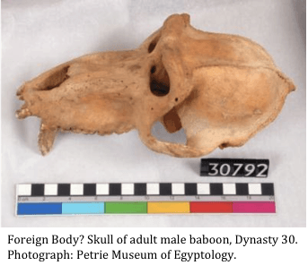
Given the wealth of figurines, statues, engravings and even mummies of baboons found in Egypt, it may seem odd that a baboon skull features as an object in our current Foreign Bodies exhibition in UCL’s North Cloisters.

Baboon SkullThe key to this puzzle is that baboons are not, and never have been, indigenous to the areas of Egypt in which their remains have been found. They were imported from Nubia and the mysterious Land of Punt for use at temples and burial sites, where their habit of stretching and ‘chattering’ was viewed as worship of the Sun God, Ra. Since these animals were sacred to Gods of wisdom and the underworld, themselves deified in the form of Babi and The Great White One, and imported at great cost – surely their lives in Egypt would be ones of luxury?

Life and death in a foreign land

The largest number of mummified baboons have been found at the tombs of Saqqarah, an arid desert environment that contrasts starkly with the natural forest and savannah habitat of baboons. In their natural environment, baboons spend most of their waking hours foraging for food in the form of leaves, seeds, fruit and insects, none of which would be possible in the desert. In fact, the environment of the temples and burial sites where baboons were kept, was so foreign to these species that most died from malnutrition, vitamin deficiencies, and fractured bones. Of around 200 mummies analysed, few had lived beyond 6-10 years, despite the natural lifespan of the sacred Hamadryas baboon being around 30 years.

Baboon environment


From a primatological point of view, this highlights just how unsustainable desert colonies of baboons were. First breeding usually occurs between 5 and 7 years – with the majority of adults dying so young, it’s unlikely that much successful breeding took place at the temples. Recent studies at UCL’s Gashaka Primate Project have shown that diet has a strong effect on reproduction in baboons, with age of menarche, infant mortality and interbirth intervals all highly dependent upon nutrition.[1] In order to sustain a desert population of baboons, the Ancient Egyptians would have required constant imports of new animals, making baboons very rare and expensive offerings to the Gods.

The lost baboons of the Petrie Museum

Baboon mummy

In 2010, oxygen isotope analyses were carried out on hairs from one of the British Museum’s baboon mummies, and researchers were able to locate the Land of Punt, by comparing markers in the ancient baboon to modern samples. 3000 years after the baboon was mummified, his homeland was located as modern day Eritreia and Ethiopia, where baboons remain today.

The baboons at the Petrie Museum date from a later period than those at the British museum, and documentation indications that they were were mummified after voyages to Punt ceased. So for now, all we can really say for certain about the Petrie Museum baboons is that they were a long way from home when they died…


References

[1] J.P. Higham, Y. Warren, J. Adanu, B.N. Umaru, A.M. MacLarnon, V. Sommer, & C. Ross: (2009). ‘Living on the edge: Life-history of olive baboons at Gashaka-Gumti National Park, Nigeria’, in American Journal of Primatology, Vol. 71 (2009), pp.293-304.


Pulling Teeth: Ovarian Teratomas & the Myth of Vagina Dentata

By Gemma Angel, on 4 March 2013

  by Gemma Angel


In preparation for our upcoming exhibition, Foreign Bodies, several members of the engagement team went to visit UCL Pathology Collections, to have a look at a collection of foreign objects removed from the human body. We soon encountered a number of other specimens which resonated with the exhibition theme in various ways: From a liver infected with syphilis, to a ruptured oesophagus and the sword swallower’s sword that caused the fatal injury; to a feotus inadvertently discovered during a hysterectomy, which was performed to extract a large tumour on the uterus.

The UCL Pathology Collections comprise over 6,000 specimens dating back to around 1850, many of which have been absorbed from other London medical institutions over the past 25 years, and these are currently in the process of being re-catalogued and conserved. It is a fascinating, not to mention an educationally invaluable collection – not least because it contains many specimens that demonstrate gross clinical manifestations of diseases which are now very rare in the Western world. Some of these diseases, such as syphilis, are unfortunately making a comeback, so it seems more important than ever that medical students are able to recognise the clinical signs of these infections. Pathology collections are a highly valuable medical teaching resource; particularly since these kinds of collections are now unlikely to be expanded in the wake of the 2004 Human Tissue Act.

As with many historical pathology collections, UCL possesses its share of medical anomalies or curiosities. Fragments of preserved skin belonging to a tattooed man certainly seem to fall into the category of the anatomically curious – there is certainly nothing pathological about this specimen. One of the biggest surprises I encountered during my visit to the collections, was the revelation that the female reproductive anatomy can, and occasionally does, grow teeth.

Teratoma with Tooth and Hair

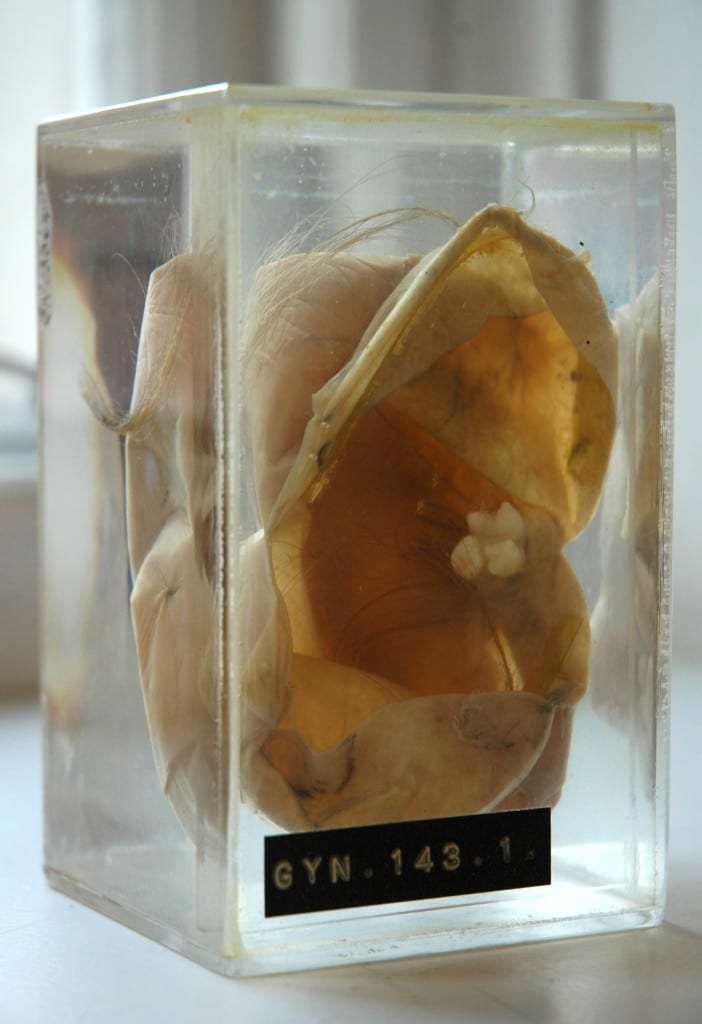
Dermoid cyst (cystic teratoma) with fully developed
tooth and hair. UCL Pathology Collections.

The specimen shown here (right) is a dermoid cyst, or cystic teratoma, which has formed inside an ovary. When I first came across it, I experienced a strong visceral reaction: I didn’t have to be a medical student to recognise that this tooth, entwined in long hair drifting in the liquid-filled vitrine, was out of place – so much so, that the sight of it provoked an immediate and simultaneous sense of revulsion and fascination. The term teratoma is derived from the Greek, tera, meaning monster, and literally means “monstrous growth”; it was easy for me to see how such biological anomalies could become the stuff of nightmares. Despite the ominous name, however, ovarian teratomas are usually benign, and arise from totipotent stem cells which are capable of developing into any type of body cell. One 1941 pathology text describes these tumours as follows:

Dermoid cysts are usually globular in shape and dull white in color. They contain structures associated with epidermal tissues, such as hair, teeth, bone, sebaceous material resembling fat … The following is a partial list of tissues which have been found in dermoids: Skin and its derivatives, sebaceous glands, hair, sweat glands, and bone, especially the maxillae containing teeth. Up to 300 teeth have been found in one cyst … Long bones, digits, fingernails, and skull have been found. Brain tissue and its derivatives, intestinal loops, thyroid tissue, eyes, salivary glands, may occasionally be found. Even rudimentary fetuses have been described, such as a pelvis with hairy pubes and a vulva and clitoris. Brains with ventricles, spinal cords and a few complete extremities, have been observed. [1]

Although teratomas can develop in almost any part of the body – including the brain, neck, bladder, and the testes in men – being confronted with a toothy tumour in the female reproductive organs brought to mind mythic archetypes of the sexually devouring and deadly woman. I was immediately struck by the parallels between this specimen and the image of the vagina dentata. I am not the first to make such an observation,[2] and whilst I am not suggesting that there is any explanatory relationship to be found between the biological phenomena and the myths, it is certainly an intriguing association. The toothed vagina appears in the creation myths and folk stories of many cultures, from Native America, Russia and Japan (amongst the Ainu), to India, Samoa and New Zealand. [3] Funk and Wagnalls Standard Dictionary of Folklore, Mythology and Legend records this entry concerning vagina dentata:

The toothed vagina motif, so prominent in North American Indian mythology, is also found in the Chaco and the Guianas. The first men in the world were unable to have sexual relationships with their wives until the culture hero broke the teeth of the women’s vaginas (Chaco). According to the Waspishiana and Taruma Indians the first woman had a carnivorous fish inside her vagina. [4]

Many 19th and 20th century European interpretations linked the motif to Freudian concepts of castration anxiety, in which young males are said to experience an unconscious fear of castration upon seeing female genitalia. Whilst a Freudian analysis is undoubtedly culturally and historically specific, many vagina dentata legends explicitly articulate male fears of castration in the act of normal sexual intercourse, and warn of the necessity of removing the teeth from women’s vaginas, in order to transform her into a nonthreatening and marriageable sexual partner. A particularly telling collection of stories comes from India, in which the ferocious sexual appetites of beautiful young women are tamed and ‘made safe’ to men through the violent breaking of the teeth hidden inside their vaginas. [5]

Lloyd, Charles Augustus, d 1930. Lloyd, Charles A fl 1880s-1912 (Photographer) : Maori wood carving of the goddess Hine-nui-te-po, and Maui. Original photographic prints and postcards from file print collection, Box 14. Ref: PAColl-6585-10. Alexander Turnbull Library, Wellington, New Zealand. http://natlib.govt.nz/records/22708288

Māori wood carving of the goddess Hine-nui-te-pō and Māui.
Photograph by Charles Augustus Lloyd, c.1880s-1912.
Alexander Turnbull Library, Wellington, New Zealand.

The toothed vagina motif is not exclusively associated with male fears of the ‘castrating female’, however. In some traditions, the terrible power of the vagina dentata lies principally not in fears of the sexual act, but in its associations with death. The Māori legend of Māui and Hine-nui-te-pō is particularly interesting in this respect. Hine-nui-te-pō was the goddess of death and gatekeeper of the underworld, whom the trickster demigod Māui sought to kill in order to win immortality for humankind. When Māui asks his father what his ancestress Hine-nui-te-pō is like, he responds by pointing to the icy mountains beneath the fiery clouds of sunset. He explains:

What you see there is Hine-nui, flashing where the sky meets the earth. Her body is like a woman’s, but the pupils of her eyes are greenstone and her hair is kelp. Her mouth is that of a barracuda, and in the place where men enter her she has sharp teeth of obsidian and greenstone. [6]

Undeterred by his father’s grave warnings, Māui sets off on his quest with a gathering of bird companions. He proposes to kill Hine-nui-te-pō by entering her vagina and exiting through her mouth whilst she is sleeping, thus reversing the natural passage into life via birth. Māui finds the great goddess sleeping “with her legs apart” such that they can clearly see “those flints that were set between her thighs”, and he transforms himself into a caterpillar in order to crawl through her body. But his bird companions are so struck by the absurdity of his actions, that they laugh out loud and wake Hine-nui-te-pō from her slumber. Angry at Māui’s impiety, she crushes him with the obsidian teeth in her vagina; thus Māui becomes the first man to die and seals the fate of all humankind, who were ever after destined to die and be welcomed into the underworld by Hine-nui-te-pō. In this version of the myth, the vagina dentata appears as an inverse manifestation of the generative, life-giving powers of woman, which Māui attempts to subvert – he endeavours to overcome the forces of life and death, and therefore “by the way of rebirth he met his end.” [7]

Ovarian Dermoid Cyst

X ray of a dermoid cyst, showing a cluster of teeth in the pelvic cavity.

The mythical theme of the vagina-with-teeth can in most cases be read as an attempt to render the potentially dangerous sexuality of women nonthreatening to patriarchal power, through heroic acts of “pulling the teeth”. Some authors have even suggested a correspondence between this mythic construct and practices of clitoridectomy and ‘female circumcision’ in some cultures. [8] Whilst there can be little correlation between ancient stories and the observation of biological phenomena such as dermoid cysts, the removal of these peculiar tumours and their retention in pathology collections nevertheless reminds us of the remarkable complexity and diversity of human understandings of the body, and their wider cultural significance. For those readers interested in the practical removal of teratomas such as those discussed here, a demonstration of the surgical procedure can be viewed in this educational film (contains scenes of graphic live surgery).


References:

[1] Harry Sturgeon Cross and Robert James Crossen: Diseases of Women, St. Louis (1941), p.685.

[2] See, for example, Bruce Jackson: ‘Vagina Dentata and Cystic Teratoma’, in The Journal of American Folklore, Vol. 84 No. 333 (July-Sept 1971), pp.341-342. Available on JSTOR: http://www.jstor.org/stable/539812

[3] Verrier Elwin: ‘The Vagina Dentata Legend’, in British Journal of Medical Psychology, (1943) Vol. 19, pp. 439-453.

[4] Maria Leach (ed): Funk and Wagnalls Standard Dictionary of Folklore Mythology and Legend, Volume 2 J-Z (1950), p.1152.

[5]  Verrier Elwin: ‘The Vagina Dentata Legend’, in British Journal of Medical Psychology, (1943), Vol. 19, pp.439-453. A particularly illustrative example of one of these stories is recounted by Elwin on pp.439-440:

There was a Baiga girl who looked so fierce and angry, as if there was magic in her, that for all her beauty, no one dared to marry her. But she was full of passion and longed for men. She had many lovers, but – though she did not know it – she had three teeth in her vagina, and whenever she went to a man she cut his penis into three pieces. After a time she grew so beautiful that the landlord of the village determined to marry her on the condition that she allowed four of his servants to have intercourse with her first. To this she agreed, and the landlord first sent a Brahmin to her  – and he lost his penis. Then he sent a Gond, but the Gond said, “I am only a poor man and I am too shy to do this while you are looking at me.” He covered the girl’s face with a cloth. The two other servants, a Baiga and an Agaria, crept quietly into the room. The Gond held the girl down, and the Baiga thrust his flint into her vagina and knocked out one of the teeth. The Agaria inserted his tongs and pulled out the other two. The girl wept with the pain, but she was consoled when the landlord came in and said he would now marry her immediately.

[6] Antony Alpers: Maori Myths and Tribal Legends, Pearson Education, New Zealand (1964), p.67.

[7] Ibid, p.70.

[8] See for example, Jill Raitt: ‘The “Vagina Dentata” and the “Immaculatus Uterus Divini Fontis”‘, in Journal of the American Academy of Religion, Vol. 48 No. 3 (Sept. 1980), pp.415-431. Available on JSTOR: http://www.jstor.org/stable/1462869

[analytics-counter]

Musical Apes: Can Baboons Play the Harp?

By Gemma Angel, on 11 February 2013

Suzanne Harvey #2  by Suzanne Harvey


Excuse me, is that baboon playing a harp?

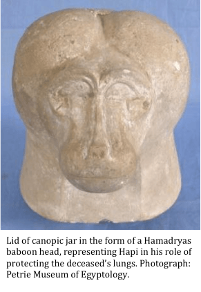
This is a question that I long to be asked when I’m working at the Petrie Museum of Egyptology. Whilst there are many examples of baboon figurines in the collection, my favourites have always been the selection from Amarna, in which the animals are shown performing acrobatics, drinking beer and playing the harp. Baboons appear throughout Egyptian mythology, and the majority of the figurines at the Petrie and elsewhere depict baboons sitting in a realistic manner, rather than performing any elaborate tricks. These figurines are often found at burial sites. Hapi, an Egyptian God of the underworld, is depicted with the head of a Hamadryas baboon and is said to protect the lungs of the deceased.[1] For this reason, it is common to find the baboon head of Hapi as a lid of canopic jars containing lungs. So, if baboons are typically viewed as sacred animals used in funerary reliquary, why is that baboon playing a harp?

Monkeys of several species were kept as pets in Ancient Egypt, so it is possible that they were trained to perform tricks. Having studied olive baboon infant tantrums, I know from experience that they are athletic animals who are often keen to throw themselves around – but acrobatics seems a stretch, and whilst I’m sure they could be trained to pluck harp strings, I doubt it would be easy listening. Since my own biological and behavioural approach does little but rule out possibilities, there must be another explanation for the existence of these unusual figurines. The Petrie Museum attracts a lot of visitors who are either professional Egyptologists or well-read enthusiasts of the subject. So, in my first ever research engager micro crowd-sourcing exercise, I’ve compiled some of their theories here:


1. The figurines were crafted at a time when the Pharaoh Akhenaten brought in monotheism, demanding that his subjects worship only one God, the Sun God. Therefore, worship of the baboon God was forbidden, and these less serious depictions of baboons may have become fashionable – Suggested by a retired German doctor who researches the beginnings of monotheism in ancient cultures as a hobby.

2. They are part of a culture of fantastical animal stories used for entertainment, and would have been high status decorations in a wealthy household – Suggested by an American Professor of art, interested in representations of animals in Egypt.

3. As baboons, particularly alpha males, could be seen as the reincarnation of dead ancestors in the form of the baboon deity Babi (not to be confused with the baboon-headed deity, Hapi…) the statues may show baboons engaged in activities that dead relatives enjoyed – Suggested by a UCL masters student studying ancient writing.

Any or all of these theories may be relevant, but overall, it seems that perspectives from art, theology and graphology lead to more interesting interpretations of this object than my own biological anthropology approach. For anyone interested in cross-disciplinary or multiple interpretations of museum objects, our upcoming exhibition Foreign Bodies will be on display in UCL’s North Cloisters, with additional featured objects in all 3 of the UCL Museums from March 18th – featuring (amongst others) my favourite baboon harpist.


References:

[1] Hans Kummer: In Quest of the Sacred Baboon. (1995), Chichester: Princeton University Press.

Tattooing in Ancient Egypt Part 2: The Mummy of Amunet

By Gemma Angel, on 10 December 2012

  by Gemma Angel


In my previous post, I wrote about the possible connection between objects in the Petrie Museum, and ancient Egyptian tattooing practices. One of the greatest challenges in reconstructing the body modification practices of ancient peoples is in interpreting the fragmentary remains of material culture found at excavation sites. As archaeologist Geoffrey Tassie writes:

The use of many artefacts can only be inferred from their context and association, and tattoo needles are no different, although, if sufficiently well-preserved, scientific analysis of their tips may identify traces of blood or the pigment used to create the tattoo.[1]

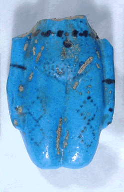
In the absence of any such scientific testing, uncertainty remains as to whether the 7 prick points in the Petrie collection were used for tattooing. However, the decorative markings on a collection of blue faience figurines are less ambiguous. Although ancient Egyptian textual records make no mention of tattooing, there is nevertheless a considerable amount of iconographic evidence for the practice, which includes the engraved markings on faience figurines such as those on display in the Petrie Museum. Interestingly, these “tattooed” figures are invariably female, suggesting that tattooing was practiced exclusively by women.[2]

Blue faience figurine fragment,
showing tattoo markings on the
abdomen and thighs.
Image © UCL Museums & Collections

Faience figurines dating from the Middle Kingdom traditionally known as “Brides of the Dead”[3], frequently display a series of dotted geometric tattoo patterns, running in horizontal bands across the lower abdomen. Occasionally, the thighs are also decorated, as can be seen in the example shown (left). There are many examples of footless faience figurines such as these in museum collections around the world. According to Robert Bianchi, dependent upon their context, these figurines maybe interpreted ‘as guarantors of the deceased’s procreative abilities on analogy with those of the goddess Hathor’, who both represented fertility, childbirth and love, and welcomed the dead into the next life. Faience figurines are often found in tombs, interred with the dead in order to ensure resurrection.[4]

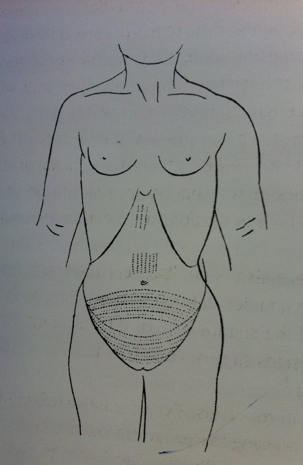
Tattooing practice in ancient Egypt is further supported by the discovery of a number of tattooed mummies in the late 19th and early 20th centuries. The most famous of these was discovered in Deir el-Bahari by French Egyptologist Eugène Grébaut in 1891. Dating from Dynasty XI (c.2134-1991 BC), a female mummy identified as Amunet, a Priestess of the goddess Hathor at Thebes, was found to have a number of tattooed markings on her body, which show striking correspondence with the patterns depicted on Middle Kingdom faience figurines. A design consisting of multiple diamond shapes composed of dots, are tattooed on the middle of her right thigh, similar to those engraved on the faience figure pictured above. As well as tattoos on her left shoulder and breast, and on her right arm below the elbow, Amunet also bore extensive tattooing over her abdomen: A series of dots and dashes forming an elliptical pattern of rows covers almost the entire abdominal wall in the suprapubic region (see sketch below right).

Drawing showing tattoo markings attributed
to the mummified remains of Amunet.
From Fouquet (1898), p.278

A further 2 female mummies, described as ‘Hathoric dancers in the court of King Mentuhotep,’ were excavated from pits located very near to the tomb of Amunet in 1923.[5] These women both bore similar body-markings to those of Amunet, in particular over the abdomen, which may suggest that these tattoos served fertility purposes:

Tattoos on the abdominal part of the female body would have become particularly notable when the woman became pregnant – the patterns would expand, forming an even more symbolically interesting pattern, like a web or netting design.[6]

The mummy of Amunet was unearthed at the height of the “Golden Age” of Egyptology, when the discovery of mass burials of mummified royalty and clergy became a source of popular fascination. As “Egyptomania” swept across Europe, some artists sought to commemorate the “great discoveries” of European explorers and scientists. For instance, the painting below, by French artist Paul Dominique Philippoteaux, depicts an historical event: The unwrapping of a mummy discovered at Deir el-Bahari, the same site where Amunet was buried. Although the mummy pictured dates from Dynasty XXI (c.970 BC) in the Third Intermediate Period, many of the men present in this scene were also involved in the excavation of Amunet. The eminent Dr. Daniel Fouquet takes centre stage, demonstrating to his learned audience of colleagues and lady spectators, as he unveils the mummified body of the “Priestess”, known as Ta-usa-ra. Mr. Grébaut, the leader of the expedition, also appears in the painting, second from left and wearing a fez.[7]

“Examination of a Mummy – The Priestess of Ammon” (1891)
Oil on canvas, by Paul Dominique Philippoteaux.
Photo credit: Peter Nahum at The Leicester Gallery, London.

In 1898, Fouquet wrote an article on “medical tattooing” practices in Ancient Egypt and the contemporary era, in which he describes the tattooed markings on the female mummies found at the Deir el-Bahari site. He speculated that the tattoos and other scarifications observed on the bodies may have served a medicinal or therapeutic purpose:

The examination of these scars, some white, others blue, leaves in no doubt that they are not, in essence, ornament, but an established treatment for a condition of the pelvis, very probably chronic pelvic peritonitis.[8]

Photograph showing the
tattooed abdomen of one of
female mummies found at
the Deir el-Bahari site,
possibly Amunet.

Whilst it is clear that the white scars Fouquet refers to are likely scarifications, the blue marks must be interpreted as tattoos – but whether or not they were primarily medicinal markings, or served a more ritual and symbolic function is uncertain. Based on the iconographic and material evidence of human remains, it certainly seems that some women in Ancient Egypt marked themselves as sexual beings; as Robert Bianchi writes:

The priestess Amunet and the figurines…have an undeniably carnal overtone. The eroticism that is undoubtedly associated with Egyptian tattoo of the Middle Kingdom correlates with the prevailing religious attitude that linked physical procreation with the loftier aspirations of resurrection in the Hereafter.[9]

Amunet’s mummified remains now lie in the Egyptian Museum of Antiquities, Cairo.


Did tattooing really have a medicinal purpose in the Ancient world? Check back for my next post on the history of tattooing as a therapeutic practice – and the health risks involved in becoming tattooed prior to modern antisepsis.


References:

[1] Geoffrey Tassie, ‘Identifying the Practice of Tattooing in Ancient Egypt and Nubia’, in Papers from the Institute of Archaeology, Vol.14 (2003), p86.

[2] According to Tassie, there is only one exception in the archaeological record – a Dynasty XII stele (a standing stone tablet) from Abydos:  ‘This depicts a figure, which is said to be male, with marks coming down over the chest. As the stele is extremely worn it is hard to distinguish whether the marks indeed represent tattoos.’ (Ibid, p.88).

[3] Robert Bianchi, ‘Tattooing and Skin Painting in the Ancient Nile Valley’, in Celenko, T. (ed.) Egypt in Africa, (1996), Indianapolis University Press, p.81.

[4] Ibid, pp.82-82.

[5] Tassie (2003), p.90.

[6] Ibid, p.91.

[7] Philippoteaux’s painting includes a plaque inscribed (in French) with the names of the sitters, as well as an historical description of the scene. From left to right: Marquis de Reverseaux (Ministre de France au Caire); Mr. Eugène Grébaut (Directeur Génerale du Service des Antiquities); Dr. Daniel Fouquet (Médecin au Caire); E. Brugshe Pacha (Conservateur du Musée); Mr. Georges Daressy (Conservateur adjoint du Musée) – pictured taking notes; Mr. H. Bazil (Secrétaire complable du Musée); Mr. J. Barois (Secrétaire Génerale du Ministére du Travaux Publies); Mr. U. Bouriant (Directeur de la Misien Archéologique française au Caire).

[8] Daniel Fouquet, ‘Le Tatouage Medicale en Egypte dans l’Antiquite et a l’Epoque Actuelle’, in Archives d’Anthropologie Criminelle, Tome 13 (1898), p.271.  Available online at Criminocorpus. Translated from the French: L’examen de ces cicatrices, les unes blanches, les autres bleues, ne laisse aucun doute dans l’espirit, il s’agit la non d’un ornement, mais bien d’un traitement institué pour une affection du petit bassin, très probablement une pelvi-péritonite chronique.

[9] Bianchi (1996), p.82.

[10] See also: Carolyn Graves-Brown, Dancing for Hathor. Women in Ancient Egypt, (2010), London  New York: Continuum Books.

[analytics-counter]