X Close

Researchers in Museums

Home

Engaging the public with research & collections

Menu

Archive for the 'Case #2: External Invaders' Category

Tattoos That Repel Venomous Creatures! The Tragic Tale of Prince Giolo

By Gemma Angel, on 27 May 2013

Gemma Angel by Gemma Angel


The tattooed body has been an object of spectacle and a source of fascination in Europe for at least 4 hundred years. Tattooed natives captured by European explorers were transported to Europe and put on display as curiosities or ‘sights’ from as early as the middle of the 16th century. In 1566, a tattooed Inuit woman and her child were kidnapped by French sailors and put on display in a tavern in Antwerp, The Netherlands. 10 years later, the sometime pirate and seaman Martin Frobisher returned to England from his voyage to Baffin Island in northeastern Canada with a native man whom he had abducted; this unfortunate individual caused such a stir in London, that Frobisher returned from his second voyage to the region with 3 more Inuit captives, who drew equally fascinated crowds when he landed in Bristol. Sadly, all 3 of his human cargo died shortly after their arrival on British shores, succumbing to common European illnesses against which they had no natural immunity.

A similar fate befell the Miangas islander named Jeoly, who became popularly known as ‘Prince Giolo’ when he arrived in England in 1691. Perhaps the most famous of all the tattooed ‘curiosities’ exhibited in Britain, Jeoly was purchased as a slave by the buccaneer-adventurer William Dampier in Mindanao, the Philippines, in 1690. Having failed in his ambitions to discover unexploited spice and gold wealth in the Spice Islands, Dampier returned to England broke, with only his diaries and his ‘Painted Prince’ to show for travels. On his arrival home, Dampier sold Jeoly on to business interests, and later published his journals under the title A New Voyage Around the World, in 1697. In these diaries, Dampier describes Jeoly’s elaborate tattoos in some detail:

He was painted all down the Breast, between his Shoulders behind; on his Thighs (mostly) before; and the Form of several broad Rings, or Bracelets around his Arms and Legs. I cannot liken the Drawings to any Figure of Animals, or the like; but they were very curious, full of great variety of Lines, Flourishes, Chequered-Work, &c. keeping a very graceful Proportion, and appearing very artificial, even to Wonder, especially that upon and between his Shoulder-blades […] I understood that the Painting was done in the same manner, as the Jerusalem Cross is made in Mens Arms, by pricking the Skin, and rubbing in a Pigment. [1]
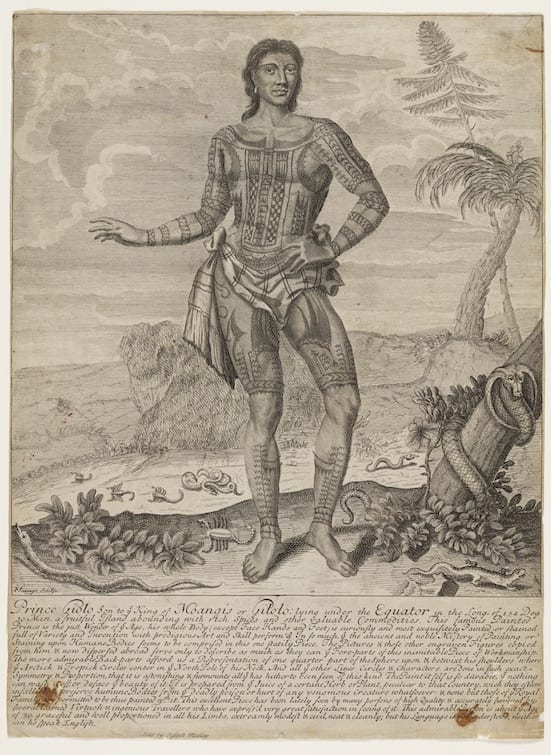
Prince Giolo, 1692

Playbill advertising ‘Prince Giolo’ in London, 1692.
Etching by John Savage.
Image courtesy of the Mitchell Library, State Library of New South Wales, Australia.

Jeoly was put on display ‘as a sight’ at the Blue Boar’s Head Inn in Fleet Street in June 1692. A number of copies of the playbill advertising his public appearances survive (pictured above). The original advertisement includes a detailed etching of Jeoly by John Savage, showing the tattoos over the front of his body, arms and legs, which resemble traditional Micronesian tattoos of the Caroline and Palau Islands. [2] As well as this striking image, a somewhat embellished story of his life was printed beneath the illustration. Interestingly, this accompanying text ascribes potent protective and healing powers to Jeoly’s tattoos, claiming that his people believed them to be a defense against ‘venomous creatures’:

The Paint it self is so durable, that nothing can wash it off, or deface the beauty of it: It is prepared from the Juice of a certain Herb or Plant, peculiar to that Country, which they esteem infallible to preserve humane Bodies from the deadly poison or hurt of any venomous Creatures whatsoever.

Whilst tattooing was considered to possess magical, protective and medicinal properties in many cultures, it is more than likely that the stories claiming that Jeoly’s tattoos repelled venomous creatures were dreamed up by his exhibitors, rather than having any genuine basis in his own native belief system. Dampier himself remarked upon the ‘Romantick stories’ which circulated in England about Jeoly’s origins, openly ridiculing the marketing campaign:

In the little printed Relation that was made of him when he was shown for a Sight in England, there was a romantick Story of a beautiful Sister of his a Slave with them at Mindanao; and of the Sultan’s falling in Love with her; but they were Stories indeed. They reported also that this Paint was of such Virtue, that Serpents, and venomous Creatures would flee from him, for which reason, I suppose, they represented so many Serpents scampering about in the printed Picture that was made of him. But I never knew of any Paint of such Virtue: and as for Jeoly, I have seen him as much afraid of Snakes, Scorpions, or Centapees, as my self. [3]

In the lower foreground of the illustration, a variety of reptiles and scorpions can be seen fleeing from Jeoly’s feet, his tattoos apparently acting as some kind of aposematic deterrent. Tragically however, Jeoly’s tattoos could not protect him from the foreign infections that he was exposed to in England; he died of smallpox in Oxford sometime in 1693. Although his grave is not marked, and his name does not appear in the Parish register, Jeoly is thought to be buried in St Ebbe’s Churchyard. After his death, a fragment of his tattooed skin was removed and preserved for the Anatomy School collections at Oxford University by the surgeon Theophilius Poynter. This skin fragment was recorded in a list of ‘Anatomical Rarities’ in the Appendix of John Pointer’s 4 volume catalogue for his Musaeum Pointerianum, the cabinet of curiosities he left to St. John’s College Oxford in 1740. [4] Although the skin did not survive, having been lost by the early 20th century, this appears to be the first documented instance of the collection and preservation of tattooed human skin as an anatomical curiosity in England.

Jeoly’s tragic story of enslavement, forced re-location to Europe, public exhibition for profit, fatal illness, and the preservation of his tattooed skin for display as an anatomical rarity, speaks of the foreign body on multiple levels. From the 16th century onwards, the tattooed body of the native became a powerful symbol of foreignness, that could reliably draw curious European crowds and turn a profit for unscrupulous entrepreneurs; but the consequences for displaced foreigners like Frobisher’s Inuits and Dampier’s ‘Painted Prince’ were grave indeed. Exposed to invisible and deadly foreign bodies such as measles and smallpox, they died far from home, unable to fight off common European illnesses against which they had no natural defences.


References:

[1] William Dampier, A New Voyage Around the World, ed. N. M. Penzer (London: Adam & Charles Black), 1937, p. 344.

[2] See Tricia Allen, “European Explorers and Marquesan Tattooing: The Wildest Island Style” in D.E. Hardy (ed) Tattootime Volume V: Art from the Heart, (1991) pp. 86-101; also Kotondo Hasebe, “The Tattooing of the Western Micronesians” in The Journal of the Anthropological Society of Tokyo Vol. XLIII No.s 483-494 (1928), pp. 129-152 (in Japanese).

[3] Dampier, A New Voyage Around the World, p.346.

[4] Geraldine Barnes “Curiosity, Wonder and William Dampier’s Painted Prince“, Journal for Early Modern Cultural Studies, Vol. 6, No. 1 (2006), p. 32 & 43.

[analytics-counter]

Painted Skins & Butterfly Wings

By Gemma Angel, on 1 April 2013

Gemma Angelby Gemma Angel


When I first began my doctoral research into tattoo preservation three and a half years ago, I assumed that tattoo collections such as those held by the Science Museum in London were rare. Whilst collections of inked human skin are most definitely unusual, I was soon surprised and intrigued to discover that such objects exist in almost every museum archive, university anatomy department, or pathology collection that I have visited over the course of my research. The largest collections, of which the Wellcome Collection is the major exemplar, are dry-preserved and date from the 19th century – similar collections can be found across Europe, and the MNHN in Paris has a collection of 56 tattoos which are very similar to those in the Wellcome collection. Historically, these collections may be medical, anthropological, or criminological in origin.

In anatomy and pathology collections, tattoo specimens tend to be wet-preserved and date more recently, usually from the early part of the 20th century anywhere up to around the 1980s. But why are these objects preserved in anatomy and pathology departments at all? There is of course nothing pathological about tattooed skin in itself – so it seems strange that specimens like the one pictured below are displayed alongside other pathological skin specimens such as cutaneous anthrax, fibromas, keloids and glanders. What, if anything, can be learned from these tattoos in medical terms? Or are these striking collections of decorated human skin merely objects of curiosity? Often, the simple answer is that they are a little bit of both…

Tattooed human skin specimen. UCL Pathology Collections. Photograph © Gemma Angel.

Tattooed human skin specimen. UCL Pathology Collections.
Photograph © Gemma Angel.

The collection of tattoos pictured here are a case in point. These particular tattoos belonged to one individual, whose very brief case notes have been recorded and retained along with the specimen in UCL Pathology Collections. The notes provide an intriguing glimpse into the life of the individual to whom the tattoos belonged, as well as revealing something of the clinical interests and collecting practices of the doctor who preserved them:

From a man aged 79 years who had earned his living for many years as the Tattooed Man in a circus. His entire body, except for the head and neck, hands and soles of his feet, was covered with elaborate tattoo designs. He died of peritonitis due to a perforation of an anastomatic ulcer … In tattooing, fine particles of pigment are introduced through the skin, taken up by histiocytes and become lodged in the tissue spaces of the dermis. Pigment also passes to the regional lymph glands via the lymphatics. In this case, all the superficial lymph nodes were heavily pigmented.

It is clear from these brief comments that the nature and extent of this man’s tattoos were indeed of anatomical interest to the medical practitioner: The tattooed man had been so extensively tattooed that gradual migration of ink particles resulted in the collection of pigment in the lymph glands. This demonstrates that although tattoo ink is trapped permanently under the skin following healing, it does actually travel within the body over time, filtering into the body’s tissue drainage system, and collecting in the lymph glands. Whilst this is certainly an interesting anatomical observation, it is not the pigmented lymph glands that the doctor has chosen to preserve, but rather the tattooed skin itself. Without these accompanying case notes, we would never have known that this man’s tattoos had exerted any effect on another of the body’s organs and systems at all.

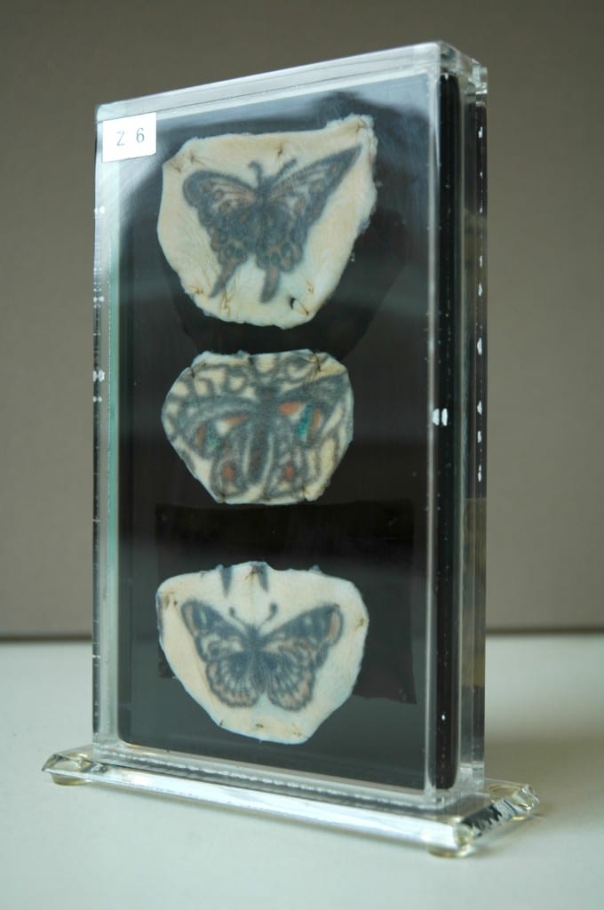
Reverse panel of tattooed human skin specimen Z6,  showing tattoos of a butterfly and a flying fish.UCL Pathology Collections.Photograph © Gemma Angel.

Reverse panel of tattooed human skin
specimen Z6, showing tattoos of a
butterfly and a flying fish.
UCL Pathology Collections.
Photograph © Gemma Angel.

It would be equally impossible to know whether or not these were the only tattoos he possessed – or indeed, if they all necessarily belonged to the same person. There are strong stylistic similarities between the butterfly motifs, suggesting the work of a single tattooist, or perhaps that the individual motifs were part of a larger design. But just how large or complex the design may have been, we certainly cannot tell just by looking at these 5 small tattoos. We know that they belonged to a 79-year-old man, who made his living as a Tattooed Man, only because the doctor tells us so. He or she also tells us that his body was covered in tattoos – yet only 5 small pieces have been preserved. Five carefully selected motifs, chosen by the doctor from an already complete collection, which provided the livelihood and told the life story of one unnamed man. What selection criteria did the pathologist adopt when deciding which tattoos to preserve, and which to consign to the grave? The manner in which the specimens have been excised and mounted are strikingly reminiscent of a lepidopterist‘s collection of butterflies – could this reflect the personal collecting interests of the pathologist, or perhaps even the Tattooed Man himself? Both the pathologist and Tattooed Man alike chose these butterflies – did they also share a passion for lepidoptery?

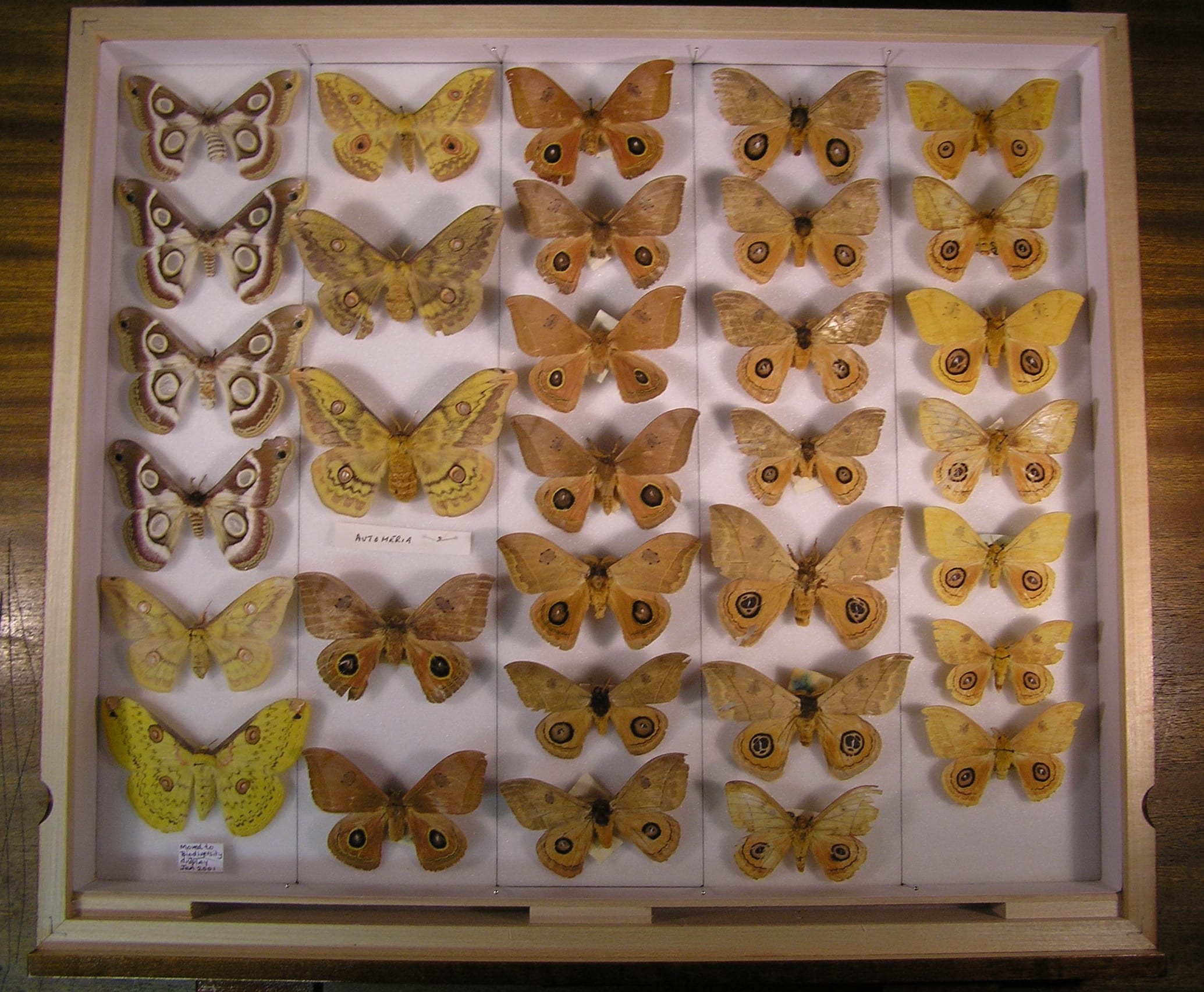
Many people will be familiar with the kind of insect specimen displays that are a staple of natural history collections – the old 19th century museum cases containing neat rows of pinned and mounted moths and butterflies, neatly organised according to subspecies and morphological characteristics. The tattooed butterflies share some remarkable similarities with these entomology collections; they are arranged one above another, and “pinned” to a support with small surgical stitches. Unusually for specimens found in pathology collections, this support is a slightly translucent black. This appears to be a deliberate choice on the part of the pathologist – the black perspex provides a contrasting ground for the display of tattoos on opposite sides of the vitrine, such that they do not visually detract from one another. These aesthetic choices suggest a nuanced interest in the collection and display of these specimens, which goes far beyond a straightforward medical interest in the anatomy of the tattoo. From the limited case notes and analysis of the specimen itself, we can learn something about the pathologist’s interest in the tattoo, but we are still no closer to being able to answer the fundamental question – why collect tattoos at all?

Butterfly display at UCL's Grant Museum. Photograph © UCL, Grant Museum of Zoology.

Lepidoptera display at UCL’s Grant Museum. Photograph © UCL, Grant Museum of Zoology.

There is no part of the body able to register the history of a life lived so much as the skin: wrinkles, scars and lines all map out our lives on the surface of our bodies as we age. The tattoo reinforces this unique capacity of the skin to record the traces of our experience, in the conscious act of permanently inscribing memory in skin. The pathologist, uniquely acquainted with death by virtue of their specialism, is perhaps best positioned amongst medical professionals to appreciate the peculiar relationship of the tattoo with mortality. It is a trace of the subjectivity of the deceased that is capable of outliving them, akin to a photograph or written memoir. From this point of view, it no longer seems surprising that tattoos are so often found in pathology collections; perhaps the pathologist who collected the Tattooed Man’s butterflies simply wished to preserve a small part of a colourful and remarkable life.


[analytics-counter]

Toxic Tattoos: Mercury Based Pigments in the 19th and 20th Centuries

By Gemma Angel, on 4 February 2013

  by Gemma Angel


In January this year, myself and fellow Research Engager Sarah Chaney went to visit the UCL Geology Collections, to see if there were any mineral or rock samples in the collection that would fit in with our upcoming cross-collections exhibition, Foreign Bodies. Neither of us being geologists, we didn’t have particularly high expectations – how interesting can rocks be, really? As it turned out, the answer to that question is – very! We spent a fascinating hour in the Rock Room, where we quickly realised that there were many specimens that could be interpreted as foreign bodies in one way or another: The fossilised forms of plants and animals in rock; a rusted nail fused into a lump of lava; and perhaps the ultimate foreign body, a beautifully patterned fragment of meteorite.

One particular sample drew my attention – a surprisingly heavy lump of purplish-red rock with pretty pink and bright red veins (pictured below). When I asked if I could have a closer look, I was told that I would have to wear gloves to handle this piece of rock, as it was in fact toxic. The rock sample was cinnabar, the common ore of mercury. I am well aware of the toxicity of mercury from my own research – gloves are also required when I’m handling preserved tattooed human skins as part of my work at the Science Museum archives. It is speculated that one of the substances used in the dry-preservation process of human skin is mercuric sulphide, and many of the specimens betray the typical orange-red staining that this chemical causes. But there is another unexpected connection between mercury and my research. Cinnabar has been used to make bold red pigments since antiquity – and this pigment was also historically used in European tattooing.

Cinnabar ore and powder (8.5% Hg) sample, in the UCL Rock Room.
UCL Geology Collections.


Red mercuric sulphide occurs naturally, and has been manufactured for use as a pigment since the early Middle Ages. The pigment was referred to interchangeably as vermilion or cinnabar, although vermilion became the more commonly used term by the 17th century. [1] Vermilion is now the standard English name given to red artists’ pigment based on artificially produced mercuric sulphide. [2] Since the toxic effects of mercury were historically well known, it might seem strange that cinnabar was used in tattooing at all. In fact, mercury has been used in medicine to treat a range of ailments throughout history, most notably syphilis. In European tattooing, red pigments were not commonly used pre-20th century, with red inks tending to be used sparingly for small areas of embellishment.

Most cinnabar was mined in China and by the mid 19th century, Chinese vermilion was generally considered to be the purest form, producing a superior hue to the European variety. The cinnabar ore on which vermillion production depended was costly; as a result, European vermilion was often mixed with inexpensive materials including brick, orpiment, iron oxide, Persian red, iodine scarlet, and minium (red lead). Whilst these additives also produced a bright red pigment, their relative impermanence made it an inferior choice for artists’ colours.

This may explain why there is marked variability amongst preserved tattoos containing red inks, in terms of both permanence and vibrancy of colour: The more commonly available and cheaper European variety of vermilion used by some 19th century tattooists likely contained additives which reduced colour saturation, and made the pigment more susceptible to light-degradation over time. The Wellcome Collection possesses only a handful of tattoos containing red dye, and most of these are very degraded, such that little colour is visible. In these cases, the red has often faded far more dramatically than the black ink used in the same tattoos. However, there are one or two preserved specimens containing exceptionally bright ink, which has lost none of its vivid red colour, an example of which can be seen below.

Tattooed human skin with bold red pigment, likely cinnabar.
Science Museum object no. A687. Photograph © Gemma Angel,
courtesy of the Science Museum London.


Since heavy mineral pigments do not generally lose saturation over time, it is possible to speculate that the bold red ink seen here very likely contains a high concentration of cinnabar, although it is impossible to know for certain without physical testing. There are, however, historical references to the use of mercury-based pigments in tattooing, most of which can be found in 20th century medical journals. As may be expected, these sources focus on the toxic effects of cinnabar-based tattoo pigments. In particular, mercury dermatitis in tattoos was sometimes reported during the early-mid 20th century, often many years after the tattoo was acquired by the patient.

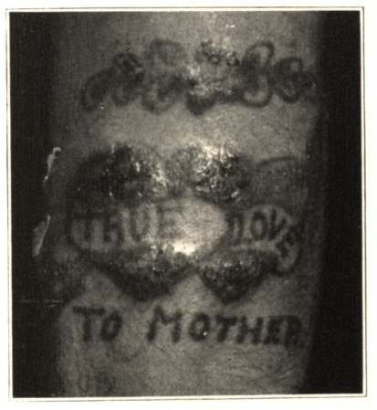
In 1930, one such case appeared in the Archives of Dermatology and Syphilology, written by Dr. Paul Gerson Unna. His patient, a 63-year-old man who had been tattooed in his youth, suddenly developed itching, swelling and blistering in the red portions of the tattoo, following a mercury-based treatment for haemorrhoids. Three years later, Dr. D. B. Ballin reported a case in which a young male patient had developed itching, swelling and oozing in the red portions of a tattoo, 2 years after he had been tattooed. The patient was treated by the removal of the affected areas using a dermal punch, and the tattooed skin samples were sent for histological testing; however, the resultant scar tissue in the punched areas later developed the same reaction.

Photograph from Ballin’s 1933 report,
Cutaneous Hypersensitivity to Mercury from Tattooing
Caption reads: “Forearm of patient showing sensitivity
to mercury as a result of tattooing.”

Throughout the 1940s and 50s, cases of mercurial sensitivity and dermatitis in red tattoos appear sporadically in the medical literature, [4] though the apparent causes of the onset of symptoms vary. According to Keiller and Warin:

In some cases the use of mercurial applications elsewhere has led to the development of sensitivity and the red areas of the tattoo have subsequently become swollen. Other cases are reported in which the sensitivity has developed spontaneously. [5]

Interestingly, there were also reports of the apparent ‘positive’ effects of cinnabar tattoo pigments in cases of cutaneous syphilis during the early 20th century. It was observed that the red portions of a tattoo were seldom effected by syphilis sores – even in cases where adjacent areas of skin tattooed in black ink were engulfed by the infection.


References:

[1] R. D. Harley: Artists’ Pigments c.1600-1835: A Study in English Documentary Sources, (1982) Butterworth Scientific, p.125.

[2] Rutherford J. Gettens et. al. : ‘Vermilion and Cinnabar’, in Studies in Conservation, Vol. 17 No. 2. (May 1972), p.45. Available on JSTOR: http://www.jstor.org/stable/1505572

[3]  D. B. Ballin: ‘Cutaneous Hypersenistivity to Mercury From Tattooing’, in Archives of Dermatology and Syphilology, Vol. 27, No.2 (February 1933), pp.292-294.

[4] See, for example: Howard I. Goldberg: ‘Mercurial Reaction in a Tattoo’, in Canadian Medical Association Journal, Vol. 80 (Feb. 1 1959), pp.203-204. Available online: http://www.ncbi.nlm.nih.gov/pmc/articles/PMC1830587/ ; also R. A. G. Lane et. al.: ‘Mercurial Granuloma in a Tattoo’, in Canadian Medical Association Journal, Vol. 70 (May 1954), pp.546-548. Available online: http://www.ncbi.nlm.nih.gov/pmc/articles/PMC1825326/

[5] F. E. S. Keiller & R.P. Warin: ‘Mercury Dermatitis in a Tattoo: Treated With Dimercaprol’, in The British Medical Journal, Vol. 1, 5020 (Mar. 23, 1957), p.678. Available on JSTOR: http://www.jstor.org/stable/20361174

[6] For more on the history of tattooing and skin disease, see Gemma Angel: ‘Atavistic Marks and Risky Practices: the Tattoo in Medico-Legal Debate 1850~1950’, in J. Reinarz & K. Siena (eds.) A Medical History of Skin: Scratching The Surface, Pickering Chatto, (2013) pp.165-179.

[analytics-counter]

Tattooing in Ancient Egypt

By Gemma Angel, on 19 November 2012

  by Gemma Angel


Of all the objects on display in the Petrie Museum of Egyptology, there are two particular groups of items that immediately jumped out at me when I first began to look closely at the collections. These objects are grouped closely together in one of the densely packed cabinets towards the back of the gallery, near the sarcophagi – but no connection is explicitly made between them in the displays. This is not surprising, given that the first collection of objects – a series of tapered and sharpened metal implements – are described in the museum catalogue as “prick points” of indeterminate use:

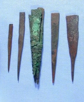
Tool set identified as a “stock of seven prick points” for removing thorns, bronze; three bound together with thread, a fourth with thread round; three separate; cleaned and treated. No information on findplace within Madinat al-Ghurab; purpose and date uncertain.[1]

Seven “prick points” on display in the Petrie Museum,
possibly used for tattooing.
Image © UCL Museums & Collections

The prick points were excavated by Petrie at Gurob in 1880, alongside cosmetic items and fine pottery ware, which is suggestive of a cosmetic use. [2] When I first saw these objects, they brought to mind an article that I had read by Charlotte Booth, titled Possible Tattooing Instruments in the Petrie Museum. Booth carried out a close material analysis of the prick points, and found that they were made by folding the corners of a flat rectangle of bronze inwards at one end, and then beaten into a smooth finish. [3] Each of the 7 prick points were manufactured in this fashion. Interestingly, 3 of them are bound together with thread, which has become permanently fused to the surface by corrosion (pictured left: 3rd from right). Petrie believed that these objects were in fact tweezer points used for removing thorns:

Slips of bronze were made with long sharp points, and kept sometimes in a bunch of half a dozen; but they were not fastened to the tweezers. [4]

However, as Booth points out, although the points are very sharp, they do not appear to be fine enough for removing thorns from the skin. If they had served such an everyday purpose as Petrie suggested, then surely similar instruments would be more commonly found amongst toiletry objects in ancient tomb excavations? Could they perhaps have served a different, more specialised purpose?

A modern tattoo needle bundle, showing
a large number of fine needles in a
round arrangement, used for shading.
Needles may also be arranged in ‘flat’
or ‘magum’ formations.

To the eye of a tattooist, it is easy to see a resemblance between these ancient bronze points and modern tattoo needles, as they share a number of similarities: Tattoo needles are not hollow like hypodermic needles, as some people assume, but are actually solid. Groupings of very fine needles are bound together in a bundle, much like the Egyptian prick points, except that modern needles are soldered in place onto a stainless steel needle bar, rather than bound together with thread. Modern tattoo needles may be grouped in bundles of anything between 3 and 7 individual needles for line work, and as many as 14, 16 or 18 for shading and colouring. The needles operate by drawing ink up into the narrow spaces between them via capillary action as they’re repeatedly dipped into a small inkwell. This remarkably simple yet effective technology hasn’t changed in hundreds – perhaps thousands – of years. Writing on tattooing in Upper Egypt in the 1920’s, Winifred Blackman describes tattoo instruments consisting of 7 needles fixed to the end of a stick used by the Fellahin; [5] if these needles were indeed similar to those in the Petrie collection, this would suggest that the practice dates back at least 4,000 years in Egypt.

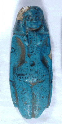
Blue faience figurine, decorated
to show hair, jewellery and tattoos.
Image © UCL Museums & Collections

Material evidence of ancient tattooing is extremely rare in the archaeological record, not least because of the difficulty in identifying artefacts and determining their original purpose. So what evidence is there that the ancient Egyptians practiced tattooing at all? The second group of objects which caught my attention at the Petrie museum provide some clues: two small blue faience figurines. Both of these objects depict nude female figures with black glazed decoration – one is broken at the waist, with only the lower portion of the torso preserved (object no. UC16724). The second figure is intact, with black detail indicating hair and what are assumed to be beads around her neck (object no. UC16725, pictured left).

Both figures are also decorated with a series of dotted lines across the abdomen, which are thought to represent tattoo markings. These markings bear striking similarity to tattoos found on ancient female mummies, which you can read more about in my next post on the Mummy of Amunet.


References:

[1] Petrie Museum online catalogue, object no. UC7790. See additional publications for more information on the origins of these objects.

[2] Charlotte Booth, ‘Possible Tattooing Instruments in the Petrie Museum’, in The Journal of Egyptian Archaeology, Vol. 87 (2001), p.172.

[3] Ibid, p.173.

[4] W.M.F. Petrie, Tools and Weapons, (London, 1917), pp.51-52.

[5] Winifred Blackman, The Fellahin of Upper Egypt, (London, 1927), pp.50-55.

[analytics-counter]