X Close

Researchers in Museums

Home

Engaging the public with research & collections

Menu

Archive for April, 2017

Question of the Week: Why do brains have wrinkles?

By ucbtch1, on 27 April 2017

The brains displayed at the entrance of the Grant Museum are mostly mammal’s brains but we can observe differences in sizes and in how smooth or wrinkly they are. The folds of a brain are called gyri and the grooves are called sulci. These morphological features are produced by the folding of the cortex, the part of our brain responsible for higher cognitive processes like memories, language and consciousness. During development, all brains start off with a smooth surface and as they grow, gyrification (the development of the gyri and sulci) occurs. It is interesting to note that the major folds are very consistent amongst individuals, meaning that development is similar sometimes even amongst species.

c_ucl_gmz_matt_clayton020

The brain collection on display at the Grant Museum of Zoology (Image credit Grant Museum of Zoology).


It has been assumed that the wrinkles in brains correlate with an animal’s intelligence. The reasoning behind this is that a bigger brain, and hence more neurons, need more space. The folds allow the cortex to increase its area while being packed in a confined space like our cranium. There are several factors and hypothesis of how gyrification occurs. Recently, researchers at Harvard developed a 3D gel model based on MRI (magnetic resonance imaging) images to understand how this process occurs. They found out that it all boils down to the mechanical properties of the cortex. While neuronal cells grow and divide, the increasingly bigger brain leads to a compression of the cortex and to the formation of the folds. The researchers were able to mimic the folds of the cortex and were stunned at how similar their gel model looked to a real human brain.

tumblr_o21mv85Nt01t5fphqo1_1280

Gel model of a foetal brain (Image credit: Mahadevan Lab/Harvard SEAS).


Even though most of the brains on display in the Grant museum have gyri and sulci, in nature, most animals have smooth brains. In general, larger brains have folds while smaller brains do not, even small mammals like rats or mice have smooth brains. In humans, a lissencephalic brain is one without gyri and sulci and is a result of a rare disorder that is characterised by mental abnormalities. From the collection of brains in the Grant Museum, there is only one lissencephalic brain—next time you visit the museum see if you can spot it. Additionally, try to find the brain coral. Because of its intricate maze–like pattern, Diploria labyrinthiformis has very similar ridges and grooves as a brain, and so is referred to as brain coral. Overall, I find looking at brains and their grooves fascinating, each species with their own pattern and each groove in a specific place. Makes me wonder how brain coral gets its patterns.

brain coral 3

Diploria labyrinthiformis also known as brain coral(Grant Museum C1084).


References:

Roth, G. and Dicke, U., 2005. Evolution of the brain and intelligence. Trends in cognitive sciences9(5), pp.250-257.

Ronan, L. and Fletcher, P.C., 2015. From genes to folds: a review of cortical gyrification theory. Brain Structure and Function220(5), pp.2475-2483.

Manger, P.R., Prowse, M., Haagensen, M. and Hemingway, J., 2012. Quantitative analysis of neocortical gyrencephaly in African elephants (Loxodonta africana) and six species of cetaceans: comparison with other mammals. Journal of Comparative Neurology520(11), pp.2430-2439.


Question of the Week: What’s that zigzag on your skull?

By Arendse I Lund, on 25 April 2017

Stan hangs out in a corner of the Grant Museum amid cases filled with insect exoskeletons and bisected animal heads. Standing at around two meters, he keeps watch through empty sockets over the animal bones, taxidermy, and jar specimens.

“Can I hold his hand?” I’ve been asked more than once. “Is he real?” comes the hesitant question. As a matter of fact, Stan is a model skeleton, the likes of which you’ve probably seen in any biology classroom. Although he’s resin and missing a joint or two he’s still a remarkably good way to explain what we’re made of once you strip all our clothes, skin, and muscles away.

One of Stan’s characteristics is a zigzagging line arching its way across his skull. Surprised by the mark, a visitor wanted to know why Stan bears this line. She might have been surprised to know that she has one too. It’s actually a feature all human skulls have. Known as the coronal suture, it’s an immovable joint that runs transverse across the skull, separating the frontal bone from the parietal bones.

Top view of a skull with coronal suture extending from ear to ear (Image: Stanford's Children Health Hospital)

Top view of a skull with coronal suture extending from ear to ear (Image: Stanford’s Children Health Hospital)

At birth, the various bones of the skull don’t quite join up, making it easier for the infant to fit through the birth canal; following the birth, the gap persists for a while and the coronal suture reflects where that separation once was. There can be “premature closing” of the suture if the bones fuse too soon and people will develop conditions such as oxycephaly—where the skull is lengthened—or plagiocephaly—where the skull is flattened.

Top view of skull casts, the left found in Beijing and commonly referred to as the "Peking man" but is actually thought to be female (Grant Museum Z2681); and the right of a Rhodesian Man found in Kabwe and known as the Broken Hill 1 skull (Grant Museum Z2684).

Top view of skull casts, the left found in Beijing and commonly referred to as the “Peking man” but is actually thought to be female (Grant Museum Z2681); and the right of a Rhodesian Man found in Kabwe and known as the Broken Hill 1 skull (Grant Museum Z2684).

If you take an “exploded skull” view then you can see how the various parts of your head all join up. We can see these sutures in other skulls than just modern humans as these skulls are formed in similar ways.

Chimpanzee skull (left, Grant Museum Z461) and Neanderthal skull (right, Grant Museum Z2020) both showing coronal sutures.

Chimpanzee skull (left, Grant Museum Z461) and Neanderthal skull (right, Grant Museum Z2020) both showing coronal sutures.

Stan has a few friends at the Grant Museum. There’s a Neanderthal skull alongside Homo erectus, Homo habilis, and Australopithecus afarensis. There’s also a human skeleton that oversees the museum up on a balcony accompanied by an orangutan, gorilla, and chimpanzee—all bearing these sutures.

Next time you see a human skull in a museum, see if you can spot the coronal suture. While knowing its name may not win you any prizes in a pub quiz, it’ll certainly impress Stan. He’ll be waiting to say hi.

Follow @Arendse on Twitter or read more of her blog posts here.

A History of Legs in 5 Objects

By Stacy Hackner, on 11 April 2017

DSC_0745by Stacy Hackner

My research focuses on the tibia, the largest bone in the lower leg. You probably know it as the shin bone, or the one that makes frequent contact with your coffee table resulting in lots of yelling and hopping around; that’s why footballers often wear shinguards. The intense pain is because the front of the tibia is a sharp crest that sits directly beneath the skin. There are a lot of leg-related objects in UCL Museums, so here’s a whirlwind tour of a few of them!

One of the few places you can see a human tibia is the Petrie’s pot burial. This skeleton from the site of Badari in Egypt has rather long tibiae, indicating that the individual was quite tall. The last estimation of his height was made in 1985, probably using regression equations based on the lengths of the tibia and femur (thigh bone): these indicated that he was almost 2 meters tall. However, the equations used in the 80s were based on a paper from 1958, which used bone lengths from Americans who died in the Korean War. There are two problems that we now know of with this calculation: height is related to genetics and diet, and different populations have differing limb length ratios.

Pot burial from Hemamieh, near the village of Badari UC14856-8

Pot burial from Hemamieh, near the village of Badari UC14856-8

The Americans born in the 1930s-40s had a vastly different diet from predynastic Egyptians, and the formulae were developed for (and thus work best when testing) white Americans. This is where limb length ratios come into play. Some people have short torsos and long legs, while others have long torsos and short legs. East Africans tend to have long legs and short torsos, and an equation developed for the inverse would result in a height much taller than he actually was! Another thing to notice is the cartilage around the knee joint. At this point in time, the Egyptians didn’t practice artificial mummification – but the dry conditions of the desert preserved some soft tissue in a process called natural mummification. Thus you can see the ligaments and muscles connecting the tibia to the patella (knee cap) and femur.

The Petrie also has a collection of ancient shoes and sandals. I think the sandals are fascinating because they show a design that has obviously been perfected: the flip flop. One of my favorites is an Amarna-period child’s woven reed sandal featuring two straps which meet at a toe thong. The flip flop is a utilitarian design, ideal for keeping the foot cool in the heat and protecting the sole of the foot from sharp objects and hot ground surfaces. These are actually some of the earliest attested flip flops in the world, making their appearance in the 18th Dynasty (around 1300 BCE).

An Egyptian flip-flop. UC769.

An Egyptian flip-flop. UC769.

Another shoe, this time from the site of Hawara, is a closed-toe right leather shoe. Dating to the Roman period, this shows that flip flops were not the only kind of shoe worn in Egypt. This shoe has evidence of wear and even has some mud on the sole from the last time it was worn.  This shoe could have been worn with knit wool socks, one of which has been preserved. However, the Petrie Collection’s sock has a separate big toe, potentially indicating that ancient Egyptians did not have a problem wearing socks and sandals together, a trend abhorrent to modern followers of fashion (except to fans of Birkenstocks).

Ancient Egyptian shoe (UC28271) and sock (UC16767.

Ancient Egyptian shoe (UC28271).

sock UC16767

Ancient Egyptian sock (UC16767).

The Grant Museum contains a huge number of legs, but only one set belonging to a human. For instructive purposes, I prefer to show visitors the tibiae of the tiger (Panthera tigris) on display in the southwest corner of the museum. These tibiae show a pronounced muscle attachment on the rear side where the soleus muscle connects to the bone. In bioarchaeology, we score this attachment on a scale of 1-5, where 5 indicates a really robust attachment. The more robust  – attachment, the bigger the muscle; this means that either the individual had more testosterone, which increases muscle size, or they performed a large amount of activity using that muscle. (We wouldn’t score this one because it doesn’t belong to a human.) In humans, this could be walking, running, jumping, or squatting. Practice doing some of these to increase your soleal line attachment site!

The posterior tibia of a tiger.

The posterior tibia of a tiger.

Moving to the Art Museum, we can see legs from an aesthetic rather than practical perspective. A statue featuring an interesting leg posture the legs is “Spinario or Boy With Thorn”, a bronze statue produced by Sabatino de Angelis & Fils of Naples in the 19th century. It is a copy of a famous Greco-Roman bronze, one of very few that has not been lost (bronze was frequently melted down and reused). The position of the boy is rather interesting: he is seated with one foot on the ground and the opposite foot on his knee as he examines his sole to remove a thorn. This is a very human position, and shows the versatility of the joints of the hip, knee, and ankle. The hip is adducted and outwardly rotated, the knee is flexed, and the ankle is everted. It’s rare for the leg to be shown in such a bent position in art, as statues usually depict humans standing or walking.

Spinario, or Boy With Thorn.

Spinario, or Boy With Thorn.

Bipedalism, or walking on two legs, is one of the traits we associate with being human. It’s rare in the animal world. Hopefully next time you look at a statue, slip on your flip flops, or go for a jog, you’ll think of all the work your tibiae are doing for you – and keep them out of the way of the coffee table.

(OK, I know that was six objects… but imagine the sock inside the shoe!)

Neanderthals: Not So Different?

By Josie Mills, on 4 April 2017

Although opinions of Neanderthals are rapidly changing within academic research groups, their image as primitive, brutish, and violent, can still be pervasive in wider spread media. This division between Homo sapiens and Neanderthals has deep roots in Europe, exacerbated by the historic tendency to see Anatomically Modern Humans (H. sapiens) as the only behaviourally complex hominin species. The first recognised Neanderthal fossil was discovered in 1856 in the Neander Valley in Germany and rapidly prompted widespread chaos in the scientific community as to where it fitted within the hominin lineage.

Much of this dialogue focused on perceived ‘primitive’ features of Neanderthal anatomy highlighting skeletal differences such as large protruding brow ridges, shorter stature, and barrel-like rib cages (if you visit the Grant Museum a selection of hominin crania are displayed showing some of these differences!). Discussion also focused on disparities in cognitive capacity and behaviour, quickly restricting Neanderthals to a species who favoured hunting over culture, and were more likely to display violence than altruism.

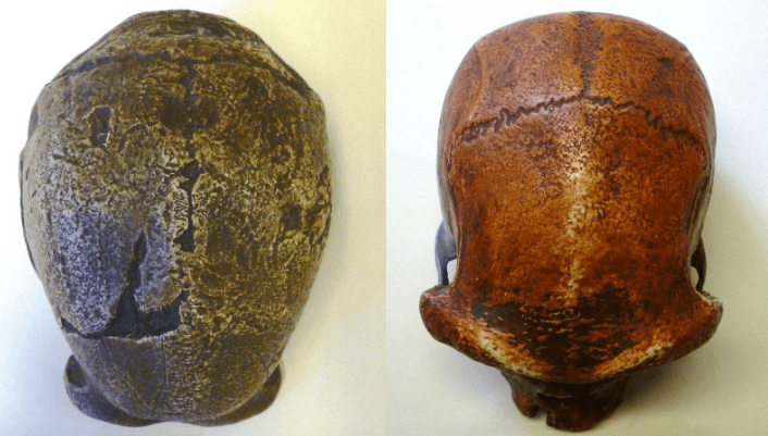
My PhD is based on unravelling aspects of Neanderthal landscape use and migration in the Western English Channel region during the Middle Palaeolithic, a period stretching from around 400 – 40,000 years ago. I am exploring behavioural complexities and reactions to environmental change through Neanderthal material culture, mainly via studying the movement of stone tools. Therefore it isn’t surprising that when I am engaging in the Grant Museum I gravitate towards the Neanderthal cast, which is a replica of the famous skull excavated from the site of La Chapelle-Aux-Saints in France.

Chappelle

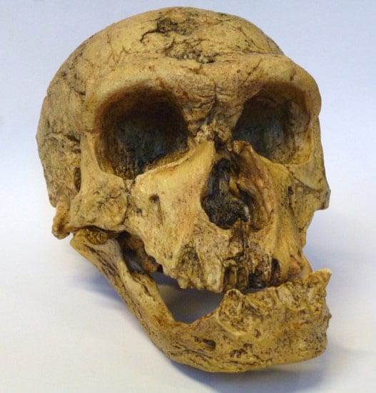
Figure 1: La Chapelle-Aux-Saints Neanderthal cast held at the Grant Museum—note the pronounced brow ridge over the eye sockets. Although the mandible and teeth look very different from Anatomically Modern Humans this is a cast taken from the skull of a particularly old individual who had advanced dental problems including gum disease! (Grant Museum, z2020)

Interestingly the most common theme in conversations I have with visitors to the Grant Museum is the shared similarities, rather than differences, between Anatomically Modern Humans and Neanderthals. It seems that what captures our imaginations now are the significance of concepts previously thought of as unique to Homo sapiens that are being gradually recognised in association with Neanderthals. Important advances in dating and DNA analysis have shown that Neanderthals and Anatomically Modern Humans co-existed in Europe for at least 40,000 years, with population groups meeting and interacting at different times. This is seen both in the archaeological record but also in the sequencing of the Neanderthal genome, which indicates that most modern people living outside of Africa inherited around 1-4% of their DNA from Neanderthals. As I mentioned, after the discovery of the first Neanderthal fossils people weren’t too keen on any evidence that threatened to topple the shiny pedestal reserved for Homo sapiens, however these advances in modern science have prompted a greater openness when exploring Neanderthal archaeology.

In order to investigate these aspects of complex behaviour, such as symbolism and art, we consider behaviours preserved in the archaeological record that appear to surpass the functional everyday need for survival. Recent discoveries have suggested that Neanderthals were making jewellery from eagle talons in Croatia and may have had more involvement than previously thought in the complex archaeological assemblages found at sites like Grotte du Renne. However evidence of these behaviours in Neanderthal populations remains rare and although this may relate to the historic viewpoint (it simply hasn’t been looked for…), empirically we just do not see it on the same scale.

Two examples I often refer to when discussing this at the museum are the recent discoveries of potential abstract art at Gorham’s Cave Gibraltar and the Neanderthal structures found underground at Bruniquel Cave. The abstract art (disclaimer: I understand that ‘art’ depends on the definition of the concept itself but that’s for another blog post!) was found at Gorham’s Cave in Gibraltar, a well-known Neanderthal occupation site. Often nicknamed ‘the hashtag’ it is a series of overlapping lines that appear to have been made deliberately by repeated cutting motions using a stone tool. The archaeologists who discovered the hashtag suggest that it was created around 40,000 years ago and that, as it was found underlying Neanderthal stone tools, it can definitely be attributed to them. They hail it as an example of Neanderthal abstract art that may even have represented a map, suggesting an elevated level of conceptual understanding. Whatever the marks represent, if they are associated with the Neanderthal occupation of the cave this is a behaviour that has not been observed elsewhere!

hashtag

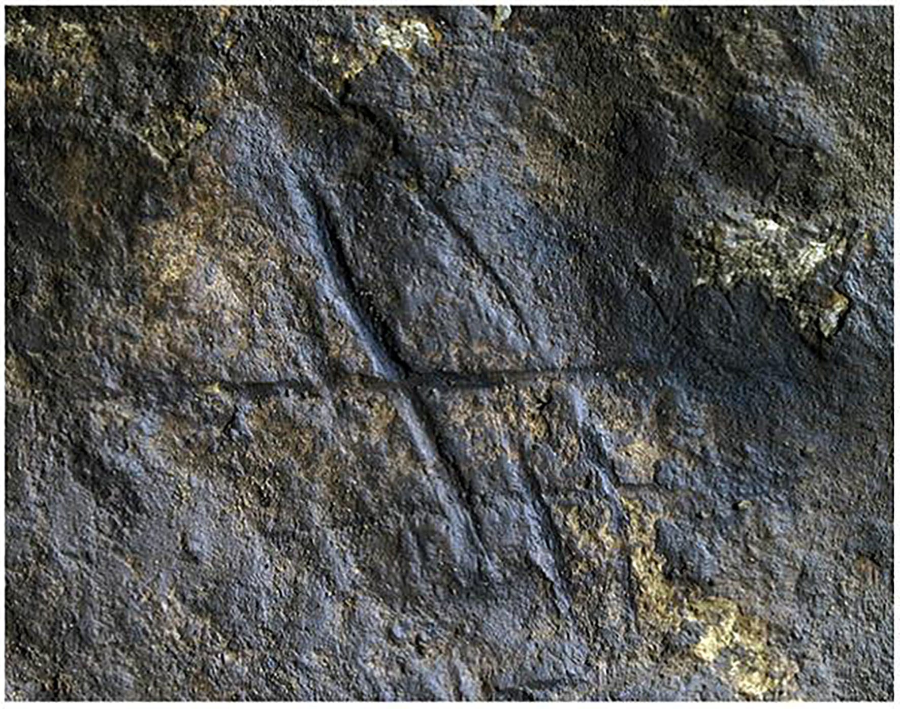
Figure 2: An image of the Neanderthal ‘hashtag’ made deliberately with repeatedly with strokes of a stone tool on a raised podium in Gorham’s Cave Gibraltar (Photo: Rodríguez-Vidal et al. 2014)

The other example that I mentioned is the site of Bruniquel Cave in southwest France, where unusual underground structures deliberately made from stalagmites have been dated via uranium series to 176,000 years old. This date firmly places the creation of the structures in a time where Neanderthals were the sole occupants of the region. The structures themselves are circular in diameter and are composed of fragmented stalagmites (all of a similar length c.34cm) with evidence of deliberately made fire. The function of these structures is not immediately obvious but as there is a distinct lack of other archaeological material in the cave it is unlikely they were used for domestic purposes. Equally their potential for functioning as shelters is unclear as they are located a whopping 336 metres from the cave entrance in an area that would not have faced the elements.

For me this location deep within the cave presents one of the key implications for Neanderthal behaviour in that no natural light whatsoever would have reached the chamber! This indicates a degree of familiarity with the subterranean world and potentially hints at the symbolic or ritual significance of the cave. Whatever the purpose of the structures, the authors of the study conclude that they represent unique evidence of the use of space, which may reflect the complex social structures of the Neanderthals who built there.

Bruniquel

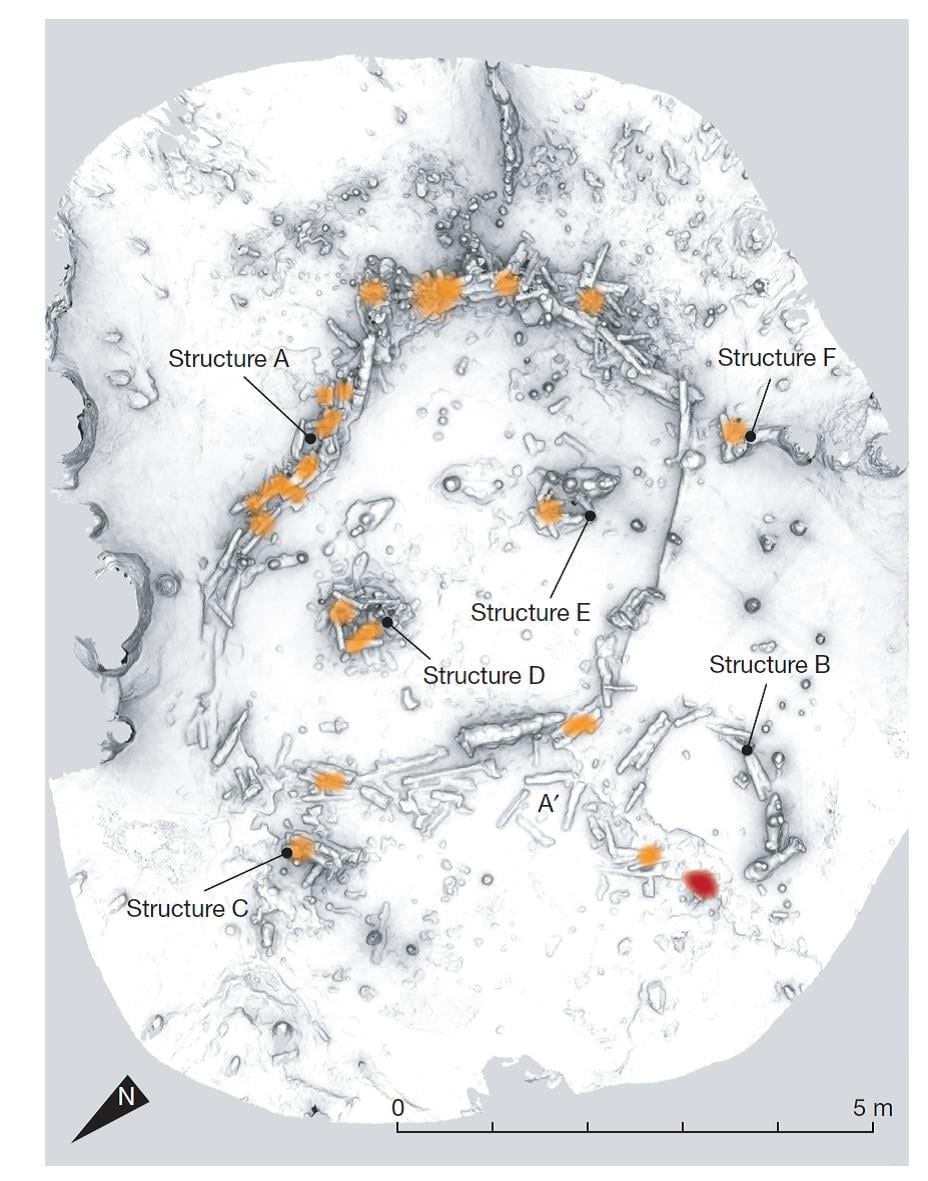
Figure 3: A schematic of the circular structures made with stalagmites deep underground in Bruniquel Cave, the orange colouration shows the areas of deliberate burning (Photo: Jaubert et al. 2016)

The inferences that are made from these Neanderthal finds are carefully considered by both the researchers concerned and the general archaeological community, disseminating the evidence and evaluating what archaeological information can be drawn from it. Overall there is something undeniably privileged to be working in a time where the complexity of Neanderthals is recognised and the potential for art, symbolism and other human characteristics is discussed!

References:

Green, R.E., Krause, J., Briggs, A.W., Maricic, T., Stenzel, U., Kircher, M., Patterson, N., Li, H., Zhai, W., Fritz, M.H.Y. and Hansen, N.F. 2010. A draft sequence of the Neandertal genome. Science 328 (5979), 710-722

Jaubert, J., Verheyden, S., Genty, D., Soulier, M., Cheng, H., Blamart, D., Burlet, C., Camus, H., Delaby, S., Deldicque, D. and Edwards, R.L. 2016. Early Neanderthal constructions deep in Bruniquel Cave in southwestern France. Nature534 (7605), 111-114

Radovčić, D., Sršen, A.O., Radovčić, J. and Frayer, D.W. 2015. Evidence for Neandertal jewelry: modified white-tailed eagle claws at Krapina. PloS one 10 (3), p.e 0119802.

Rodríguez-Vidal, J., d’Errico, F., Pacheco, F.G., Blasco, R., Rosell, J., Jennings, R.P., Queffelec, A., Finlayson, G., Fa, D.A., López, J.M.G. and Carrión, J.S., 2014. A rock engraving made by Neanderthals in Gibraltar. Proceedings of the National Academy of Sciences 111 (37), 13301-13306.