X Close

Researchers in Museums

Home

Engaging the public with research & collections

Menu

The leg bones connected to the hip bone…

By Mark V Kearney, on 28 November 2018

One of the nice benefits of working as a student engager is that during the down times when there aren’t that many people in the museum — the last 20-30 min before closing gets pretty calm — I have a little time to explore the collection. Despite my lack of formal study in biology (a strange feature of the Irish school system which allows one to obtain a degree in science without having to study biology), I have always really enjoyed my explorations in the Grant Museum of Zoology.

I’ve spent the majority of my time as an engager working at the Grant rather than the other museums and I keep coming back to the same observation when I look into the display cases: Why would animals evolve to have fibulas that seem so incredibly fragile? It’s a weird observation I know, but I’m a Physicist at heart and these mechanical aspects jump out at me.

The fibula is a very slender bone that is found in the lower leg and sits behind the tibia.

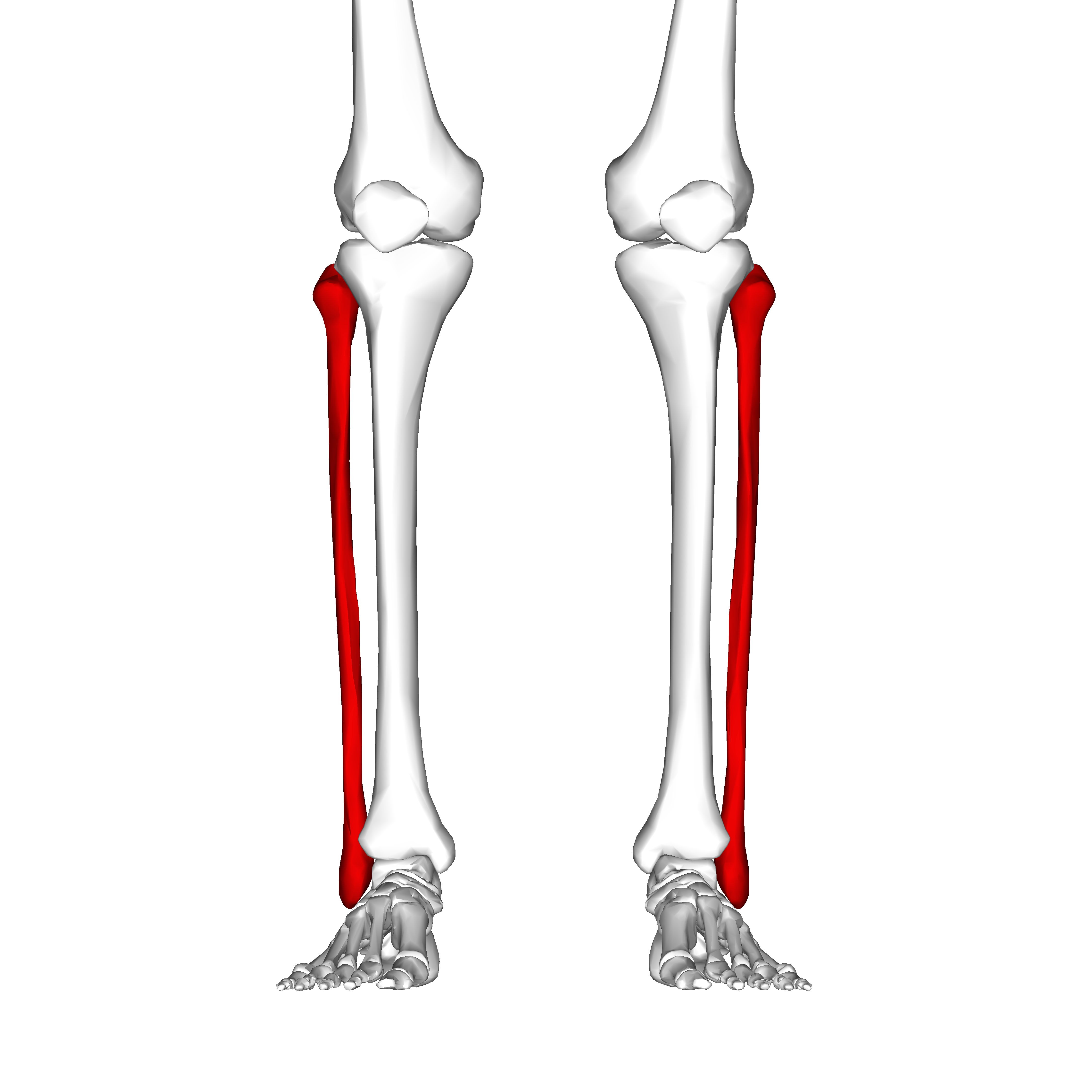
Figure 1 – Where the fibula is located in humans (Source: Wikimedia)

Now the issue I have with the fibula is this: our leg bones need to be strong enough to run on, how else would we evade some other animal that’s trying to eat us… and that goes for most other animals too (FIG.1). But why then is it so slender? What is it actually doing? Why don’t we break it all the time? And if it’s more of a hindrance to use, why haven’t we evolved to remove it?

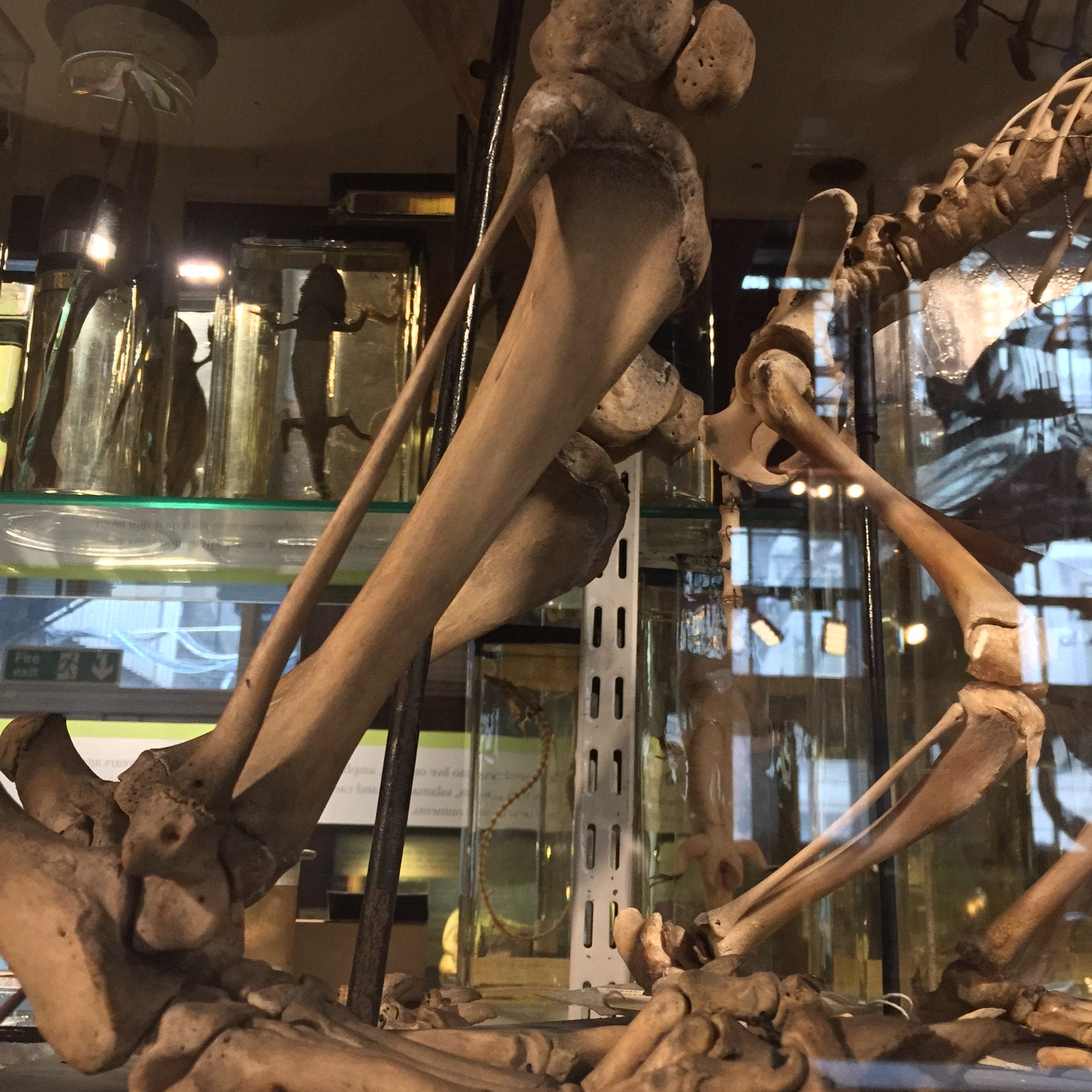
The back legs of a Brown Bear (Author’s own photo)

The Tiger on display at the grant has a tiny fibula compared to its tibia. (Author’s own photo)

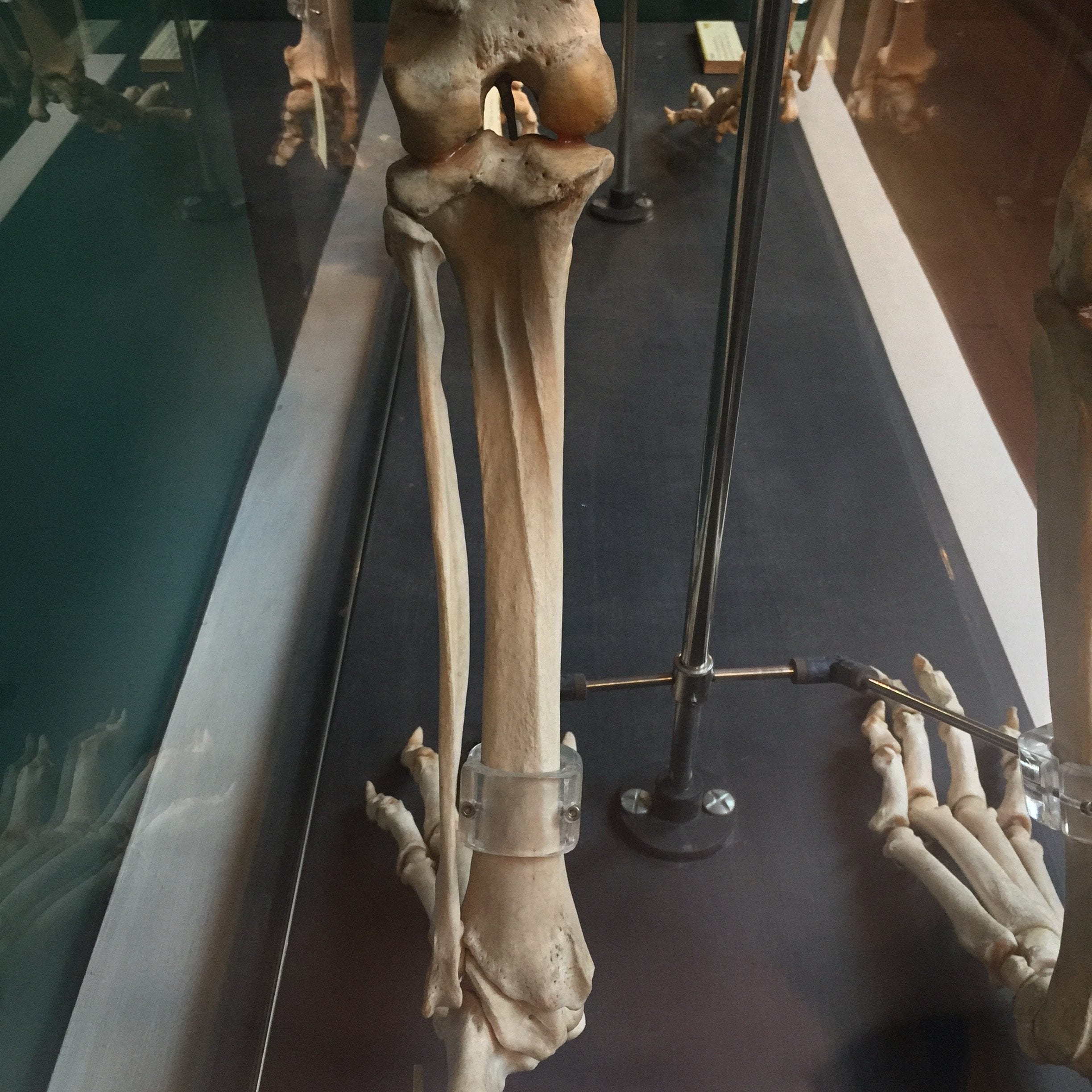
Let’s start by talking about what it does – from the reading I found for this blog it would seem that it doesn’t do very much. The fibula only takes about 10% of body weight and that is likely the answer to why it’s so slender. It doesn’t need to take as much weight as you might think; the tibia does that and that bone is multiple times the fibula’s diameter. The fibula also connects some muscles together and in the case of humans, it helps keep our ankle stable when we move.

More tiny fibula, this time from a Macaque on display at the Grant (Author’s own photo)

This Tree Shrew on display has a broken fibula… kinda proving my point that they break easily… (Author’s own photo)


Trying to find out about how often we fracture the fibula has proven more difficult than I thought. The studies that I have found don’t separate it from the tibia or sometimes they lump it in with the ankle making it hard for me to judge if it’s the tibia or the fibula that break first – though I did find one study suggesting the ‘fibula made up 12% of the tibia/fibula fracture cohort’. But that was only one reference so we won’t base everything off of that! At about 10% of fractures, that area seems to be one of the more common ones to get broken.

If the fibula doesn’t do much or if other things could do it better why do animals still have it and why hasn’t it been removed over the years via evolution? Well, it turns out that some animals are doing just that! First off, humans still have it because all tetrapods have inherited this basic form; we all ‘share the same pattern of bones in [our] limbs’. But some animals have started to evolve to do without it. For example, scientists have studied chickens and by looking their ancestors, early theropod dinosaurs, they’ve found that the fibula is now shorter and ‘splinterlike toward its distal end’.  This idea of the reduction of the fibula isn’t new at all… in fact, one reference I found is dated to 1918!

Through noticing the peculiarities of fragile fibulas my research has led me to learn more about the function and form of the skeleton, evolution, and finally landing on dinosaurs (which is always a great place to end up after a days research!) . That’s the beauty of museums, you can go in to kill some time and end up learning something fantastic!

Five years of research: a summary

By Stacy Hackner, on 3 July 2017

DSC_0745

by Stacy Hackner

A PhD often feels like an unrewarding process. There are setbacks, data failures, non-significant results, and a general lack of the small successes that (I hear) make general worklife pleasant: “I got that promotion!” “Everyone applauded my presentation!” “I moved to the desk near the window!” PhD life is one giant slog until the end, a nerve-wracking hours-long session where you’re grilled by the only people who know more about your field than you.

I survived.

Hopefully some of you have been following my research here, starting from astronauts and moving on to runners and foraging patterns. It all ties together, I promise. I recently gave a talk at the Engagers’ event “Materials & Objects” summarizing my research, which I can now tell you about in its full glory! I’m pleased to announce: I had significant findings.

The lowdown is that (as expected) there are differences in the shape of the tibia (shin bone) between nomads and farmers in Sudan. Why would this be? Well, if you’ve been following along, bones change shape in response to activity, particularly activities performed during adolescence. The major categories of tibial shape were those that indicated long-distance walking, doing activity in one place, and doing very little activity. Looking at the distribution, the majority of the nomadic males had the leg shape indicating long-distance walking, and some of the agricultural males had the long-distance shape and others had the staying-in-place shape. This makes sense considering the varying types of activity performed in an agricultural society, particularly one that also had herds to take care of: some individuals would be taking the herds up and down along the Nile to find grazing land while others stayed local, tending farms. While it’s unclear how often a nomadic group needs to move camp to be considered truly nomadic, in this case it seems like they were walking a lot – enough to compare their tibial shape to that of modern long-distance runners. These differences in food acquisition are culturally-adapted responses to differing environments: the nomads live in semi-arid grassland and can travel slowly over a large area to graze sheep and cattle, while the farmers are constrained to a narrow strip of fertile land along the Nile banks, limiting how many people can move around, and how often.

Perhaps the most important finding is the difference between males and females. In addition to looking at shape, I also conducted tests to show how strong each bone is regardless of shape, a result called polar second moment of inertia (and shortened to, unexpectedly, J). The males at each site had higher values for J – thus, stronger bones – than the females. However, the nomadic females had higher J values than some of the males at the agricultural sites! This is in spite of most females from both sites having the tibial shape indicating “not very much activity”. This shape may be the juvenile shape of the tibia, which females have retained into adulthood despite performing enough activity to give them higher strength values than male farmers. Similar results have actually been noted in studies examining different time periods – for instance, the Paleolithic to Neolithic – and found much more similarity between females than between males. Researchers often interpret this as evidence of changing male roles but female roles remaining the same, which strikes me as unlikely considering the time spans covered. I instead conclude that females build bone differently in adolescence, and perhaps there are subtleties in bone development that don’t reveal themselves as differences in shape. As females have lower rates of testosterone, which builds bone as well as muscle, they may have to work harder or longer than males to attain the same bone shape and strength. I’m using this to argue that the roles of women in archaeological societies – particularly nomadic ones – have been unexamined in light of biological evidence.

Of course, the best conclusion for a PhD is a call for more research, and mine is that we need to examine male and female adolescent athletes together to see when exactly shape change occurs. If we can pin down the amount of activity necessary for women to have bones as strong as those of their male peers, we can more accurately interpret the types of activities ancient people were performing without devaluing the work of women.

My examiners found all this enthralling, and I’m pleased to say I passed! The work of this woman is valued in the eyes of the academe.

How are Ancient Nubians Like Astronauts?

By Stacy Hackner, on 6 January 2014

Stacy Hackner_Thumbnail By Stacy Hackner

Some respected individuals (supervisors, mentors, parents) have advised me to not get distracted by the primrose paths that crop up during a PhD. These primrose paths are always deliciously exciting, offering opportunities to study wonderful new topics that one can justify as marginally related to one’s thesis and therefore potentially of use. Of all the primrose paths I’ve followed, I never expected the most relevant one to be about astronauts.

Credit: Wikipedia Commons

Sudanese pyramids. (Wikimedia commons.)

My thesis explores ancient Nubia, the region that is now northern Sudan, from roughly 3000 years ago to medieval times. Unlike their contemporaries, the Egyptians, the Nubians didn’t have a system of writing until the Meroitic period (300 BCE-400 CE), a time of Egyptianizing influence. They built small pyramids and imported Egyptian goods, attesting to the influence of their famous northern neighbors. In the absence of writing (and even in the presence of texts, as humans tend to play with the truth), archaeologists try to build a picture of the ancient society using physical evidence, including human remains. Fortunately, the dry climate and sandy soil usually result in excellent bone preservation, allowing me to identify differences in bone shape. But wait – let me back up a little.

We aren’t entirely sure how bone works. There are two types of cell responsible for bone maintenance – osteoblasts and osteoclasts. Osteoblasts build bone, and osteoclasts take it away. The body is highly responsive to changes in activity, and bone is constantly updating itself accordingly. The general principle is that your body thinks what’s happening now will happen forever. Think about when you’re running a race: it’s hard to start because your body’s been used to standing still and needs some time to amp up your heart rate and muscle contractions. When you finish the race, your heart keeps pounding for a minute or two because it hasn’t quite got the signal to stop running yet. Bone works in a similar way. In response to physical stress, bone will accumulate more osteoblasts to strengthen itself. Each step makes tiny microfractures, which tells the bone “Come on, I’m breakin’ here! Give me more strength!” and the osteoblasts pile on. In the absence of activity – during periods of prolonged sitting or lying down – the osteoclasts come in to take away unnecessary bone. “You’re not using this one, right? Then we can send the calcium somewhere else.” The thing is, scientists don’t know all the signals involved in this process. We know what happens, but not the channels of communication. I like to imagine bone cells having little conversations with each other, but clearly it’s all on a neurochemical level we haven’t yet discovered.

The concept of bone building in necessary areas is keenly presented in studies of elite athletes. In a study by Haapsalo et al (1998) of young female tennis players, the players gained significant bone mineral content in the bones of their dominant (forehand and serving) arm. When the authors looked at a control sample (girls who did not play tennis), there was minimal or no difference between their arms; there was also minimal difference between the nondominant arms of the tennis players and the controls.

Another study, by Shaw & Stock (2009), examined differences between university athletes who competed in either hockey or long-distance running. They found significant differences in the actual shape of the tibia (shin bone) due to the physical stress of these activities. The tibias of the long-distance runners were more elongated front to back while the tibias of the hockey players were more even side-to-side, showing a distinct difference in the direction of activity in these sports. Clearly, osteoclasts were being sent to the bone locations these athletes needed them most: for runners, the front, and for hockey players, the sides. It is important to point out that many of the studies investigating activity and bone growth look at adolescents, since their bones develop until the end of puberty. After that, it seems to take a lot more effort to alter bone shape and density.

Credit: wikipedia

Her bones are losing mineral content by the minute! (Wikimedia commons.)

And what about the other side of the cycle? The osteoclasts? That’s where the astronauts (and cosmonauts) come in. The constant pounding of our feet against the floor keeps our bones as strong and dense as they need to be for everyday use. In zero gravity, though, there’s no pounding, just the occasional soft push off the wall of the space station. The osteoblasts don’t have any stress to react to, and the osteoclasts assume the extra bone is useless, so it starts to be resorbed. It helps that astronauts are some of the most-studied individuals on our planet (and definitely the most studied off the planet!). During spaceflight, urine calcium output is found to increase, indicating that bone is being sapped of minerals, and post-spaceflight bone scans reveal a condition called “spaceflight osteoporosis”, similar to the osteoporosis experienced on earth – but the bone density is only lost from the legs, feet, and hips, all weight-bearing regions, including an 8% loss in four months (compared to 1% loss per year for earth-bound sufferers of osteoporosis). The upper body and head generally remain unaffected (unless one of the astronauts was a tennis player, of course). One study found that after a “long-duration” spaceflight of 4-6 months, it took up to three years for astronauts to recover the bone density they’d lost in space (Sibonga et al, 2007). It makes one really appreciate gravity.

So how do I apply this to ancient populations? The data from astronauts indicates that most of the density lost is from trabecular bone, from the internal core, rather than from the outside. This means that even if ancient bones have lost density due to age, either before death or after burial, it’s likely to have happened from the inside out and thus the external shape should remain intact. This gives me more confidence in figuring out what kinds of activities they performed during adolescence, which in most cultures was when young people started to take up adult cultural roles. I hope to compare the shape of the bones of Nubians to those of athletes and to other populations whose activities are known in order to draw a better picture of their society.

Sources

For an amusing (but factual) look at the craziness that is astronaut and cosmonaut research, check out Mary Roach’s “Packing for Mars: The Curious Science of Life in the Void” (Norton & Co, 2010).

Haapasalo, H, P Kannus, H Sievänen, M Pasanen, K Uusi-Rasi, A Heinonen, P Oja, and I Vuori. 1998. Effect of long-term unilateral activity on bone mineral density of female junior tennis players. Journal of Bone and Mineral Research 13/2, 310-319.

Shaw, CN and JT Stock. 2009. Intensity, Repetitiveness, and Directionality of Habitual Adolescent Mobility Patterns Influence the Tibial Diaphysis Morphology of Athletes. AJPA 140, 149-159.

Sibonga, JD, HJ Evans, HG Sung, ER Spector, TF Lang, VS Oganov, AV Baulkin, LC Shackelford, and AD LeBlanc. 2007. Recovery of spaceflight-induced bone loss: bone mineral density after long-duration missions as fitted with an exponential function. Bone 41, 973-978.