X Close

Researchers in Museums

Home

Engaging the public with research & collections

Menu

Archive for September, 2017

Label Detective: Are we alone in here?

By tcrnkl0, on 20 September 2017

In the first two instalments of the Label Detective series we investigated the meaning of the word cynocephalus and the impact of British eugenics on Egyptian archaeology. Now we’re moving over to the Grant Museum of Zoology to tackle the label mysteries of the animal kingdom.

Case 4

Let’s start with the Portuguese Man O’War. Here’s a picture of one floating peacefully (and extremely poisonously) in Cornwall earlier this month.

Portuguese Man O'War

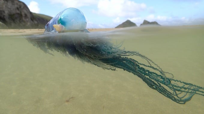
Photo credit: Corwall Wildlife Trust

And here’s the label on the specimen in the Grant Museum.

This is my bad photo. There's a better picture of the specimen underneath at the bottom of this post.

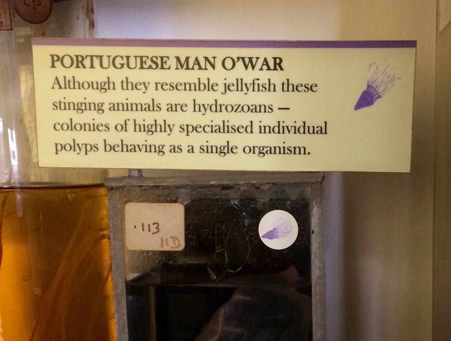
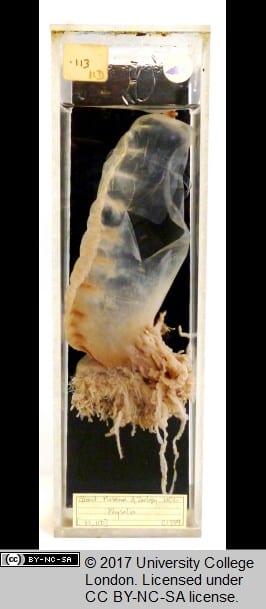
This is my bad photo. There’s a better picture of the specimen underneath at the bottom of this post.

The Label: I got curious about the Portuguese Man O’War because the label uses the word ‘colony’ here in a way that I didn’t really understand. When we talk about colonies in the animal kingdom we are usually referring to insects, like bees or ants, where lots of individuals make up a colony. But what does it mean for a colony to make an individual? In the case of the Portuguese Man O’War, a siphonophore, four different types of polyps come together to make an individual like the one pictured. Each kind of polyp has a different function. The inflated bladder, or sail of the Man O’War, helps the creature to float. Then there are reproductive polyps, eating/digestive polyps, and ones that provide the Man O’War’s stinging defence. These latter three types of polyps are themselves made up of groups of individuals called zooids. It’s multiplicities all the way down.

Case Notes: The polyps and zooids that make up a Portuguese Man O’War are genetically identical, and so specialised as to be interdependent (though the individual zooids are structurally similar to other independent species) – so in many ways it does make sense to consider them an individual. But it challenges assumptions that an individual is something entirely singular or uniform.

‘Individuals’ are rarely a closed, or self-contained system. What does this mean? Consider you and your mother. When you are a fetus, some of your cells pass through the placenta and take up residence in your mother’s body. You also get some of your mother’s cells. Even weirder, if you aren’t your mother’s first child, you not only get your mother’s cells, but cells from all your siblings as well. You don’t just have other people’s cells in your body — you also have loads of cells that aren’t human at all. Developmental genetics and embryology scholar Scott F. Gilbert says: ‘Only about half of the cells in our bodies contain a “human genome.” The other cells include about 160 different bacterial genomes. We have about 160 major species of bacteria in our bodies, and they all form complex ecosystems. Human bodies are and contain a plurality of ecosystems.’

These examples are not the only way that genetic transfer is more diverse than the Darwinian model of sexual selection (i.e. getting all of your genes from two parents). And a lot of these more varied and spectacular ways are down to bacteria. Next time on Label Detective, we’ll get into these messier models of evolution.

Status: I would say case closed, but since I’ve just spent the blog post arguing against the concept of a closed system, this seems wrong. But we’re done for now.

wwwopac.exe

Notes

This post was inspired by the book Arts of Living on a Damaged Planet, an anthology of essays by zoologists, anthropologists, and scholars that explores how environmental crisis highlights the complex and surprising ways in which all life on earth is entangled. The quote from Scott F. Gilbert comes from his contribution to this book.


Embryological Wax Models

By ucbtch1, on 14 September 2017

The Grant Museum has a number of embryological wax models on display (Images 1, 2, 4, and 5 amongst others not shown here). These models, while often ignored by visitors, are actually quite remarkable as they showcase the brilliance and mystery of embryological development. They were created to help elucidate essential questions like: how do humans and other animals form? How are a bunch of seemingly insignificant cells, with no shape other than a ball, able to grow so much and in such detail to form intricate patterns like our eyes? How can one cell transform itself into such different tissues, from hard rock bone to the jelly like liver? In order to understand how a human body is formed it is vital to study the very first stages of its creation, i.e. when we are just a bunch of cells.

Image 1. Placental mammal embryo (Z3100)

Image 1. Placental mammal embryo (Z3100)

The very first cells that are formed after fertilization are called stem cells and they start with an unlimited capacity to form any type of cells. With time, they start to differentiate and mature into specialized cells with a limited lifetime. In this process, little by little they lose their unlimited capacity until they can only form cells that are similar in lineage. In this manner, totipotent stem cells can form any body part including extraembryonic tissue like the placenta. On the next level, pluripotent stem cells (embryonic stem cells for example) are capable of forming any body part but have lost the capacity to form extraembryonic tissue. And finally, multipotent stem cells, much more restricted, can only form cells from a specific tissue or organ.

This may appear as a straightforward process, but the development of an animal is a deeply specific, delicate, and sophisticated interplay of signals and coordinated transitions. Think of it as an orchestrated dance of on and off switches leading to specialisation and exponential growth. In fact, it is so complex that we still don’t understand it entirely.

Although not all human, the wax models display the first stages of development of vertebrates and closely related animals. First, one cell divides symmetrically into two, then four, then eight and so on (Image 1 and front models of Image 2). Afterwards, cells start organizing themselves answering to chemical and physical signals and different patterns start to appear (Image 2 models in the back). Eventually, an axis emerges on which cells migrate along which will give rise to the head on one side and the body and limbs on the other (Image 4). 

Slowly but surely, we all go from looking like little worms to fully grown animals (Image 3). It is important to note that in this initial period most embryos have a very similar appearance, at least between vertebrates. These similarities tell us that a lot of the genes that govern this initial growth haven’t changed between species over time. It’s like nature is saying, “well, if it ain’t broke don’t fix it” and so these mechanisms have been conserved in different animals.

Image 2. Branchiostoma sp, Lancelet Embryo (fish-like invertebrate; T114)

Image 2. Branchiostoma sp, Lancelet Embryo (fish-like invertebrate; T114)

These early stages are crucial moments because if one little element of the spatial/temporal organization is out of place, improper organization can lead to lifelong malformations, diseases, or even the termination of the embryo. Hence the importance of understanding how this process works. We know that even in adult life, there are still stem cells proliferating and forming new tissue to a certain degree. Some organs, like the skin, have a lot of stem cells to replace old cells when they die or get injured. But other organs like the brain, have a very limited capacity to grow new cells—one of the reasons why a brain is much more difficult to fix.

Image 3. Development of the external form of the human face (LDUCZ-Z480) and development of external form of human embryo (LDUCZ-Z430)

Image 3. Development of the external form of the human face (LDUCZ-Z480) and development of external form of human embryo (LDUCZ-Z430)

Image 4. Vertebrate embryos. Image taken from https://www.ncbi.nlm.nih.gov/books/NBK9974/

Image 4. Vertebrate embryos. Image taken from https://www.ncbi.nlm.nih.gov/books/NBK9974/

So can we get back all the limitless capacity there once was in the developing embryo? Even though the genetic and molecular mechanisms governing all these changes are still somewhat elusive, researchers are using stem cells and powerful genetic tools to answer this question and decipher every single step of how a human is formed in the womb. Moreover, if we can understand the process, then we can recreate and modify it in the lab, and this is exactly what the field of stem cells and regenerative medicine is trying to do. Imagine having the capacity to grow new organs to be used for transplantation or drug testing. How about growing a brand new functioning leg or arm for amputees? Or studying the mechanisms of diseases like Parkinson’s, leukaemia, diabetes, amongst others. The benefits of harnessing the regenerative potential of cells are far-reaching.

Image 5. Development of the external form of the human face (LDUCZ-Z480).

Image 5. Development of the external form of the human face (LDUCZ-Z480).

The exciting field of stem cells and regenerative medicine has come a long way, more than a century has passed from the first time the term stem cells was used in 1906 up until the creation of genetically modified human embryos in 2017. The embryological wax models represent initial efforts of identifying how changes give rise to specific structures and ultimately how an animal comes into existence. Furthermore, the future still holds exciting breakthroughs, there is still a lot to understand about human development and the wax models are a fantastic resource to portray the morphogenetic changes we all once went through.


Resources:

Gilbert SF. Developmental Biology. 6th edition. Sunderland (MA): Sinauer Associates; 2000. Comparative Embryology. Available from: https://www.ncbi.nlm.nih.gov/books/NBK9974/

http://www.labonline.com.au/content/life-science-clinical-diagnostics-instruments/article/why-do-all-animal-embryos-look-the-same–236915402

Question of the Week: Bird vs Reptile

By uctzcbr, on 8 September 2017

Yesterday, in the Grant Museum, I was asked why reptile and bird eggs are different. I did not know that reptile and bird eggs were different, let alone why. I have now investigated this question and prepared a pretty egg-cellent answer (if I do say so myself).

Bird eggs and reptile eggs are different in a couple of ways (besides the fact that one comes out and contains reptiles and the other birds). They can differ in shape, shell, and colour. These differences are not universal across the classes and science cannot explain why all of the differences have occurred, so this answer isn’t going to be egg-xact but I will try and be as thorough as possible.

So, first, the egg shape. Reptiles have symmetrical eggs where as some birds lay eggs which are, well, egg-shaped: i.e. asymmetrical as they are tapered at one end. A popular explanation for why bird eggs are often this shape is that it prevents them from rolling off of cliffs as they, instead, roll in a circle. However an enormous study conducted at Princeton University has provided data to debunk this theory. The study, which examined almost 50,000 eggs from more than 1,400 species found a correlation between the egg shape and wing shape in bird species. This points to the egg shape to more likely being a product of a bird’s flight adaptation than where they nest. You can read the study here.

The second distinction is in the shell – birds lay eggs with hard shells where as some reptile species lay soft-shelled eggs. Why might be the case? Well, if a mother cannot lay a hard-shelled egg at its full size, it could lay a soft-shelled egg instead allowing the egg to eggspand (not the technical term) after laying. Additionally, a soft shell has the capacity to absorb moisture from the atmosphere and ground. For some reasons, birds have evolved such that their eggs don’t need additional moisture, which is not the case for some species of reptiles. For the opposite reason, some turtle species will lay hard shelled eggs if they also lay their eggs in wet environments as this stops the egg absorbing too much moisture.

Another reason reptiles lay soft-shelled eggs is because of the way they are incubated. Birds will sit on their eggs and use the warmth of their bodies but reptiles tend to utilise the natural heat of vegetation or the earth to incubate their eggs. As reptile eggs don’t have to be strong enough to protect the unborn contents from the full weight of its parent, they can be soft-shelled.

Finally, most reptile eggs are white in colour whereas bird eggs are lots

A pretty symmetrical looking Ostrich Egg (Ostriches, famously, not great flyers).

A pretty symmetrical looking Ostrich Egg (Ostriches, famously, not great flyers).

of different, some would say eggstraordinary, colours. As with the shape, science cannot prove a definitive answer for why this is the case. However, there is a theory which has, in part, been developed by comparing reptile and bird eggs. Bird eggs are often kept in much more conspicuous places than reptile eggs – in open nests as opposed to buried underground or hidden in a crevice. As such, bird eggs can benefit from camouflaging to their surroundings, which would require them to come in a wider variety of colours.

So, there you have it, for the most part, eggs is eggs. However, reptile and bird species have evolved different characteristics specific to their environment. If you have any more questions, come visit the Grant Museum or tweet them at us.