X Close

UCL Culture Blog

Home

News and musings from the UCL Culture team

Menu

Archive for the 'Pathology' Category

The Anthropologist, the Anatomist, and the Highwayman: Stories from student research 2020/21

By ucwehlc, on 21 July 2021

Teaching with museum objects during a global pandemic has been something of a challenge, to say the least. You can read more about how the UCL Museums have tackled it in this previous blog.

One of the real success stories has been how UCL students have continued to research objects for their projects without being able to visit the objects in real life, and with reduced access to resources in other museums, libraries, and archives. Their perseverance and ingenuity this year has allowed them to uncover compelling stories of tragedy, prejudice, and redemption in the UCL Science Collections. As the academic year comes to an end, and I add their findings to our database, I thought I would share a few with you.


Eye Colour Gauge by Rudolf Martin: The Anthropologist’s Story

Museum Studies master’s students Karolina Pekala, Helena Smith Parucker, Ailsa Hendry and Emma McKean researched this object for their Collections Curatorship module. Before their project began, we knew that the eye gauge had been designed by someone called Rudolf Martin, and that it had been owned by either Francis Galton or Karl Pearson, both of whom were instrumental in establishing the world’s first Eugenics Department at UCL.

Black metal box containing 16 glass eyes of different colours set into an aluminium backing

Rudolf Martin’s Eye Colour Gauge LDUGC-365. Copyright UCL Culture


The object itself has an unsettling look to it, even before we consider its links to the history of eugenics. It was designed by Swiss anthropologist Rudolf Martin and manufactured between 1903 and 1907. The students examined Martin’s background and his views on the developing field of eugenics in the early 20th century. They concluded that Martin himself was not actively involved in eugenic research, being more interested in developing methods for accurately measuring humans. However, he was well aware what other researchers were using his methods and tools for, and he supported racially biased anthropological research.

This particular eye gauge was used by Karl Pearson and Margaret Moul in eugenic research on Jewish school boys in London in the 1920s. A later version of the eye gauge was used in German research in the Tarnów Ghetto in Poland in the 1930s.

This story has a tragic sting in the tail for the Martin family. Rudolf Martin died in 1925, so he did not live to see eugenics lead to the horrors of the Holocaust. His second wife Dr Stefanie Martin-Oppenheim survived him, but as she was Jewish, she was imprisoned in Theresienstadt Concentration Camp, where she died in 1940.

You can read more about eugenics, anthropology, medicine, and the Holocaust in this United States Holocaust Memorial Museum online exhibition.


Obtaining Specimens for the UCL Anatomy Museum: The Anatomist’s Story

Museum Studies students at UCL complete a practical placement in a museum as part of their degree. As objects were off limits in 2020/21 our student placements this year were all about our digitised archives. Archival material has a reputation for being a little dull, but it is often the source of the most fascinating insights into our collections. Nicky Stitchman’s project tracked the development of the museums at UCL from 1826 to 1926 using the UCL calendars and committee meeting minutes.

When students visit the UCL Pathology Museum at the Royal Free Hospital we often discuss the ethics of keeping and using specimens of human remains for teaching and study. Nicky’s research demonstrates that this was not always a matter of concern in medical teaching. In 1854 the rules of University College Hospital stated that

“No specimens of disease removed from patients, or from persons who may die in the Hospital, may be taken from the Hospital until after consultation with the Curator of the Museum of the College, for the purpose of determining whether such specimens shall be preserved in the Museum of Anatomy.”

And it was the duty of Physicians’ Assistants and House-Surgeons

“To deliver to the Curator of the Museum of Anatomy of the College according to Regulation § 54, all specimens of disease removed from patients, whether living or deceased, in their respective departments, and to give him previous notice of all post-mortem examinations.”

So, doctors had to inform Professor Sharpey (the Curator of the Anatomy Museum) whenever they did a post-mortem just in case he wanted any specimens for the museum. No mention is made of patient consent or the ethics of displaying the dead.


Sepia image of the UCL Anatomy Museum in the 19th century

UCL Anatomy Museum when Professor Sharpey was the curator. Image courtesy of UCL Special Collections, ref: UCLCA/7


These days the UCL Pathology Museum no longer actively collects human remains, and institutions that do so have to adhere to the rules of the Human Tissue Authority, obtaining informed, written, witnessed consent from patients.

You can read more about Nicky’s discoveries in this blog where she shows a little love for Assistant Curators, the unsung heroes of the UCL Museums.


Phrenological Head Cast: The Highwayman’s Story

UCL is home to a truly remarkable collection of heads. These life and death masks were collected by Phrenologist Robert Noel in the 19th century to explore ideas of genius and criminality. Phrenology is the long-disproven idea that the shape of someone’s head reflects their intelligence and personality. It is fair to say that Robert Noel’s collection tells us at least as much about Victorian theories of race, class, gender, crime, and mental health as it does about the personalities of his subjects.

Bachelor of Arts and Sciences student Iris Perigaud-Grunfeld wrote her Object Lessons project on head number 41, Babinsky the highwayman. Robert Noel was convinced that Babinsky was a Robin Hood character who stole for good but misguided reasons, and concluded that his head was not of the ‘criminal type’. This cast was taken from life in Prague in 1845 when Babinsky was in prison for robbery. Later Babinsky was released for good behaviour and became a gardener at a monastery.

Plaster cast of a man's head. His eyes are closed an he is wearing a neck scarf.

Plaster life cast of Vaclav Babinsky’s head. LDUSC-Noel-41. Copyright UCL Culture

Iris’ research uncovered details that Robert Noel had missed, and filled in the gaps about what happened after Babinsky died. After his release from prison notorious highwayman Vaçlav Babinsky became a genuine folk hero in Bohemia and Germany, with songs and novels written about him. He even featured in a Czech TV show, and recently Radio Prague International produced an English language podcast about his life which is well worth a listen.

Black and white photograph of an elderly man with muttonchop whiskers

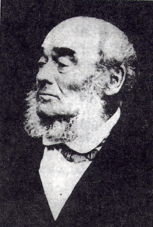
Vaclav Babinsky as an old man. Image in the public domain.

Knowing Babinsky’s full name has also allowed us to find out more about his crimes. He was convicted of attacking a government official, and there is some suggestion that this charge was for biting the town mayor, which makes for a suitably colourful episode in the life of a famous highwayman. However, he was also convicted of at least 2 violent robberies and involvement in a murder, which shows our folk hero in a very different light. Would Robert Noel have interpreted Babinsky’s head differently if Babinsky had been executed for his crimes and never had the chance to turn his life around?


Whether our students next year are working with objects in person, or if they are working with digitised archives, I cannot wait to see what they uncover in the collections.

Hannah Cornish is Curator of Science at UCL Culture

The Unsung Heroes of UCL Museums and Collections

By ucwehlc, on 6 July 2021

During 2020 and 2021 while UCL was closed due to the coronavirus pandemic, UCL Culture’s curatorial team worked with students from the Institute of Archaeology’s MA Museum Studies on our first-ever virtual work placements. These projects, which included archive transcription, documentation and object label writing, provided opportunities for the students to gain practical curatorial skills to prepare them for their future careers while undertaking valuable work towards better understanding the collections.

This blog was written by Nicky Stitchman, UCL MA Museum Studies

As part of my MA in Museum Studies, I undertook a work placement with Hannah Cornish, Science Curator at UCL. My brief was to discover the different locations of UCL’s museums and teaching collections from the university’s origins in 1828. Ferreting out information from primary and secondary sources and finding maps that showed the movement of the various museums as the university expanded was fascinating, but I found myself drawn to people behind the museums. I am not talking here about the headliners – the Flinders Petries or Robert Grants of this world – but rather the curators, assistant curators and demonstrators who would have done most of the day-to-day tasks such as cataloguing, labelling, teaching, and physically moving the artefacts and objects within the collections. 

James Cossar Ewart at the Grant Museum 

J Cossar Ewart was the first professional, rather than professorial, curator of both the Anatomical Museum and Comparative Anatomy/Zoology Museum between 1875-1878. He was appointed after the retirement of William Sharpey (curator of the Anatomical Museum) and the death of Robert Grant (professor of comparative anatomy). In the official records, it was Professor Lankester, Grant’s successor, who refitted and rearranged the Museum of Zoology over this period but Ewart was instrumental in making the zoological preparations and was also known to have helped organise and take the subsequent practical classes that Lankester introduced to UCL. 

Black and white image of the head and shoulders of James Cossar Ewart

James Cossar Ewart worked at the Grant Museum 1875-1878. Image in public domain

After Ewart, there was a change in the running of the two largest museums at UCL at that time, with a separate curator appointed for the Museum of Anatomy, while the Zoological Museum (now the Grant Museum of Zoology) was titularly run by the Head of Department with a curatorial assistant.

Shattock and Stonham: Anatomy and Pathology

Mr Samuel Shattock succeeded Ewart as the Curator of the Anatomical and Pathological Museum. He had originally shown up in the records as a Mr Betty which caused me some confusion at the time, until I discovered that he had decided to change his name to prevent the extinction of the Shattock family name!  Shattock never qualified as a physician but dedicated his life to pathological medicine. He was responsible, alongside Dr Marcus Beck, for a descriptive catalogue of the surgical pathology preparations at UCL. His successor Charles Stonham also worked with Marcus Beck on Part II of this catalogue and then in 1890 produced another medical pathology catalogue, which can be found online at the Wellcome Collection. In the preface to this catalogue, Dr Barlow and Dr Money acknowledge the work of Charles Stonham, and state that it is ‘to him the preparation of this work is almost entirely due’.

Black and white image of Charles Stoneham in military uniform sitting on a wooden chair holding a riding crop

Charles Stonham Curator of the UCL Anatomical and Pathological Museum (copyright UCL CC By 3.0)

Stonham was also responsible for the division of the pathology from the anatomy collection and its rehousing within the museum. He is also remembered on the UCL Roll of Honour as not only was he an instrumental figure in the London Mounted Brigade Field Ambulance, but he volunteered as a member of the Royal Army Medical Corps during WWI and died on service in January 1916 aged 58. 

Margaret Murray and the Petrie Museum

The dedication of many assistant curators at UCL is very clear. The redoubtable Margaret Murray (pictured here), who worked alongside Flinders Petrie for many years, was effectively in charge of the museum’s collections during Petrie’s many excavations in Egypt and the Levant. It should also be remembered that the Petrie Museum of Egyptology and the Department itself was founded on the collection and endowment of Mrs Amelia Edwards, who favoured UCL due to the early admission of women into the College.

Black and white image of Margret Murray wearing a lace shawl, seated and holding a book

Egyptologist Margaret Murray (Image in public domain)


Edith Goodyear and the Geology Museum

One of the first women to be involved in UCL’s museums was Edith Goodyear who was appointed as the Assistant in the Geology Museum in 1904 and subsequently remained in the department until the Second World War. Edith worked alongside Professor Edmund Garwood, reorganising the museum, teaching, and researching papers. A room in the Lewis Building was named for her, along with the First Year Student prize within Earth Sciences. It is also worth noting that in an age of inequality, the 1916/17 council minutes show Edith was paid £150, the same as her male colleague Dr J Elsden. 

Keeping it in the family: Mary and Geoffrey Hett

1917 also saw the appointment of Mary L Hett as Assistant in the Zoology Dept on the same salary of £150, where she remained until she took up the post as Professor of Biology at the Hardinge Medical College, Delhi.  She had followed her brother Geoffrey S Hett to UCL where he held the post of Curator of the Anatomical Museum from 1907-1910. Like his sister, he had a great interest in the natural world and was an authority on both British birds and on bats. Geoffrey became an ENT Specialist and during his time at UCL completed valuable research on the anatomy of the tonsils. Like his predecessor, Charles Stonham, he also served in World War I and used the skills learnt at UCL to treat head, and in particular, nasal injuries during this period. 

The stories of the men and women who studied and worked at UCL museums over the years are many and various, and these are just a sample of those whom I have met in my research for the Mapping UCL Museums Project. We may never be able to give the Curators and Assistant Curators the recognition that their work and dedication deserve but in introducing these few to you, I hope to have redressed the balance very slightly in their favour!  

How do you move an Auto-Icon?

By Katie Davenport-Mackey, on 26 March 2020

Until recently, the ‘Auto-Icon’ of the eccentric English philosopher – comprising Bentham’s skeleton and wax head – sat in a wooden box in the corner of the Wilkins Building, dressed in 18th century clothing and holding Bentham’s favourite walking stick known as ‘Dapple’.

Bentham is UCL’s most popular museum exhibit, attracting visitors from all over the world. His new home provides greatly enhanced preservation conditions, better visitor access and a place at the centre of the student community.

We asked Christina McGregor, Head of Collections Management at UCL Culture what is involved in conserving and moving Jeremy Bentham.

(more…)

Specimen of the Week 385: UCL skulls

By ucwehlc, on 12 July 2019

This blog was written by Lily Garnett, volunteer at the UCL Pathology Museum

UCL.16.009 and UCL.16.018 are skulls. Like any human face, although they have the same features, they look different. They have both been processed for medical teaching, with cuts and hooks to reveal the inner workings of the cranium.

In terms of provenance, the history of these two individuals remains a mystery. The UCL prefix to their number indicates that they were found within the collection and their history prior to this is unknown. Before accurate plastic models became available in the 1990s, real human bones were used for teaching.

But where do you get a skeleton from?

Skulls UCL.16.009 and UCL.16.018

Skulls UCL.16.009 and UCL.16.018

(more…)

Specimen of the Week 383: Fracture of the odontoid process

By ucwehlc, on 14 June 2019

This blog was written by Lily Garnett, volunteer at the UCL Pathology Museum

These human remains tell a true story of murder, attempted suicide and the understanding of mental health in 1855.

It is rare for human remains within pathology collections to bear the name of the individual they were once a part of in life. In historic collecting this is most probably due to a collector’s medical interest in a pathology as a ‘specimen’, disassociating it from the reality that it was once part of a person with feelings, thoughts and family. Moreover, in the past the maintaining of deceased individuals often went unmonitored. Until as late as a public enquiry in 1999, with a focus being on the Alder Hey Hospital in Liverpool, the vast majority were unaware that hospitals were retaining patients’ organs without consent. This ultimately led to the Human Tissue Act 2004, which is monitored by the Human Tissue Authority. Now, any institution holding human remains that are less than 100 years old since the death of an individual legally require a license.

D.15 Fracture of the odontoid process caused by a bullet

D.15 Fracture of the odontoid process caused by a bullet (bullet visible top left)

(more…)

Specimen of the week: Trichobezoar

By Katie Davenport-Mackey, on 29 March 2019

Our blog this week is from Subhadra Das, Curator of Science Collections at UCL Culture.

Today’s specimen of the week comes from UCL Pathology Collections. The Collections are displayed at the UCL Pathology Museum at the Royal Free Campus of the UCL Medical School in Hampstead. The museum includes a medical teaching collection of nearly 3,000 specimens of human remains illustrating the history of disease. To open up these specialist medical displays to a wider audience, we’ve developed a trail of 10 specimens of well known diseases. As the museum only opens to the public for special events, we’re sharing the trail as part of the Specimen of the Week series.

This week’s specimen is a trichobezoar — a mass of undigested hair from the stomach and large intestine of a young girl. As with most of the specimens in UCL Pathology Collections, we know little about the person the specimen comes from beyond their sex and their age, but this rare condition provides an interesting window into the practice of medicine, and its cultural significance extends into the realms of magic. (more…)

Specimen of the Week 376: Carcinoma of the breast

By Subhadra Das, on 8 March 2019

Today’s specimen of the week comes from UCL Pathology Collections. The Collections are displayed at the UCL Pathology Museum at the Royal Free Campus of the UCL Medical School in Hampstead. The museum includes a medical teaching collection of nearly 3,000 specimens of human remains illustrating the history of disease. To open up these specialist medical displays to a wider audience, we’ve developed a trail of 10 specimens of well known diseases. As the museum only opens to the public for special events, we’re sharing the trail as part of the Specimen of the Week series.

All of the entries for the UCL Pathology Collections Top 10 Medical Trail have been written by Nazli Pulatmen, who worked with us for her MA Museum Studies placement in the summer of 2018.

Today’s specimen of the week is an advanced example of breast cancer. The specimen shows how the cancerous tissue has expanded to such an extent that it has completely misplaced and replaced the healthy tissue within the breast. The patient reported that the lump began at about the size of a pea and developed to this enormity over the course of 10 months.

Image of a cross-section of breast tissue showing malignant cancerous growth

Breast.25.1: Carcinoma of the breast. The cancerous tissue appears solid compared to the normal, fibrous breast tissue surrounding it.

(more…)

Specimen of the Week 373: Coal worker’s pneumoconiosis

By Subhadra Das, on 11 January 2019

Today’s specimen of the week comes from UCL Pathology Collections. The Collections are displayed at the UCL Pathology Museum at the Royal Free Campus of the UCL Medical School in Hampstead. The museum includes a medical teaching collection of nearly 3,000 specimens of human remains illustrating the history of disease. To open up these specialist medical displays to a wider audience, we’ve developed a trail of 10 specimens of well known diseases. As the museum only opens to the public for special events, we’re sharing the trail as part of the Specimen of the Week series.

All of the entries for the UCL Pathology Collections Top 10 Medical Trail have been written by Nazli Pulatmen, who worked with us for her MA Museum Studies placement in the summer of 2018.

This week’s specimen is a lung belonging to a person who worked in soft coal mines in Wales for almost 50 years and died aged 62 from haemoptysis – coughing up blood in laymen’s terms.

A cross-section of human lung showing coal worker’s pneumoconiosis tuberculosis

LDUCPC-RFH.J32.1 Lung in coal worker’s pneumoconiosis tuberculosis

(more…)

Specimen of the Week 370: Alcoholic Fatty Liver

By Subhadra Das, on 7 December 2018

Today’s specimen of the week comes from UCL Pathology Collections. The Collections are displayed at the UCL Pathology Museum at the Royal Free Campus of the UCL Medical School in Hampstead. The museum includes a medical teaching collection of nearly 3,000 specimens of human remains illustrating the history of disease. To open up these specialist medical displays to a wider audience, we’ve developed a trail of 10 specimens of well known diseases. As the museum only opens to the public for special events, we’re sharing the trail as part of the Specimen of the Week series.

All of the entries for the UCL Pathology Collections Top 10 Medical Trail have been written by Nazli Pulatmen, who worked with us for her MA Museum Studies placement in the summer of 2018.

This extreme case of alcoholic fatty liver was taken from a 30-year-old patient who died from liver failure. According to the patient’s clinical history, he consumed on average 1 to 2 bottles of vodka each day for 15 years.

A section of liver showing fatty liver disease

The liver of a 30-year-old who died from liver failure

(more…)

Specimen of the Week 366: Acute Lobar Collapse

By Subhadra Das, on 9 November 2018

Today’s specimen of the week comes from UCL Pathology Collections. The Collections are displayed at the UCL Pathology Museum at the Royal Free Campus of the UCL Medical School in Hampstead. The museum includes a medical teaching collection of nearly 3,000 specimens of human remains illustrating the history of disease. To open up these specialist medical displays to a wider audience, we’ve developed a trail of 10 specimens of well known diseases. As the museum only opens to the public for special events, we’re sharing the trail as part of the Specimen of the Week series.

All of the entries for the UCL Pathology Collections Top 10 Medical Trail have been written by Nazli Pulatmen, who worked with us for her MA Museum Studies placement in the summer of 2018.

This week’s specimen is the collapsed lung of a small child. As with all our specimens from children, this blog comes with a warning that its content relates to child death.

A child's lungs and windpipe, obstructed by a kernel of corn.

RESP.C.8: Acute lobar collapse caused by obstruction by a foreign body.

(more…)