Specimen of the Week: Week 146
By Dean W Veall, on 28 July 2014
Dean Veall here. This week it is I who am bringing you specimen of the week and I have the great pleasure of bringing you specimen 146! Huzzah. But can it really have been seven whole weeks since I last shared a specimen with you? In my role of Learning and Access Officer I have several hats I wear, (these hats pale in comparison to the hats worn by Joe Cain during our Film Nights) so more like caps then. Naturally they are of the flat variety, or as we call them back home Dai Caps, reflecting my heritage, politics and social status as a ‘working class hero’ (who works in the arts and cultural sector!?). When I take off my more showy Dai Cap I wear for our evening events for adults that showcase UCL research I put my more hardier Dai Cap I wear during the day for our Schools learning programme. This week’s specimen of the week is one that I use heavily in our sessions we run for primary schools here in the Museum. It is one that inspires a myriad of questions from the pupils, most frequent being that old favourite “Is it alive?” and a new kid on the block “But why is it moving?”. To find out the answers to these questions and more read on. This week’s specimen of the week is……….
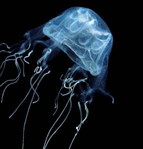
**The Box Jellyfish**
1) This box jellyfish belongs to the genus Chriropsalmus, part of the group of cnidarians (the C is silent) known as Cubozoa. In total there are over 10,000 different species of cnidarians that can be found (mostly) in our oceans split between the Anthozoa that include corals and the Medusozoa, that include jellyfish. Technically this specimen is not a ‘true’ jellyfish, those are species that belong to the group Scyphozoa and can be distinguished by the shape of the bell, being square when viewed from above whilst the true-bies have a rounded bell. By all accounts cubozoans are the most advanced of jellyfish with active movement, performing copulation and hunting prey.
2) Chriropsalmus anatomy. A fleeting look at this specimen may reveal little as it looks like a mass of soft tissue, but this specimen has been bisected to reveal its anatomy. I would just like to pause a moment and contemplate that achievement, imagine the skill, patience and delicacy required to bisect a jellyfish without damaging any of the important features! The bear paws I am currently typing this blog with are finding it difficult to hit the individual keys without smashing them. Amazing. What has that bisection revealed? In the centre of the bell is a structure called manubrium, which is a tube that connects the four part mouth at the base to the of the tube to the stomach. Should the prey be alive when pushed inside the bell the manubrium will extend and reach for it with the mouth expanding to engulf it. At the four corners of the bell are fleshy muscular pads called pedalia, we can see two on this specimen, attached to each pedalium are one or more tentacles. The other soft tissues surrounding the manubrium are the gonads. These jellyfish pair up to mate with males putting his tentacles inside the bell of females, passing packets of sperm to her with fertilisation taking place inside the female.
3) A jellyfish with eyes. At the base of each corner of the bell on muscular stalks are two sensory structures called rhopalia, more than just sensory organs though these are actually ‘true’ eyes complete with retinas, corneas and lenses enabling them to see specific points of light rather than just distinguishing between light and dark. They evolved independently to vertebrate eyes. The structure of these eyes are remarkable considering these animals do not have brains, they can look inwards to the the mouth and outwards. Actively moving through the water these jellyfish can display complex movements such as directional swimming they are able to know whether they are upside down, sideways or rightside up and can correct themselves. This behaviour is due to organs inside each rhopalia called statocysts that can determine orientation.
4) Deadliest. Definitely one for Deadly 60. The cubozoans include one of the deadliest animals on our planet. Known as the sea wasp (Chironex fleckeri), this cubozoan is found around the coast of Australia and has killed 63 people between 1884 and 1963. Sea wasps have tentacles that can be up to 3m in length and covered with millions of stinging cells. A recent study revealed why these animals are so deadly, when stung a molecule called porin punches holes in red blood cells and causing potassium to leak out of them resulting in cardiac arrest. [1]
5) Museum art. This animal features in a brilliant series of images we have here in the Museum of our specimens and uniquely is the only invertebrate. This picture and the others in the series were drawn over 10 years by much cherished Grant Museum friend Janet Knell. Janet volunteered in the Museum every day for those 10 years (except Wednesdays) and was often seen by our visitors hard at work on her latest specimen until she sadly passed away this time last year. This specimen is my favourite of all the ones she drew, it was a particularly challenging one for Janet as I remember but she managed to capture the brilliance of the specimen. A reproduction postcard of the work is on sale in the Museum.
6) Just for the record the answers to the questions highlighted at the beginning were: “No, it’s not alive” and “It’s moving because the vibrations of you walking around the table cause the fluid to move which moves the jellyfish”. I’m not sure that last explanation is believed by the pupils who encounter the specimen though.
Dean Veall is Learning and Access Officer at the Grant Museum of Zoology
[1] Yanagihara, A.A. Shohet, R.V. (2012) Cubozoan Venom-Induced Cardiovascular Collapse Is Caused by Hyperkalemia and Prevented by Zinc Gluconate in Mice. PLoS ONE 7(12): e51368. doi:10.1371/journal.pone.0051368
 Close
Close


![Box jellyfish [Janet Knell, May 2013]](https://blogs.ucl.ac.uk/museums/files/2014/07/Janet-Box-jellyfish-May-2013-218x300.jpg)