 Close
Close

Specimen of the Week 244: The historic wax flatworm
By Tannis Davidson, on 17 June 2016

LDUCZ-D44 Fasciola hepatica
Since its inception in 1828, the Grant Museum of Zoology collections have always been used for teaching. This continues in the present day and the Museum welcomes students from across UCL for a wide variety of specimen-based practicals, course work and research projects.
Today we maintain detailed lists of specimens which are used in classes but I’ve often wondered what the early object-based teaching practicals looked like and which specimens were used.
Fortunately, the Museum has some relevant archives which have identified an extraordinary specimen that had been used in teaching at UCL 130 years ago. It is not only one of the oldest specimens in the collection, but also one of the most beautiful.
Take a journey back in time with this week’s Specimen of the Week…
**The wax model of flatworm Fasciola hepatica**
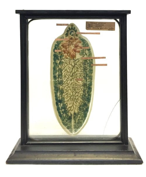
LDUCZ-D44 Fasciola hepatica model
This specimen is a wax-on-glass model of the flatworm Fasciola hepatica – the common liver fluke. It was made in the 1880’s by Rudolf Weisker at his Leipzig model factory, the Institute for Scientific Wax Image Moulding. The model is listed in the 1890 Museum catalogue but it also appears in two (of only five) photographs from 1887 of a UCL zoology practical using Grant Museum specimens:
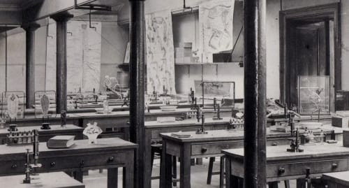
Lab set up for the ‘Longer Course in Practical Zoology’, UCL 1887.
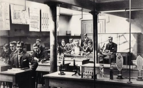
Lab with students. ‘Longer Course in Practical Zoology’, UCL 1887.
The class was the popular ‘Longer Course of Practical Zoology’ taught by E. Ray Lankester – the Museum’s curator, UCL’s chair of zoology, and all-around professor extraordinaire. In both images, the wax model can been seen alongside its now-lost twin (D44 shows the generative system of F. hepatica; the second model showed the alimentary tract and nervous system). As pictured, the models are on their original stands and not the black protective frame which houses D44 today.
Among the microscopes and chart illustrations there are mounted skeletons and skulls as well as preserved specimens either in jars or in test tubes. Here we have the zoological laboratory which Lankester developed and was to be the standard until the end of the First World War when genetics and biochemistry began to compete with anatomical studies 1.
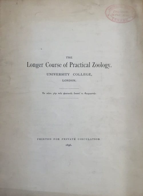
Longer Course in Practical Zoology specimen list 1896
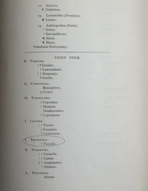
Fasciola hepatica wax model in Longer Course in Practical Zoology specimen list 1896
Amazingly, the Museum still has a copy of the course list for the ‘Longer Course’ in 1896. The specimens are listed under each of the three terms with D44 making its appearance in the third term.
So why use model instead of a real specimen? While F. hepatica is a relatively large tapeworm (it can been seen with the naked eye), individual worms measure only 2.5 cm x 1.3 cm. The use of a model to show the small anatomical features of the tapeworm therefore makes it possible to overcome physical limitations.
Tannis Davidson is the Curatorial Assistant at the Grant Museum of Zoology
References
- Lester, Joseph. 1995. E. Ray Lankester and the Making of Modern British Biology. British Society for the History of Science. Alden Press. Oxford.
2 Responses to “Specimen of the Week 244: The historic wax flatworm”
- 1
-
2
Specimen of the Week 286: The Notebook Models | UCL Museums & Collections Blog wrote on 7 April 2017:

[…] Grant Museum originally had both of these models (read more about the reproductive system model here), as indicated by the accession register, two images from 1887 (showing the models in the zoology […]
I am trying to find a photo of Florence Buchanan (UCL Zoology student from 1886 – 1890) and wondered whether it is known who the female students in these photos are? We are collating material for an exhibition of prestigious female contributors to the field of physiology and so far have had no luck sourcing an image of Florence, who was a notable physiologist at Oxford.
Best wishes,
Victoria Bullett