Conserve it! Part II – Research and Investigation
By Nick J Booth, on 27 February 2013
The second instalment of the Conserve it! blog, written by Miriam Orsini, details the research and analysis that conservators have to undertake before they even begin to work on objects. Particularly exciting for me is the photo towards the end of this post showing one of the X-Ray tubes glowing green under UV light! I had no idea they could do this…
Probably one of the most exciting things conservators must do before they start conserving an object is researching and analysing the object itself. This is the moment when the object starts to talk to you and tells you its story. In this post we are going to share some of the pretty amazing stories which these X-Ray tubes have told us.
We started with some preliminary research using the internet and academic literature to find out more about what kind of X-Ray tubes we were dealing with, and to try to understand their functioning and to date them. We realised that the tubes represented four stages in the history of the manufacture and design of X-Ray tubes, from earlier examples to more modern models.
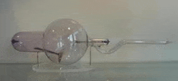
My tube (above) is an early example of an X-Ray tube known as Jackson Tube or Focus Tube. This particular example was produced by a company based in London, Newton & Co. The presence of the company’s name and address, Fleet Street London, inscribed on the metal plate contained in the tube, led us to think that the tube was made before 1930, when the company moved from Fleet Street to Wigmore Street. In addition, according to the literature, the Focus Tube, which was developed by Herbert Jackson in 1896, a year after Wilhelm Conrad Röntgen discovered the existence of X-rays, was only in use until the 1920s, meaning it probably dates from much earlier. What represents a significant development in the design of the Jackson Tube, and differentiates it from early examples, is its cathode. Its cup-shape has the function to focus the electron beam on the anode, which is made of a small platinum foil mounted at 45° to the cathode, so that the source of X-Rays becomes limited in size, allowing for major definition. A similar example to this tube can be found in a display in the Medical Physics Building at UCL.
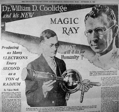
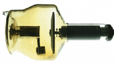
Leslie’s tube (above) is a Coolidge Tube. Its invention in 1913 by William Coolidge is believed to be one of the most sensational advances in radiology. Technically speaking, this is due to the fact that Coolidge replaced the cold cathode with a heated filament cathode, as a source of electrons, operating under a high vacuum, and substituted the platinum target with a tungsten one. This had the upshot that unlike early tubes, such as the Jackson tube, of uncertain performance, the Coolidge Tube was able to produce highly reliable X-Rays, and therefore, sharper images. Indeed, modern X-Ray tubes are still based on this design! As far as we know, it is possible that Leslie’s tube was manufactured before 1930, when Coolidge tubes started to be made completely cylindrical, without the spherical bulb.
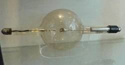
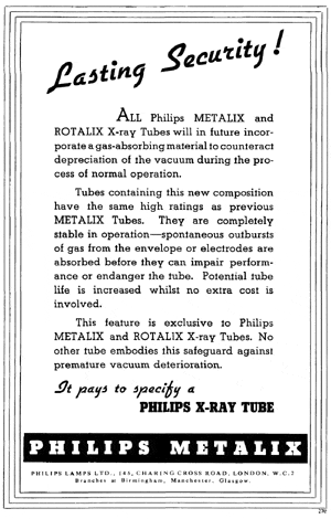
Louise’s Tube (above) is a more modern model of an X-Ray Tube. Although we cannot be certain, due to the fact that some elements are missing, this is presumably a Rotalix produced by Philips in the 50s. The great innovation of this tube is its rotating anode, the aim of which was to guarantee simplicity of operation and safety of both the operator and patient.
Kate’s preliminary research led to her consulting a specialist to find out more about her X-Ray Tube (right). After contacting a curator based in the United States she discovered that what we believed to be a fourth X-Ray Tube, is in fact likely to be a valve tube instead. She also contacted a radiologist for a further opinion, who confirmed the curator’s view. A valve tube is part of the X-Ray tube set up and supplies the X-Ray tube with energy.
Many interesting questions about the X-Ray tubes could only be half answered through literature research, so we also decided to carry out some analysis to help us confirm our hypotheses. First of all we decided to examine the tubes under UV light, since the fluorescence of the glass can be indicative of the type of glass used to make them. None of the glasses showed fluorescence, except for one, the Jackson Tube, which to our great amazement fluoresced bright green! This confirmed what we had suspected: the tube is possibly made of uranium glass, which appears to be consistent with some early X-Ray tubes. However, the amazement was then followed by a little concern due to the possible radioactivity of the glass, so we decided to measure the radiation levels using a Geiger Counter – the time between the clicks reassured us that there was nothing to worry about!
As a result of their use, X-Ray tube glass tends to change colour. The resulting colouration is usually due to the depositing of impurities on the inside surface of the glass. While the Jackson Tube shows a purple colouration, the other three tubes have taken on a yellow-amber colour. To attempt to determine the nature of the impurities, we decided to analyse the objects using a Portable X-Ray Fluorescence Analyser (pXRF). Although we could not achieve definite answers, we were able to establish that the deposits are of a metallic nature. Although we will not sample any of the X-Ray tubes in order to carry out further analysis of the impurities on the glass surfaces, Kate will analyse some glass splinters belonging to her valve tube, using the Scanning Electron Microscope. Aside from the glass, XRF analysis was also carried out on other components of the tubes, allowing us to identify their composition, helping us to confirm what we had suspected following our preliminary research.
The next Conserve it! Blog post will show the progress made in the conservation of the tubes. Some impressive reconstruction is going on!
 Close
Close


