X Close

UCL Culture Blog

Home

News and musings from the UCL Culture team

Menu

Specimen of the Week 392: Hide of the Dragon: Achaemenid Scale Armour

By edwin.wood, on 6 March 2020

Two sets of corroded iron scales laid out in ordered rows

UC74787 Some of the scales from the armour, each pierced by two holes. Copyright UCL Petrie Museum

Scale armour is a form of defensive garment that is made by attaching small scales to a fabric or leather undergarment in an overlapping pattern. The examples in the Petrie collection are all of metal, either copper alloy (Bronze or Brass) or iron. However, examples of rawhide scales are known from sites in Egypt, notably Tutankhamun’s tomb (Dean 2017). This type of armour is one of the earliest forms to be developed and provides a flexible but effective defence that can be easily repaired if it becomes damaged. The armour is effective against a range of attacks, protecting from projectiles, cuts and blunt force impact. When combined with a stiffened or padded undergarment the protective quality of the armour is increased.

(more…)

Specimen of the Week 388: Lunar Orbiter Images

By ucwehlc, on 9 August 2019

In honour of the 50th anniversary of the Apollo 11 moon landing this month we have something extra-terrestrial for you.

UCL is home to an amazing collection of images taken from spacecraft; our specimen of the week is nothing less than the whole of the planet Earth as seen from the Moon

UCL's print of Lunar Orbiter 1 Photograph 1117, the Earth viewed from lunar orbit. Copyright NASA 1966, public domain

UCL’s print of Lunar Orbiter 1 Photograph 1117, the Earth viewed from lunar orbit. Copyright NASA 1966, public domain

(more…)

Specimen of the Week 385: UCL skulls

By ucwehlc, on 12 July 2019

This blog was written by Lily Garnett, volunteer at the UCL Pathology Museum

UCL.16.009 and UCL.16.018 are skulls. Like any human face, although they have the same features, they look different. They have both been processed for medical teaching, with cuts and hooks to reveal the inner workings of the cranium.

In terms of provenance, the history of these two individuals remains a mystery. The UCL prefix to their number indicates that they were found within the collection and their history prior to this is unknown. Before accurate plastic models became available in the 1990s, real human bones were used for teaching.

But where do you get a skeleton from?

Skulls UCL.16.009 and UCL.16.018

Skulls UCL.16.009 and UCL.16.018

(more…)

Specimen of the Week 383: Fracture of the odontoid process

By ucwehlc, on 14 June 2019

This blog was written by Lily Garnett, volunteer at the UCL Pathology Museum

These human remains tell a true story of murder, attempted suicide and the understanding of mental health in 1855.

It is rare for human remains within pathology collections to bear the name of the individual they were once a part of in life. In historic collecting this is most probably due to a collector’s medical interest in a pathology as a ‘specimen’, disassociating it from the reality that it was once part of a person with feelings, thoughts and family. Moreover, in the past the maintaining of deceased individuals often went unmonitored. Until as late as a public enquiry in 1999, with a focus being on the Alder Hey Hospital in Liverpool, the vast majority were unaware that hospitals were retaining patients’ organs without consent. This ultimately led to the Human Tissue Act 2004, which is monitored by the Human Tissue Authority. Now, any institution holding human remains that are less than 100 years old since the death of an individual legally require a license.

D.15 Fracture of the odontoid process caused by a bullet

D.15 Fracture of the odontoid process caused by a bullet (bullet visible top left)

(more…)

Specimen of the Week 381: What Lies Beneath

By Tannis Davidson, on 17 May 2019

Mystery ceramic pots

Mystery ceramic pots

The fluid store room for the Grant Museum is perhaps an unlikely setting for a hair-raising tale. In it, rows of metal shelves are neatly arranged holding jars of preserved zoology specimens arranged by taxonomy. Order and classification dictate the placement of specimens, and as a whole, the contents of the store are visible, documented and accessible.

Apart from the bottom shelf of the last row. Although a numbered (legitimate) location, it is a wildcard area which houses several large ceramic pots of unknown content. No one knows exactly how and when the pots came to be a part of the Grant Museum collection and because it is impossible to see into them, no one knows what they hold. A few clues exist – the occasional faded label or a more modern post-it note – but as the pots have not been opened and investigated in living memory, their contents are a mystery.

What follows is the first-hand account of the opening of two of the pots. This is a true story.

**WARNING  Graphic images below **

(more…)

Specimen of the Week 380: Malignant melanoma of the eye

By Subhadra Das, on 10 May 2019

Today’s specimen of the week comes from UCL Pathology Collections. The Collections are displayed at the UCL Pathology Museum at the Royal Free Campus of the UCL Medical School in Hampstead. The museum includes a medical teaching collection of nearly 3,000 specimens of human remains illustrating the history of disease. To open up these specialist medical displays to a wider audience, we’ve developed a trail of 10 specimens of well known diseases. As the museum only opens to the public for special events, we’re sharing the trail as part of the Specimen of the Week series.

In honour of Skin Cancer Awareness Month this May, today’s specimen of the week is a malignant melanoma of the eye.

Melanoma is a cancer — an uncontrolled growth of cells — that usually develops in the pigment-containing cells of the skin. The main cause of melanoma is over-exposure to ultra-violet light rays, either directly from sunlight or artificial sources like tanning beds.

Melanoma most commonly occurs in people with lighter skin, particularly white people living in tropical or subtropical climates. The highest instances of melanoma are in Australia and New Zealand.

If detected early, a melanoma can be surgically removed before it spreads to other parts of the body. Sadly, that wasn’t the case here.

(more…)

Specimen of the week: Trichobezoar

By Katie Davenport-Mackey, on 29 March 2019

Our blog this week is from Subhadra Das, Curator of Science Collections at UCL Culture.

Today’s specimen of the week comes from UCL Pathology Collections. The Collections are displayed at the UCL Pathology Museum at the Royal Free Campus of the UCL Medical School in Hampstead. The museum includes a medical teaching collection of nearly 3,000 specimens of human remains illustrating the history of disease. To open up these specialist medical displays to a wider audience, we’ve developed a trail of 10 specimens of well known diseases. As the museum only opens to the public for special events, we’re sharing the trail as part of the Specimen of the Week series.

This week’s specimen is a trichobezoar — a mass of undigested hair from the stomach and large intestine of a young girl. As with most of the specimens in UCL Pathology Collections, we know little about the person the specimen comes from beyond their sex and their age, but this rare condition provides an interesting window into the practice of medicine, and its cultural significance extends into the realms of magic. (more…)

Object of the week 378: Tess Jaray, Always Now

By Nina Pearlman, on 22 March 2019

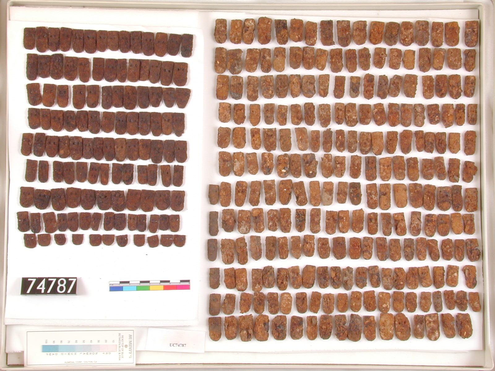
Today’s entry has been written by Viktoria Espelund. Viktoria gained her MA in History of Art From UCL and has worked with the UCL Art Museum team throughout her studies as a volunteer and later in a professional capacity as well.  We are thrilled to see her knowledge and expertise gain a wider audience through her recent contribution as a writer to a current exhibition at the Barber Institute of Fine Art in Birmingham. Her experienced is shared in this post.

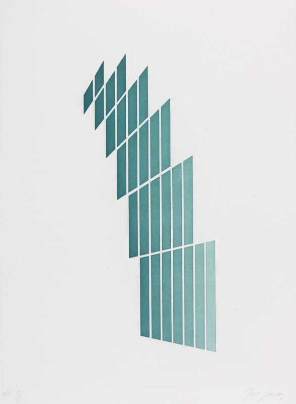
Always Now (LDUCS 7876) is a print by the British painter and printmaker Tess Jaray RA (b. 1937, Austria).  This print is one of nine works that are held of Tess Jaray in UCL Art Museum’s collections and is incredibly special to me, being the first ever work I saw by the artist now showing new work at the Barber Institute in Birmingham. The aquatint derives from a painting completed in 1982. Whereas the painting is painted with a soft colour palette of lilacs and blues against a cream-coloured background, the print has been executed in bright turquoise on paper.

A turquoise geometric print by Tess Jaray 1982 from UCL Art Museum's collections number 7876

Tess Jaray, Always Now, 1982, aquatint, UCL Art Museum LDUCS 7876

Tess Jaray has been an influential figure in the British art world since the 1960s. As both a Senior Royal Academician and an Honorary Fellow of the RIBA, her career spans more than fifty years, throughout which time she has produced a vast body of work. Jaray arrived with her family to the UK as an infant in 1938 as part of the flight of Jewish refugees from the Nazis. Aged sixteen she embarked on her journey as an artist and enrolled first at Saint Martin’s School of Art in London later at the Slade School of Fine Art (1957-1960), studying under the likes of William Coldstream, Bartos Dos Santos and Ernst Gombrich. Jaray has further been a great influence on younger British artists through her writing and the thirty years spent teaching at the Slade (1968-1999).  (more…)

Specimen of the Week 377: The Lobster Claw

By Graham Isted, on 15 March 2019

Hello and welcome to the next instalment of Specimen of the Week. This week’s specimen is a mighty claw (LDUCZ-H671) from the lobster species Homarus gammarus, also known as the European or common lobster. Lobsters are great, whether you like them to be freely going about their lives at the bottom of the sea or perhaps prepared by a chef on a dish with some butter. Either way, I am fairly certain there are aspects of their lives you are unaware of and this blog will hopefully either make you hungry for more knowledge or perhaps just dinner.

Lobster claw LDUCZ-H671 Homarus gammarus

(more…)

Specimen of the Week 376: Carcinoma of the breast

By Subhadra Das, on 8 March 2019

Today’s specimen of the week comes from UCL Pathology Collections. The Collections are displayed at the UCL Pathology Museum at the Royal Free Campus of the UCL Medical School in Hampstead. The museum includes a medical teaching collection of nearly 3,000 specimens of human remains illustrating the history of disease. To open up these specialist medical displays to a wider audience, we’ve developed a trail of 10 specimens of well known diseases. As the museum only opens to the public for special events, we’re sharing the trail as part of the Specimen of the Week series.

All of the entries for the UCL Pathology Collections Top 10 Medical Trail have been written by Nazli Pulatmen, who worked with us for her MA Museum Studies placement in the summer of 2018.

Today’s specimen of the week is an advanced example of breast cancer. The specimen shows how the cancerous tissue has expanded to such an extent that it has completely misplaced and replaced the healthy tissue within the breast. The patient reported that the lump began at about the size of a pea and developed to this enormity over the course of 10 months.

Image of a cross-section of breast tissue showing malignant cancerous growth

Breast.25.1: Carcinoma of the breast. The cancerous tissue appears solid compared to the normal, fibrous breast tissue surrounding it.

(more…)