X Close

Medical Physics and Biomedical Engineering Teaching

Home

Highlights of the teaching going on in UCL Medical Physics and Biomedical Engineering

Menu

Fibreless Diffuse Optical Tomography summer internship

By rmapapg, on 31 October 2018

by Aman Ganglani

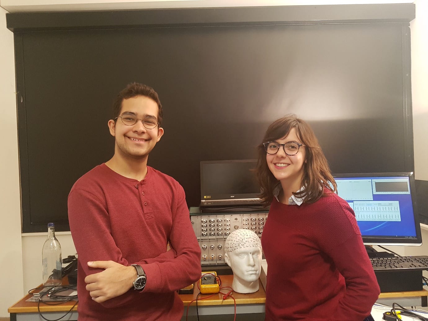
Over the summer I was lucky enough to be given a leading role in an 11 week research project working with Dr. Robert Cooper, Dr. Hubin Zhao and Dr. Sabrina Brigadoi on a fibreless Diffuse Optical Tomography (DOT) system.

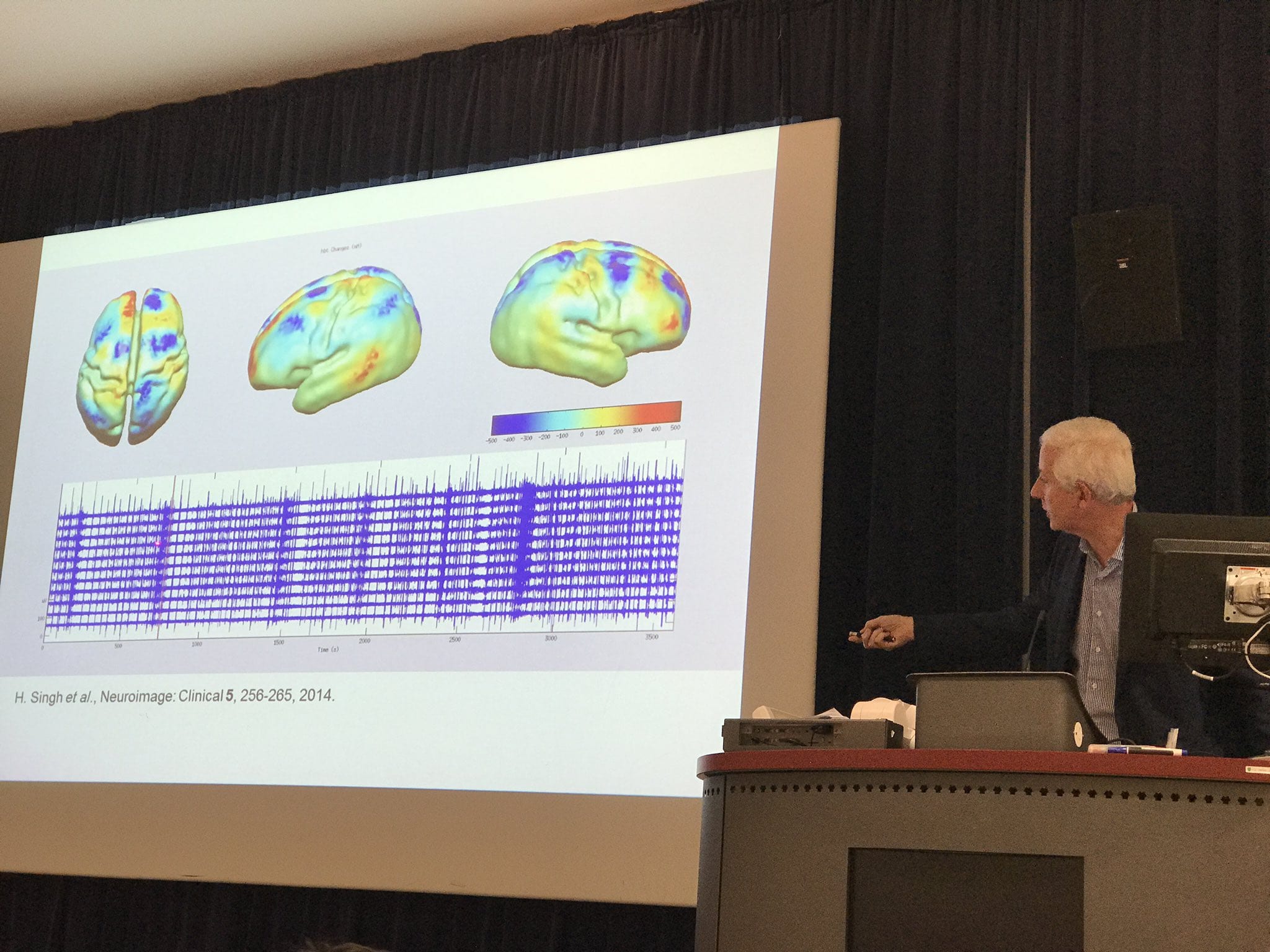
DOT is a novel imaging technique that has a wide variety of applications in neuroscience and clinical research. Specifically, using DOT to investigate neonatal brain development is a very important focus. Cerebral haemodynamic patterns in brain injured neonates is not well understood. Complications at this vital stage of development can result in critical danger to the patient and long-term disabilities. Further investigation into these complex haemodynamic signals is necessary to better understand the underlying physiology and to develop DOT into a novel imaging tool that could help diagnose and treat compromised infants [1].

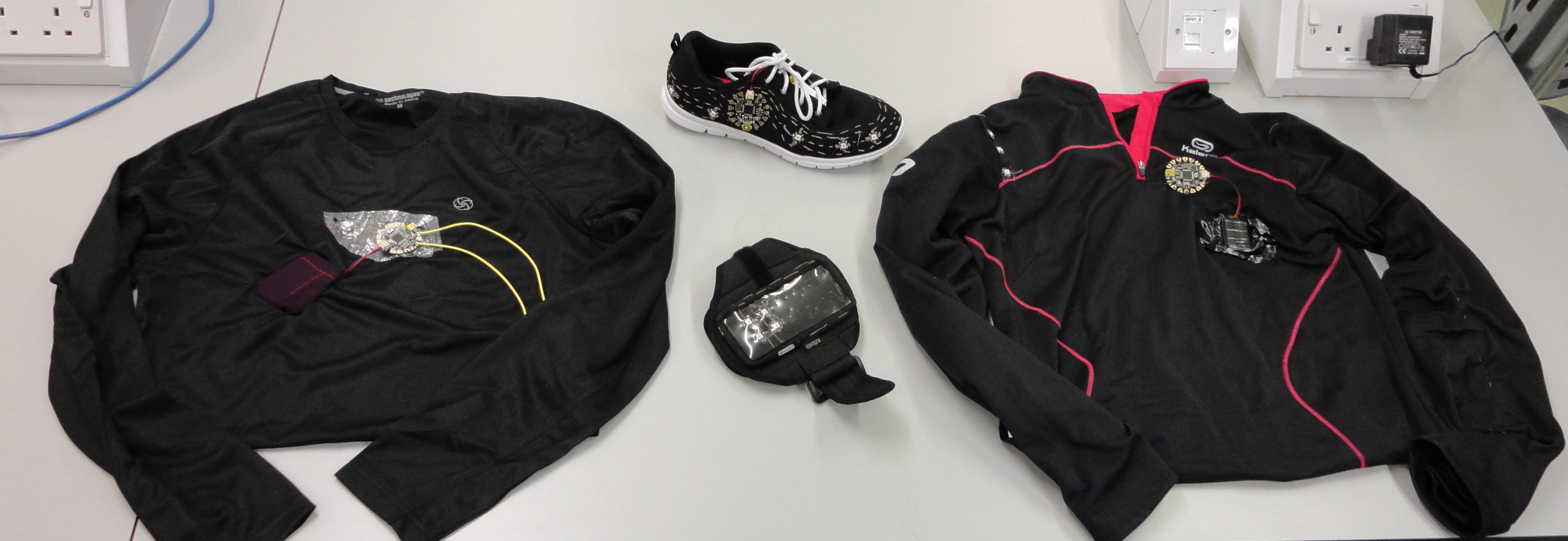
Current DOT technology is limited by bulky fibres which limit comfort, movement and ease of use. Transforming the current DOT systems into wearable, fibreless devices is a vital step in the development of DOT technology [2]. This advance will enable long term clinical application of DOT, improve data quality and make the instruments viable in a wider range of applications. However, transforming the bulky wearable DOT modules into a fibreless wearable comes with challenges. This investigation aimed to minimize motion artifacts with fibreless systems. While there are post processing methods of tackling motion artifacts [3], they alone are not enough for fibreless systems. Our novel approach has been to develop the application of motion sensors specifically for this type of movement and add them to the current fibreless systems to build the world’s first dataset of fibreless DOT and 9 axis (3x accelerometer, 3x gyroscope and 3x magnetometer) motion data.

Testing the motion sensors

Testing the motion sensors

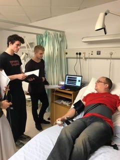
We decided to focus our investigation on the effect of forced induced movement on DOT data. We introduced two 9 axis motion sensors along with 2 DOT fibreless modules each containing a 9 axis motion sensor. Our experiment paradigm consisted of controlled head, eyebrow and full body movements.

After many considerations, we purchased two Razor 9DoF motion sensors containing the MPU_9250 Invensense chip which is the same sensor used on the DOT modules. I was able to match the operating conditions of the chip with the DOT chip by writing my own code in the Arduino IDE and MATLAB. This would ensure data acquired from both sensors could be accurately compared.

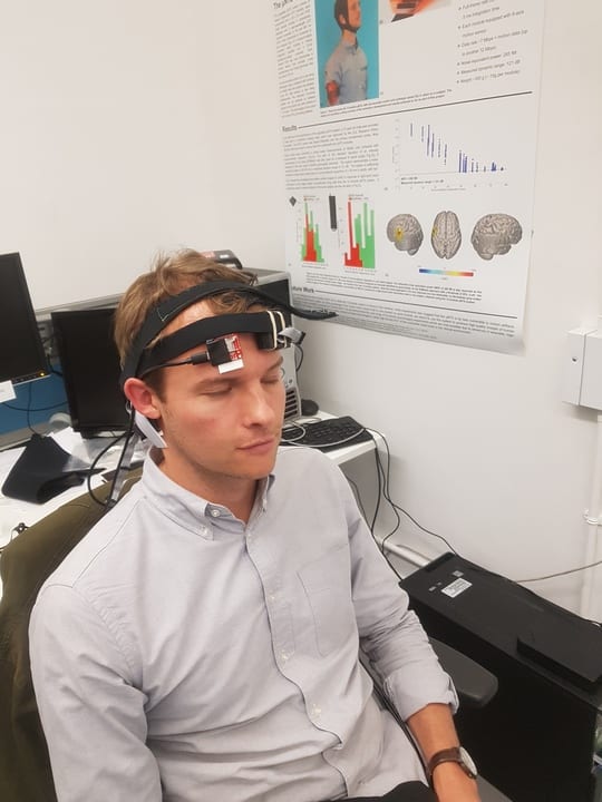
Due to the DOT modules being standalone devices, I also had to figure out a way of mounting everything (the two DOT modules and the two motion sensors) in a comfortable way. After a lot of experimentation and time in the Institute of Making, I managed to build my own headgear system which kept all the sensors completely independent of each other. The DOT modules were secured using separate sewn velcro and rubber band straps while the motion sensors used adhesive tape placed directly on the scalp.

Design of the headgear system

Design of the headgear system

Finally, all this preparation was for the experiment paradigm itself. We eventually decided we wanted to investigate the effects of eyebrow movement (this has not been explored and previous pilot studies showed large eyebrow related artifacts), the effect of scalp movement compared to head movement and induced head movement along with walking and designed a paradigm accordingly.

Within each block, chirp noises of decreasing lengths were used. When the subject hears the chirp, they must move their head throughout the chirp, this way we can control the speed of movements. Varying the speed of these movements is useful because is allows us to look at relationships between the sizes of motion artifacts and the speed of movement. The speaking section was done with words of varying syllables for the same reason. The timing of eyebrow movement was controlled by a simple tone sound rather than a chirp, because it is difficult for people to control the speed of eyebrow raises.

Dr. Cooper just before a pilot study

Dr. Cooper just before a pilot study

Additionally, I created a MATLAB script which would efficiently run everything with one press of a button using parallel computing. This massive streamlining of the whole experimental procedure will make studies synchronised and far easier to run. Our aim in the first term is to use my experiment to run multiple studies on a variety of healthy adult volunteers.

Our initial conclusions show that more investigation into 9 axis data with fibreless systems is clearly justified. I was able to help publish a poster which was presented at the fNIRS 2018 conference in Japan titled ‘Investigating the Benefits of Integrated Motion Sensing and Wearable, Modular High Density Diffuse Optical Tomography’, of which I was an author.

This has been an incredible experience. In 11 short weeks, we have managed to build and execute an experiment paradigm which has never been done before. I have been exposed to real research and have obtained a publication under my name by having my work presented at a conference. I will continue to work with the team throughout the first term.

I would like to thank Dr. Zhao, Dr. Brigadoi and Dr. Cooper for their never-ending patience and commitment. They have exposed me to world-leading research and have given me an excellent insight into the life of an academic.

Our poster at fNIRS 2018 in Tokyo

Our poster at fNIRS 2018 in Tokyo

1 Cooper RJ, et al. Transient haemodynamic events in neurologically compromised infants: a simultaneous EEG and diffuse optical imaging study. Neuroimage (2011).
2 Danial Chitnis, et al. Functional imaging of the human brain using a modular, fibre-less, high-density diffuse optical tomography system. Biomed Opt Express. 10 (2016)
3 Cooper, R. J. et al. A Systematic Comparison of Motion Artifact Correction Techniques for Functional Near-Infrared Spectroscopy. Front. Neurosci. 6, (2012)

Investigating neurovascular coupling in infants with seizures using EEG and DOT

By rmapapg, on 19 October 2017

By Aman Ganglani

Over the summer I was lucky enough to undertake an eight-week research project in the Biomedical Optics Research Lab (BORL) with the research group focusing on diffuse optical tomography.

Using data acquired from simultaneous electroencephalography (EEG) and diffuse optical tomography (DOT) measurements from the NTS system (developed by Gowerlabs), we looked at the relationship between cerebral blood flow and seizures in neonatal infants with hypoxic ischemic encephalopathy (HIE).  This work follows a previous investigation by the group titled ‘Mapping cortical haemodynamics during neonatal seizures using diffuse optical tomography: A case study’ [1]. The data analysed in this study is from the same patient as the previous investigation.

Figure 1 - The GowerLabs NTS system

Figure 1 – The GowerLabs NTS system

Most of my work was done in Matlab. By employing various statistical tests and by creating my own scripts with the help of the department I further understood the relationship between neonatal seizures and cerebral haemodynamics. Through my own investigation and discussions with the DOT team in formal weekly meetings, I was able uncover patterns and identify further research topics.

A lot of my work was concentrated on the relationship between oxy/deoxy haemoglobin (HbO and HbR respectively) and the spikes on the EEG which indicated seizure like activity. I was able to demonstrate the HbO signal leading the EEG signal through cross correlation tests and visual inspection. I was also able to show evidence of the ‘initial dip’ phenomenon and understood why it remains to be controversial given how it was not seen in every seizure (the ‘initial dip’ phenomenon is observed when there is a dip in HbO prior to a seizure). We also found other time correlations with HbR which warrants further investigation. I ran various other statistical tests such as t-tests to validate my results. Along with this, I also looked at the derivative to investigate if a sudden change in the EEG correlates to a sudden change in haemoglobin levels. Given how I previously found a time lag between the haemoglobin signals and the EEG signal, it came as no surprise that there was no direct correlation without a time lag. I also identified further research topics such as investigating the phase difference between the HbR and HbO signal along with further statistical tests that could be employed on more datasets. All of my code was commented on to specifically allow for other people to continue my work.

Along with the analysis work I was also able to visit the Evelyn Perinatal Imaging Centre at Rosie hospital in Cambridge. This was where the patients were scanned with EEG and DOT, and I could see how the devices built in the university were tailored to a hospital environment. By then attending meetings with neoLAB. I understood some of the challenges faced by engineers to connect their products with hospital staff. I was also able to do some brief work on image reconstruction which gave me an exciting scope to the future of DOT imaging.

I would like to thank the Engineering department for this amazing opportunity to be a part of this phenomenal project. Everyone at BORL has been incredibly friendly and approachable. A special thanks to Ms Dempsey, Dr Cooper and Dr Hebden for their never-ending support. This opportunity has given me a fantastic insight into the world of research and I look forward to being a part of it.

Figure 2 - Laura Dempsey and me with the NTS kit.

Figure 2 – Laura Dempsey and me with the NTS kit.

[1] H.Singh, R.Cooper, C.Lee, L.Dempsey, A.Edwards, S.Brigadoi, D.Airantzis, N.Everdell, A.Michell, D.Holder, J.Hebden, T.Austin. (2014). Mapping cortical haemodynamics during neonatal seizures using diffuse optical tomography: A case study. NeuroImage: Clinical. 5

Functional Near Infrared Spectroscopy Conference

By rmapapg, on 27 September 2017

By Nick Wood, MEng student

I was luckily able to attend the fNIRS UK conference free of charge on the 7/09/17.

For those of you who are not familiar with fNIRS, functional near infrared spectroscopy and its subgroup DOT, diffuse optical tomography, are exciting brain imaging technologies. They provide functional imaging by measuring the haemodynamic response associated with neuron activity and can be safely used at a patient’s bed side.

The proceedings opened with the first keynote speech delivered by our own head of department Jem Hebden, this interesting talk explained the some of the key differences between fNIRS and DOT and several of the challenges and benefits of designing DOT systems. The long history of the development of this technology at UCL was also explained.

Jem Hebden describing optical brain imaging

Jem Hebden describing optical brain imaging


Eight other talks followed illustrating the wide-ranging capabilities of this technology; varying from using to evaluate human computer interfaces, developing an optical biomarker of brain mitochondrial function and assessing the plasticity in the neural representation of language.

I found the second keynote talk of the day by Mr Daniel Leff particular interesting, the talk covered how fNIRS could be used to not only monitor the patient but could also be used to track the surgeon. By monitoring surgeons, the neurological differences between trainee, registrar and consultant surgeons could be spotted when undertaking different tasks, as true mastery of a skill was obtained the mental exertion required to perform this task was greatly produced. This could therefore be used as an assessment for consultant status, allowing a move away from the traditional training system of becoming a consultant after a certain amount of years of training. Ensuring people only reach the pinnacle medical role once they have truly mastered their surgical skills.

The conference closed with a humorous interactive session on the attendee’s thoughts for the future of the technology and possible talking points for the 2018 Tokyo meeting.

Key topics in fNIRS for the next few years

Key topics in fNIRS for the next few years

Overall, I had an interesting day learning about additional uses and developments of fNIRS/DOT and would like to say a thank you to all the speakers, sponsors and the programme/organisation committees for making the conference possible and free of charge.

Visiting the Stanmore Royal National Orthopaedic Hospital

By rmapapg, on 1 March 2017

by Madeline Lok, Emma Ponting and Sarita Meekul

On Friday 27th of January, our 2nd year Biomedical Engineering class got the opportunity to visit the Stanmore Royal National Orthopaedic Hospital. The purpose of the trip was for us to gain an understanding of how the clinical environment works and how devices we may help to create in the future fit into real people’s lives.

rhoh3The day began early with a long journey on the tube to Stanmore, on the outskirts of London where the class met. After a short taxi ride to the hospital, we were met by Professor Hart at the London Implant Retrieval Centre (LIRC). Prof Hart is the director of research and development at LIRC and a consultant orthopaedic surgeon at the hospital. He gave us a warm welcome and introduced us to some of the PhD students working there. At LIRC they recover and study knee and hip replacement implants that have been removed from patients to better understand why they failed. We were shown the processes these implants go through once they arrive, from cleaning to being scanned for wear and deformation and got to hold some actual implants. It was very interesting to see how something that we learn about in lectures actually looks and feels in real life.

rnoh1Then the lovely people of LIRC kindly provided us with lunch with their team. This was a good chance to chat to the people who work in the hospital and get a better insight into the kind of jobs available that we might be interested in once we graduate.

After lunch, we were split into smaller groups; some went to watch the surgeries while other went to sit-in with the doctors and their patients for real consultations. We swapped after 1.5 hours.rnoh4

We were brought to the surgery area to be able to see a real operation taking place. Before entering the operation theatres, we changed into scrubs. We were then separated into pairs and entered different theatres. Among all of us, we saw a range of operations including hip replacements, knee replacements and one ankle-foot correction surgery. We were told that the ankle-foot surgery is one of the most complicated and delicate procedures. The doctor that took us in even made a joke about how he avoided it. They used x-rays during that operation so we had to wear a lead apron to protect ourselves from the radiation. Some others of us saw the removal of implants, which was completely opposite of what the others saw. It was interesting to be able to discuss our different experiences at the end of the day when everyone was together.

Within the operating theatre, the surgeons explained to us what and how they were performing this surgery. Most of us were surprised by the atmosphere in the operating room. It was very relaxed with music playing in the background. The anaesthetist was reading his Kindle; the surgeons were even able to have a conversation and joke about their family while operating on the patient. It was surprising to see how calm and confident they were. Due to the time constraints, none of us were in there for the whole process which was a pity because we all really enjoyed it.

rnoh2The consultation sessions were an eye-opening experience too. We had the opportunity to sit through a few consultations with an orthopaedic specialist doctor, and see how they interact with their patients. All patients had very different reasons to be there, so we got to see various cases, the medical images used, and procedure followed. After sitting through the consultations, we now have a better understanding of what doctors go through when seeing patients and it definitely is a very difficult job. It’s not all smiles, hellos, reassurance and prescribing treatments as some people would think. In reality, they are potentially the ones who would be telling you how you would live out the next 10-20 years of your life (in our case, with hip/knee replacements, constant rehab, medication and so on). They have to always maintain professionalism and courtesy no matter how their patients react to whatever they tell them; even answer questions about their other concerns whether or not it is related to the real reason they came in for the consultation in the first place. The most important take-away I had from the session was that the doctors should let the patients leave with the best reassurance they can provide.

We all had a great day and learnt a lot about working in a clinical setting and working with patients. We would like to say a massive thank you to a ll of the people at Stanmore hospital who helped in making this day happen! What a day and what an experience!

Thinking clinical at the Learning Hospital

By rmapapg, on 3 December 2016

By Julian Henty

Clinical Engineering knowledge and skills were put to the test recently during a visit to UCLH’s Learning Hospital. Second year Biomedical Engineering students were given the chance to test various medical devices, such as ventilators, bedside monitors and suction machines, examine the workings of an in-house automated blood pressure machine, and observe vital signs measurement from a real ‘patient’ using a virtual bedside monitor.

Despite making good use of the department’s electronics lab for experimentation with transducers, software driven data acquisition, and aspects of electrical safety, the Clinical Engineering module fulfills its practical aims by providing a hands-on session with real hospital equipment and actual clinical measurements in a realistic clinical environment. The Learning Hospital has a ‘theatre’, complete with operating table and appropriate medical devices, and a ‘ward’ with two beds, nurses’ station and a medical gas supply.

The virtual non-invasive blood pressure (NIBP) device comprised a PC, control board and recycled components from a disassembled standard NIBP device. LabVIEW software measured the photoplethysmogram (PPG) amplitude distal to the cuff during inflation/deflation, which provided feedback to control the air pump and release valve. The software had an illustrative screen showing each step in the process of taking a real measurement. Safety aspects of NIBP measurement could also be demonstrated.

learninghospital

The patient waits anxiously for his results

The virtual bedside monitor displayed real-time graphs of the ECG, PPG, NIBP and chest movement while the ‘patient’ lay on a bed. LabVIEW software demonstrated how heart rate may be extracted from the ECG, PPG, or NIBP measurements, and respiratory rate from the ECG or chest movement. The software could also manipulate the ECG to demonstrate both noise and other lead measurements, and simulation of low amplitude due to pericardial/pleural effusion, pneumothorax, obesity or loss of viable myocardium.


With thanks to Dimitros Airantiz, Billy Dennis and Paul Burke

Regaining Control

By rmapapg, on 18 January 2016

By Rebecca Yerworth

Just before Christmas, DSCN2511 the second year Biomedical Engineering students spent a week in the lab designing and building a device to replace a computer mouse for a hypothetical client who had no hand. The devices picked up electrical activity in the muscle of the arm and translated these into cursor movements and clicks – or at least that was the theory.

Regaining controlThe students’ knowledge of electronics, anatomy and problem solving were all put to the test as they built and tested their circuits. They discovered the delights of bread-boarding moderately complex circuits – and the importance of keeping your ‘spaghetti’ colour coded! Of equal importance was realising that some muscle groups are easier to control independently that others – and that what most of us do routinely, without consciously thinking about it, takes a lot more physical and mental effort when being relearnt.

All the groups successfully detected and recorded electrical activity from muscles. Detecting muscle activity from multiple muscle groups with a sufficiently clear signal to control a mouse pointer is much more challenging, but everyone managed this, at least intermittently. In amongst the hard work, it was good to see the moments of fun and hear the cries of delight as the first hand-free mouse clicks appeared.

Pebble in the Pond

By rmapapg, on 2 December 2015

By Alan Cottenden

Congratulations to the victorious Biomedical Engineering team who managed to transport their pebble the length of the assault course they had designed and built – involving a catapult, a lift, numerous slides and prodigious quantities of string and sticky tape – and deposit it in a bucket at the finishing line with fewer “interventions” (that is, manual interferences to help it on its way!) per meter of travel than either of their two rival teams. The pictures show the creators of the assault course’s four sections admiring their handiwork while savouring the taste of victory!

pebble_in_the_pond_winning_team_1

Team Catapult

pebble_in_the_pond_winning_team_2

Team Vertical

pebble_in_the_pond_winning_team_3

Team Cup

pebble_in_the_pond_winning_team_4

Team Balloon

Clinical engineering visit to Royal National Orthopaedic Hospital

By rmapapg, on 13 November 2015

By Nishat Ahmed and Bindia Venugopal

On Wednesday the 11th of November, we were up at the crack of dawn, pumped and ready to go to the Royal National Orthopaedic Hospital in Stanmore. After missing trains due to tube closures and our taxi rides arriving a half hour late, we finally managed to reach the hospital in time to attend the Multi-Disciplinary Team meeting.

We found the meeting very interesting, watching the consultant surgeons and nurses discuss real case studies of patients. They collaborated well to work out the best way to rehabilitate patients, whether this was through further surgery or simply giving them advice and support.

Later on we headed to the operation theatres, adhering to hospital dress code we threw on our scrubs, hair nets and masks beforehand! Since we were only allowed three students at a time in the theatres, we split into groups and then went off to watch various operations taking place. The first surgery we watched involved attaching a metal plate to a fractured tibia bone to aid its healing process in a way that was ingenious! It was fascinating watching the surgeon screw the bone together and then brace the join with a metal plate. The screws held the fracture under compression, this meant it was forced to combine together rather than slide apart, and the metal plate stopped it from twisting.

The second surgery we went to was an extremely rare case where the surgeons ended up dislocating the hip bone in order to remove a benign tumour from inside the bone. They sawed the hip bone in half as bone-to-bone healing worked best compared to tendon-to-bone healing. The challenge was in trying to avoid damaging the femoral head to get to the tumour.

After this we had a little tea break and then made way to our next surgery! This was a spinal surgery where the patient had a twisted spine due to being paralysed for 10 years. They operated with a diathermy machine which uses electricity to cut through the skin and muscles as this reduces blood loss. Although we only saw the surgery for 10 minutes we learnt how vital it was to keep the fluids in the patient regulated. This job was monitored by the anaesthetist, who informed us about the patient and the precautions which needed to be taken. Two neurophysiologists were monitoring electrical activity in the spinal cord to ensure that it wasn’t damaged by the surgery.

Scrubbed up

After an insane experience watching all the surgeries, we went to have lunch which was provided by the lovely team at Stanmore. In the afternoon we got a tour around the BME department at the hospital and learnt about all the weird and wonderful things they collect and experiments they run! In fact, we found out that they have over 6000 failed hip replacements from 25 different countries in their labs to study and analyse. They conduct experiments to research why implant failure happens in some patients the way it does, especially those with metal on metal implants. They use tools for metrology which measures the exact size of the ball and socket implants with crazy precision! This information is then used to work out the amount of corrosion that happened in the body when the implants were inserted.

Overall, we had an amazing and truly valuable experience. The entire team were extremely friendly and helpful! We loved that we could ask questions and interact with the staff so well. It was remarkable to see the transition from a real-life patient problem to actually seeing the solution executed in the surgeries. It was also encouraging to see how the hospital carries out their own research which can then be implemented to the surgery procedures in only a few years’ time.

On behalf of our whole BME department, we thank you for this experience Professor Hart and RNOH!

“Oh God this is so cool! Do we really have to stick to our budget?!”

By rmapapg, on 9 November 2015

By Jenny Griffiths

Scenarios are a highlight of our new biomedical engineering programme. In a scenario, all lectures stop and students spend the whole week working on a group project where they solve a biomedical engineering problem. Last week, our second year students worked with Jenny Griffiths to build articles of smart clothing. Their brief was to design and build an item of clothing to monitor a marathon runner’s wellbeing and give an alert to inform the runner and all those around them to prevent injury. Students were encouraged to be creative and develop their own solutions as long as their device met the design brief and was safe.DSC00656

Jenny provided the students with a range of components, mainly centered around the Adafruit Flora wearable arduino. We gave them sensors including temperature and pressure sensors, accelerometers, GPS, UV and light sensors and stretchy conductive rubber. Outputs included buzzers, vibration motors, Bluetooth connectivity and programmable RGB LEDs, but they were only allowed to use up to £40 for materials. The task built upon electronics modules which students took last year, and a clinical engineering module which includes lectures on transducers which the students are taking at the moment.

We put the students into random groups and let them loose!

shoesTwo groups chose to design their own sensors from scratch to monitor electrolyte concentration in sweat. They quickly learnt how challenging it is to build a robust sensor! They sewed their home-made sensors into running shirts with conductive thread and used the arduino to control LEDs based on the resisitivity of the sensor. Another group built an arm band to monitor skin temperature. They learnt that packing 10 LEDs, a microcontroller, batteries and an temperature sensor into a package the size of a iphone can lead to wiring complexities. The winning group instrumented a running shoe with pressure-sensitive pads to measure gait continuously during the running cycle. They sewed their Flora onto the shoe and daisy-chained LEDs around the shoe with conductive thread. They went shopping to find low-cost trainers which fitted a team member and also gave them something additional to write about in their sustainability analysis.


Range of smart clothing

Students enjoyed the scenario, some saying this was the first time they’d ever worked as a team under pressure. They were ambitious and undaunted by such an open-ended task. Despite one team doing a complete redesign at the beginning of Day 4 out of 5, project management and budgeting were good even when students were tempted to go over budget (see  title of post!). All worked hard and Jenny had fun leading it, with great support from Eve, the lab technician. All enjoyed the occasional punctuations from smoking components and whoops of success. There’s now competing demand for the clothing, with students wanting to take them home to show family and friends and us wanting to hang onto them to entice prospective students in UCAS visits to join us next year.