X Close

Medical Physics and Biomedical Engineering Teaching

Home

Highlights of the teaching going on in UCL Medical Physics and Biomedical Engineering

Menu

Archive for the 'visits' Category

Study Abroad in America – Northwestern University

By rmapapg, on 30 March 2017

image1By Patrycja Dzialecka

Time flies fast. It is such a cliché, of course, but here I am, starting my third and last quarter at Northwestern, finding it hard to believe my third year of university spent abroad is going to finish so soon. The quarter system at NU, similar to the term system we have at UCL, definitely keeps everyone constantly busy and insensitive to the passing of time, with each quarter filled with homeworks, projects and exams.

As an exchange student I am allowed to take any classes I like and the school itself is certainly an intellectually stimulating environment. I have really enjoyed this freedom of choice –  I was able to do a lot of programming, an amazing mechatronics class, and a biomedical robotics class full of guest lectures from field experts working at Rehabilitation Institute of Chicago (RIC), the best rehabilitation hospital in the US, affiliated with Northwestern. My own design project this year involved cooperating with clinicians at RIC in order to develop a system for quantitative evaluation of Parkinson’s patients with the use of Kinect; some of the BME classes also take place there.

The BME department itself at NU is quite big and focuses not only on mechanical and electrical engineering but also on biological concepts such as regenerative engineering or drug transport. Students have a lot of flexibility in choosing classes and often do non-engineering ones as well, including literature or dance. And BME as a major is well respected – people here definitely know what it is (unlike often in the UK) and seem to be impressed whenever they hear about it because it is so broad in its science scope.

Dimage2
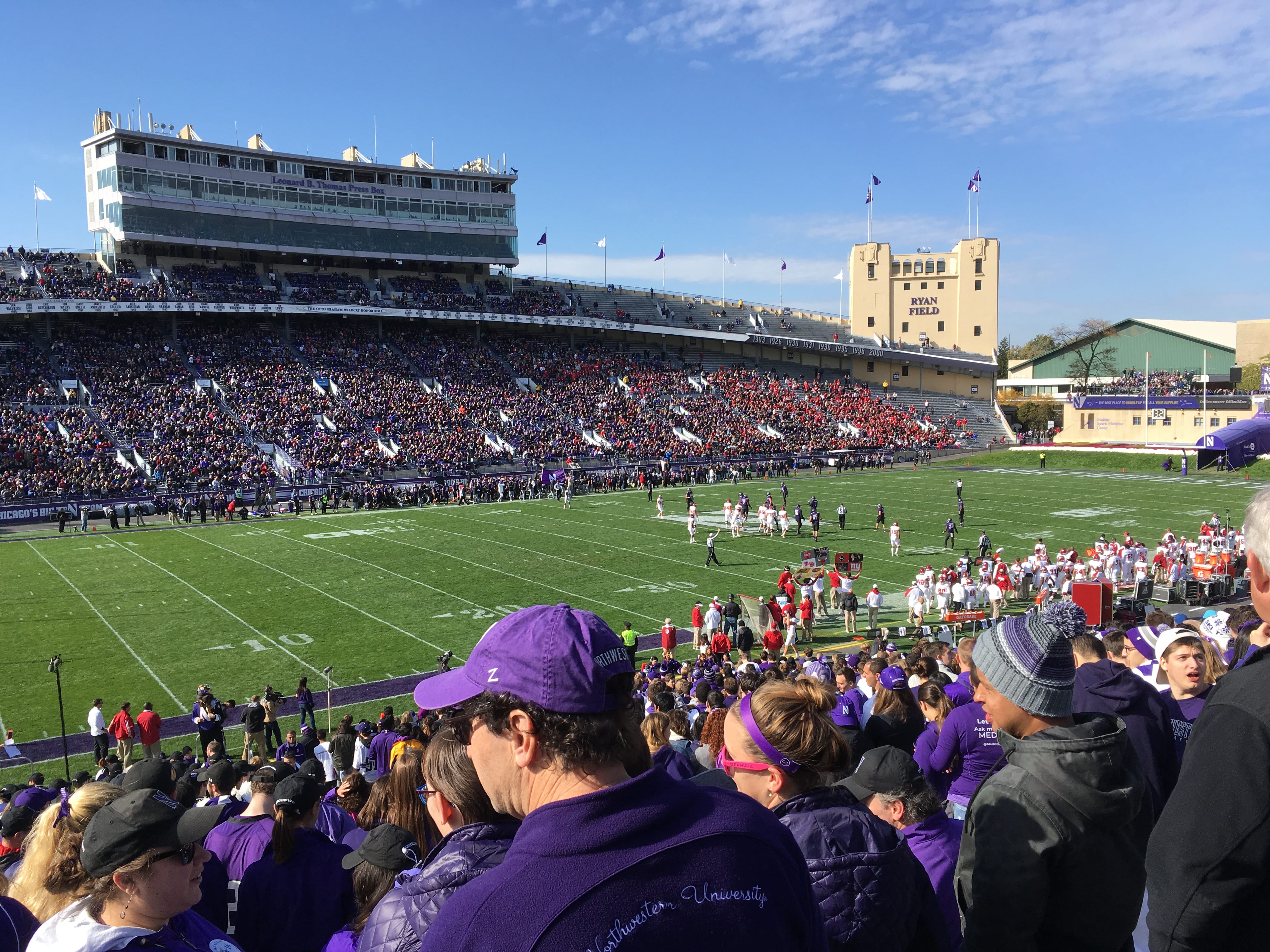
espite a lot work, life can still be enjoyable! One of the things that make me most happy here is the amazing campus area, which is very spacious and green (I just can’t wait for spring!) and literally lies on L ake Michigan (not that many universities have their own sailing club and a dock). The main campus is located in Evanston, a northern suburb of Chicago, and the second one with RIC and the school of medicine is around an hour away in Chicago downtown. Because of this, most of the student life revolves around Evanston campus – the whole university experience is quite different from the one we have in central London. The campus life, along with university sports (which are taken very seriously!), is probably what really brings people together and gives this great sense of community and pride in the school one can feel at American universities.

image3Student life is quite different here and takes some time getting used to, but it does have its own perks. Sometimes I miss London with its big-city, hectic lifestyle, but at this point I think I will soon miss the American life too. Spring and summer will hopefully not make me fall in love with Chicago too much, as I have heard it does get really lovely here. For the time being, I am planning on not worrying too much and just enjoying these last warm months here to the fullest!

Visiting the Stanmore Royal National Orthopaedic Hospital

By rmapapg, on 1 March 2017

by Madeline Lok, Emma Ponting and Sarita Meekul

On Friday 27th of January, our 2nd year Biomedical Engineering class got the opportunity to visit the Stanmore Royal National Orthopaedic Hospital. The purpose of the trip was for us to gain an understanding of how the clinical environment works and how devices we may help to create in the future fit into real people’s lives.

rhoh3The day began early with a long journey on the tube to Stanmore, on the outskirts of London where the class met. After a short taxi ride to the hospital, we were met by Professor Hart at the London Implant Retrieval Centre (LIRC). Prof Hart is the director of research and development at LIRC and a consultant orthopaedic surgeon at the hospital. He gave us a warm welcome and introduced us to some of the PhD students working there. At LIRC they recover and study knee and hip replacement implants that have been removed from patients to better understand why they failed. We were shown the processes these implants go through once they arrive, from cleaning to being scanned for wear and deformation and got to hold some actual implants. It was very interesting to see how something that we learn about in lectures actually looks and feels in real life.

rnoh1Then the lovely people of LIRC kindly provided us with lunch with their team. This was a good chance to chat to the people who work in the hospital and get a better insight into the kind of jobs available that we might be interested in once we graduate.

After lunch, we were split into smaller groups; some went to watch the surgeries while other went to sit-in with the doctors and their patients for real consultations. We swapped after 1.5 hours.rnoh4

We were brought to the surgery area to be able to see a real operation taking place. Before entering the operation theatres, we changed into scrubs. We were then separated into pairs and entered different theatres. Among all of us, we saw a range of operations including hip replacements, knee replacements and one ankle-foot correction surgery. We were told that the ankle-foot surgery is one of the most complicated and delicate procedures. The doctor that took us in even made a joke about how he avoided it. They used x-rays during that operation so we had to wear a lead apron to protect ourselves from the radiation. Some others of us saw the removal of implants, which was completely opposite of what the others saw. It was interesting to be able to discuss our different experiences at the end of the day when everyone was together.

Within the operating theatre, the surgeons explained to us what and how they were performing this surgery. Most of us were surprised by the atmosphere in the operating room. It was very relaxed with music playing in the background. The anaesthetist was reading his Kindle; the surgeons were even able to have a conversation and joke about their family while operating on the patient. It was surprising to see how calm and confident they were. Due to the time constraints, none of us were in there for the whole process which was a pity because we all really enjoyed it.

rnoh2The consultation sessions were an eye-opening experience too. We had the opportunity to sit through a few consultations with an orthopaedic specialist doctor, and see how they interact with their patients. All patients had very different reasons to be there, so we got to see various cases, the medical images used, and procedure followed. After sitting through the consultations, we now have a better understanding of what doctors go through when seeing patients and it definitely is a very difficult job. It’s not all smiles, hellos, reassurance and prescribing treatments as some people would think. In reality, they are potentially the ones who would be telling you how you would live out the next 10-20 years of your life (in our case, with hip/knee replacements, constant rehab, medication and so on). They have to always maintain professionalism and courtesy no matter how their patients react to whatever they tell them; even answer questions about their other concerns whether or not it is related to the real reason they came in for the consultation in the first place. The most important take-away I had from the session was that the doctors should let the patients leave with the best reassurance they can provide.

We all had a great day and learnt a lot about working in a clinical setting and working with patients. We would like to say a massive thank you to a ll of the people at Stanmore hospital who helped in making this day happen! What a day and what an experience!

Thinking clinical at the Learning Hospital

By rmapapg, on 3 December 2016

By Julian Henty

Clinical Engineering knowledge and skills were put to the test recently during a visit to UCLH’s Learning Hospital. Second year Biomedical Engineering students were given the chance to test various medical devices, such as ventilators, bedside monitors and suction machines, examine the workings of an in-house automated blood pressure machine, and observe vital signs measurement from a real ‘patient’ using a virtual bedside monitor.

Despite making good use of the department’s electronics lab for experimentation with transducers, software driven data acquisition, and aspects of electrical safety, the Clinical Engineering module fulfills its practical aims by providing a hands-on session with real hospital equipment and actual clinical measurements in a realistic clinical environment. The Learning Hospital has a ‘theatre’, complete with operating table and appropriate medical devices, and a ‘ward’ with two beds, nurses’ station and a medical gas supply.

The virtual non-invasive blood pressure (NIBP) device comprised a PC, control board and recycled components from a disassembled standard NIBP device. LabVIEW software measured the photoplethysmogram (PPG) amplitude distal to the cuff during inflation/deflation, which provided feedback to control the air pump and release valve. The software had an illustrative screen showing each step in the process of taking a real measurement. Safety aspects of NIBP measurement could also be demonstrated.

learninghospital

The patient waits anxiously for his results

The virtual bedside monitor displayed real-time graphs of the ECG, PPG, NIBP and chest movement while the ‘patient’ lay on a bed. LabVIEW software demonstrated how heart rate may be extracted from the ECG, PPG, or NIBP measurements, and respiratory rate from the ECG or chest movement. The software could also manipulate the ECG to demonstrate both noise and other lead measurements, and simulation of low amplitude due to pericardial/pleural effusion, pneumothorax, obesity or loss of viable myocardium.


With thanks to Dimitros Airantiz, Billy Dennis and Paul Burke

Students visit the UCLP Centre for Neurorehabilitation Symposium

By rmapapg, on 22 December 2015

By Ashkan Pakzad and Samuel Gunning

The Department of Medical Physics and Biomedical Engineering generously funded two students to attend a Neurorehabilitation Symposium organised by UCL Partners. The students were Ashkan Pakzad (MSci Medical Physics) and Samuel Gunning (iBSc in Medical Physics).

Neurorehabilitation conference flyer

Ashkan takes up the story:

Although the conference aimed to cover neurodegenerative diseases as a whole it often revolved around support and provision for those that suffered from multiple sclerosis, a disease that encompasses most aspects of any neurodegenerative condition.

As it was my first ‘real’ conference I was already nervous about the 9 hours ahead only to find out that my experience was about to curve further; I felt like an outsider, not because I was a student amongst clinicians bound to become consultants but because I was a physicist. The speakers all had the idea that they were informing clinicians rather than intruding academics but none the less I managed to keep up with their abbreviated jargon.

A wide range of speakers from top international consultants to graphic designers with no official qualifications in science working with clinics had a slot. It all started off with a few words from a retired prestigious academic, giving his take and definition on rehabilitation for neurodegenerative disease as he urged that such cases needed to not be over simplified and not be redefined into something new and that these diseases often should not even be considered as a disease but to take them as they come and to refrain from a systemic approach.

One particular speaker that I enjoyed had highlighted the need to conduct research with care as large budgets have been wasted in the field due to indirect questionnaires that measured outcomes too broadly and thus those studies had given imprecise results to critically compare techniques therefore leading to unsustainable conclusions. In fact, he claimed that much was to be learnt from experiments in physics coupled with rigorous statistical analysis when planning medical experiments.

Stalls were set up in between speakers offering close inspection of incontinence technology and several industry representatives were presenting their technologies as fast as possible with no time to lose. Overall this opportunity has been an eye opener just presenting the great wealth of knowledge delivered intensely and often ready digested thanks to talented speakers whom all have a different perspective to offer.

Samuel continues after lunch:

The symposium was approached from a greater clinical perspective after lunch. Whereas before lunch had been focused primarily on the technology that is currently circuiting, as demonstrated in Helen Paterson’s lecture on Augmentive and Alternative Communication, or the general role rehabilitation should have in neurological diseases, after the break we started to pinpoint specific conditions or symptoms which can be overcome with rehabilitation.

We began with the management of incontinence – bladder first and then bowel – issues which are quite often left untreated due to them being regarded as “private” matters. The lectures had very different structure with the first summarising the main surgical methods which would follow the failure of conservative treatments and the second concentrating on the technological interventions used, sometimes with many being used in parallel. The reason for this is, I believe, is the far more advanced care algorithm used for bowel management.

Following this we moved onto the complicated management of pregnancy in women with Charcot-Marie-Tooth disease. To a medical student this was very interesting as CMT, a hereditary sensory and motor neuropathy with peroneal muscular atrophy as well, is a perplexing condition. To then understand the methods to overcome the disease, or at least reduce the impact of some of the many symptoms attributed to CMT, was a highlight.

Moving away from disease, the next session addressed the analysis of research, specifically research into emerging rehabilitation technologies. With doctor autonomy currently growing, the ability to differentiate a potentially hugely beneficial treatment method from a false one is hugely important. The objective of the lecture, I believe, was to emphasise how an outcome was completely reliant on the measurement process and how you must first decide the outcome you want before indentifying the measurement process, a thinking point I later applied to my own research.

To finish, two support groups were identified. The key purpose of these groups is to support and to facilitate learning of how to manage oneself when struggling with a disease or symptom.

The second half of the day was much more applicable to my own learning, with such symposiums contributing to the compulsory CPD points I will one day have to accumulate. Despite this, the technological aspect was what I had expected. Having spoken to the other attendees, the majority were practicing doctors, physiotherapists and nurses, and I think the day was perfectly focused to them. For a student it was important to see how the stuff we are learning about is applied in a clinical situation as this is the eventual goal of any studying and can help focus and encourage. Furthermore, with presentations looming at the end of next term, the symposium was a valuable opportunity to identify positive presentation methods and those to avoid as well. All in all a fantastic day, filled with a wide variety of interesting topics.

Thanks to the Centre for Neurorehabilitation who agreed to offer a student discount on the registration fee.

Clinical engineering visit to Royal National Orthopaedic Hospital

By rmapapg, on 13 November 2015

By Nishat Ahmed and Bindia Venugopal

On Wednesday the 11th of November, we were up at the crack of dawn, pumped and ready to go to the Royal National Orthopaedic Hospital in Stanmore. After missing trains due to tube closures and our taxi rides arriving a half hour late, we finally managed to reach the hospital in time to attend the Multi-Disciplinary Team meeting.

We found the meeting very interesting, watching the consultant surgeons and nurses discuss real case studies of patients. They collaborated well to work out the best way to rehabilitate patients, whether this was through further surgery or simply giving them advice and support.

Later on we headed to the operation theatres, adhering to hospital dress code we threw on our scrubs, hair nets and masks beforehand! Since we were only allowed three students at a time in the theatres, we split into groups and then went off to watch various operations taking place. The first surgery we watched involved attaching a metal plate to a fractured tibia bone to aid its healing process in a way that was ingenious! It was fascinating watching the surgeon screw the bone together and then brace the join with a metal plate. The screws held the fracture under compression, this meant it was forced to combine together rather than slide apart, and the metal plate stopped it from twisting.

The second surgery we went to was an extremely rare case where the surgeons ended up dislocating the hip bone in order to remove a benign tumour from inside the bone. They sawed the hip bone in half as bone-to-bone healing worked best compared to tendon-to-bone healing. The challenge was in trying to avoid damaging the femoral head to get to the tumour.

After this we had a little tea break and then made way to our next surgery! This was a spinal surgery where the patient had a twisted spine due to being paralysed for 10 years. They operated with a diathermy machine which uses electricity to cut through the skin and muscles as this reduces blood loss. Although we only saw the surgery for 10 minutes we learnt how vital it was to keep the fluids in the patient regulated. This job was monitored by the anaesthetist, who informed us about the patient and the precautions which needed to be taken. Two neurophysiologists were monitoring electrical activity in the spinal cord to ensure that it wasn’t damaged by the surgery.

Scrubbed up

After an insane experience watching all the surgeries, we went to have lunch which was provided by the lovely team at Stanmore. In the afternoon we got a tour around the BME department at the hospital and learnt about all the weird and wonderful things they collect and experiments they run! In fact, we found out that they have over 6000 failed hip replacements from 25 different countries in their labs to study and analyse. They conduct experiments to research why implant failure happens in some patients the way it does, especially those with metal on metal implants. They use tools for metrology which measures the exact size of the ball and socket implants with crazy precision! This information is then used to work out the amount of corrosion that happened in the body when the implants were inserted.

Overall, we had an amazing and truly valuable experience. The entire team were extremely friendly and helpful! We loved that we could ask questions and interact with the staff so well. It was remarkable to see the transition from a real-life patient problem to actually seeing the solution executed in the surgeries. It was also encouraging to see how the hospital carries out their own research which can then be implemented to the surgery procedures in only a few years’ time.

On behalf of our whole BME department, we thank you for this experience Professor Hart and RNOH!