Close
Close

A breath of fresh air
By rmapapg, on 14 March 2018
By Rebecca Yerworth
How do you let steam out of you kitchen if the only window is behind the kitchen sink and, as an octogenarian you can no longer climb on the work-surface to reach the handle? We gave our second year biomedical engineers one week to find a solution.
At first it seemed trivial – just put a hook on a stick… but then they met the 87-year old client and realised that wielding anything much heavier than a cup of tea was going to be a problem, and she would like to be able to store the aid on the kitchen work surface. … and don’t forget the screw on the bottom bracket which is needed to stop the open window flapping about in the breeze.
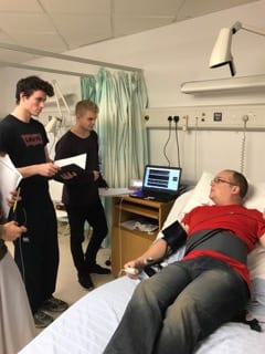
By Friday afternoon all the student teams had a device which they could use to open the window, and were waiting in trepidation for the client to test them out. “Bit heavy”, “nicely finished”, “easy to use”, were some of the comments heard. Different solutions were found to the issue of weight, from using intrinsically light bamboo to strategically shaping wooden planks to reduce their weight, to not using a pole at all – just use pulleys. Seven different ideas from seven teams.
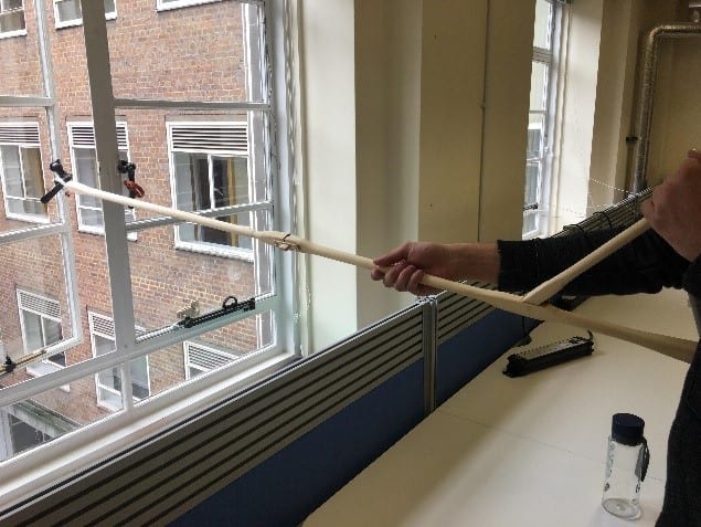
Window Opener being tested
During the week students put into practice skills they had learnt in other modules – included user-centered design, and the practical implications of material choice. They also learnt new skills as they made good use of the tools in the biomedical engineering teaching laboratory and in UCL’s MakeSpace.
This project is inspired by the work of REMAP, a national charity specialising in custom making aids for elderly and disabled people, where no suitable aid is commercially available. UCL staff and students have recently set up a REMAP-affiliated group Impactive. Volunteers from both organisations gave an inspiring talk to the students at the beginning of the week, and it sounds like they have gained some new volunteers: Biomedical Engineering students putting their skills to good use even before they graduate – well done and keep it up.