 Close
Close

How do you demonstrate gamma imaging practically to students, without a radiation hazard?
By rmapapg, on 10 January 2017
By Rebecca Yerworth
This was the challenge set to 3rd year project student Nicola Wolf. The outcome? Gamma Anna, and a paper in Physics Education. The interactive demonstration that Nicola developed is applicable to medical students, secondary school lessons, and younger children when used with an appropriate age specific work sheet.
What is Gamma Imaging? It is a medical diagnostic technique which involves injecting small amount of radioactive material (tracer) in to a patient and looking to see where it goes using a radiation detector, known as a gamma camera. The tracer is designed to be selectively absorbed into tissues of interest – e.g. radioactive glucose will accumulate in those parts of the body that are using the most energy…. Tumours have a high energy demand, so they will show up bright in the image. This is a useful tool for doctors if they want to see if a cancer has spread.
Why produce a teaching demo? It is common knowledge that well designed interactive activities increase understanding of the subject and retention of knowledge, as well as student engagement and enjoyment. However you can’t safely demonstrate real gamma imaging to students, because of the radiation hazard, quite apart from the logistics: Gamma cameras are large (room sized) and expensive. The Gamma Anna demo uses a series of analogies to explained key concepts of the imaging technique: heat, from an exothermic reaction represents the gamma radiation; tumours are represented by plaster of Paris; saline the radioactive tracer and a thermal imaging camera represents the Gamma camera. ‘Anna’ herself is a ragdoll into which the ‘tumours’ can be placed.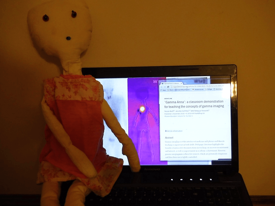
Who is the demo for? Nicola tested the demo with GCSE grade students, where it served to explain principles of radiation and showed applications of physics to medicine and engineering – a topic with the potential to motivate students, including girls, to choose STEM subjects at A’ level and beyond. She also tested it with medical students, where the focus was on improving their ability to accurately advise future patients. Gamma Anna could also be used with younger children, either at an outreach event or as play-therapy if they, or a relative, need to undergo gamma imaging. In each case age/course appropriate work sheets should be used; examples are available for download from the link above.
In conclusion… Gamma Anna is cheap, safe and easy to make and use. The largest expense being a thermal imaging camera, but mobile phone adapters are suitable, can be bought for less than £200, and are a useful resource for other demos too.
What cancer diagnostic system is best for rural China?
By rmaptst, on 23 December 2016
By Terence Leung
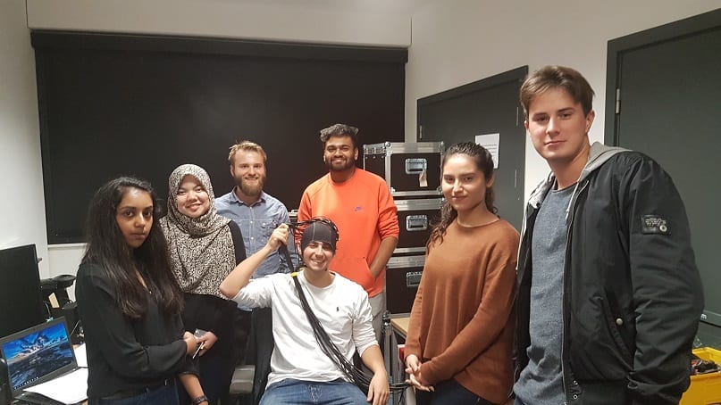
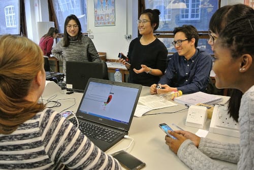
This was the challenge we set for our 1st year biomedical engineering students over the first five weeks of the first term. Welcome to the ENGS101P Engineering Challenge! They were given a (hypothetical) budget of 5 million yuan (~£583K) to design a cancer diagnostic system for the rural area of Yunnan, a mountainous region in China with annual disposable income only a third of those living in the urban areas.
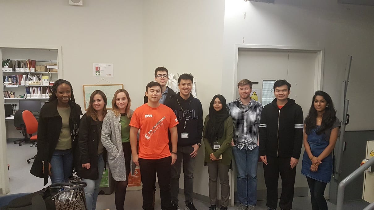
To help them decide, we gave them a talk on the cancer crisis in China, and two introductory lectures on breast and prostate cancer therapy and imaging. We also organised visits to five research labs at UCL, including X-ray Imaging Lab, Photoacoustic Imaging Lab, Diffuse Optical Imaging Lab, Ultrasound Imaging Lab at the Centre for Medical Image Computing, and Magnetic Resonance Imaging Lab at the Centre for Advanced Biomedical Imaging.
You can see what solutions they came up with in these Youtube videos.

Professor Gary Royle (UCL Professor of Medical Radiation Physics and Visiting Professor of Tsinghua University) talked about cancer crisis in China

Dr Paul Doolan, a radiotherapy and proton physicist at University College London Hospital, gave an introduction to breast and prostate cancer therapy

Miss Savanna Chung, a practising radiotherapist and PhD student in radiotherapy, gave an introduction to breast and prostate cancer imaging

Team “China Heroes” visiting Photoacoustic Imaging Lab (Host: Trung)
Team “Cancer Club” visiting Diffuse Optical Imaging Lab (Hosts: Luke and Prash)
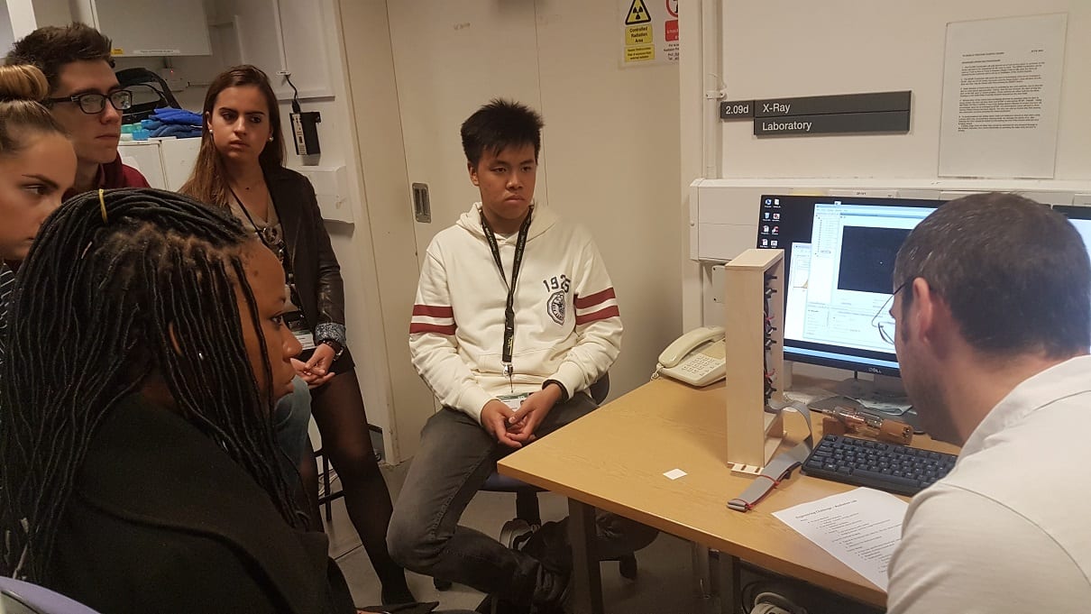
Team “CRABTech” visiting X-Ray lab (Host: Rob)

Team “Panda Cure” visiting Ultrasound Imaging Lab at the Centre for Medical Image Computing (Hosts: Yipeng and Ester)
Teams “MADLAB” and “CRABTech” visiting Magnetic Resonance Imaging Lab at the Centre for Advanced Biomedical Imaging (Hosts: Morium and Ian)
Marina Melero reports on Global Entrepreneurship challenge in Malaysia
By rmapapg, on 15 December 2016
One of our outstanding second year biomedical engineering students, Marina Melero, has returned from a visit to Kualar Lumpur. She had been invited to a meeting of the Global Entrepreneurship Community to lead a workshop on healthcare.
In her blog post, she reports on her visit and gives her thoughts on the future of technology.

Thinking clinical at the Learning Hospital
By rmapapg, on 3 December 2016
By Julian Henty
Clinical Engineering knowledge and skills were put to the test recently during a visit to UCLH’s Learning Hospital. Second year Biomedical Engineering students were given the chance to test various medical devices, such as ventilators, bedside monitors and suction machines, examine the workings of an in-house automated blood pressure machine, and observe vital signs measurement from a real ‘patient’ using a virtual bedside monitor.
Despite making good use of the department’s electronics lab for experimentation with transducers, software driven data acquisition, and aspects of electrical safety, the Clinical Engineering module fulfills its practical aims by providing a hands-on session with real hospital equipment and actual clinical measurements in a realistic clinical environment. The Learning Hospital has a ‘theatre’, complete with operating table and appropriate medical devices, and a ‘ward’ with two beds, nurses’ station and a medical gas supply.
The virtual non-invasive blood pressure (NIBP) device comprised a PC, control board and recycled components from a disassembled standard NIBP device. LabVIEW software measured the photoplethysmogram (PPG) amplitude distal to the cuff during inflation/deflation, which provided feedback to control the air pump and release valve. The software had an illustrative screen showing each step in the process of taking a real measurement. Safety aspects of NIBP measurement could also be demonstrated.
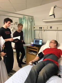
The patient waits anxiously for his results
The virtual bedside monitor displayed real-time graphs of the ECG, PPG, NIBP and chest movement while the ‘patient’ lay on a bed. LabVIEW software demonstrated how heart rate may be extracted from the ECG, PPG, or NIBP measurements, and respiratory rate from the ECG or chest movement. The software could also manipulate the ECG to demonstrate both noise and other lead measurements, and simulation of low amplitude due to pericardial/pleural effusion, pneumothorax, obesity or loss of viable myocardium.
With thanks to Dimitros Airantiz, Billy Dennis and Paul Burke
Music of Light stethoscope
By rmapapg, on 13 July 2016
By Daniil Nikitichev
What is the idea?
Photoacoustic imaging (PAI) is an emerging technique that can be used to image vasculature with high spatial resolution. The phenomenon of photoacoustics was discovered in 1880 by Alexander Graham Bell, who observed the production of sound by chopped sunlight. This observation led him to the invention of the “photophone” for transmitting the human voice. This project is centred on creating a PAI demonstration kit for teaching and public engagement activities within the University and at external Festivals and Conferences. Similar to the “photophone”, the demo involves the production of music/human voice using a simple LED torch based on photoacoustic sound generation.
What is the benefit of this teaching demo?
We developed a teaching demonstration tool to illustrate the photoacoustic effect to undergraduate and graduate students based on the experiment that led to its discovery. This demo will spark the interest of the students and increase their awareness of how this physical phenomenon can be applied to medical imaging for diagnostic purposes.
Public engagement and outreach activities at UCL would benefit from further promoting science with this tool. This demo kit provides a stimulating way of teaching pupils (secondary school students) some basic concepts of physics, and a motivation to select studies across STEM (Science, Technology, Engineering and Mathematics) subjects. On the other hand, people from different backgrounds and ages can engage with state-of-the-art research for medical imaging and diagnostic applications, since PAI is currently applied in preclinical studies based on small animal experiments.
Finally, with this demo kit, scientists and researchers will improve their public speaking and teaching skills and make their presentations more stimulating and approachable to their audience.

How did we approach our project?
The project involved the design of a simple amplification circuit connected to an Apple iPod (or a similar music playing device) through a Bluetooth connection in order to modulate the LEDs of a torch for generation of the photoacoustic effect. The generated signal was detected by a stethoscope. The demo was built at the workshop of the Department of Medical Physics and Biomedical Engineering, UCL. Several engineers from the department provided assistance on technical issues that arose during the project.
Conclusion
At the moment the total cost of the demo is several hundreds pounds due to the cost of the stethoscope. Further work will be performed in reducing the cost and size of the demo. At the moment the demo is available for use to any member of the department for teaching and demonstration purposes. Furthermore, Cambridge University decided to build similar demo based on our design.
D. I. Nikitichev, W. Xia, E. Hill, C. A. Mosse, T. Perkins, K. Konyn, S. Ourselin, A. E. Desjardins, T. Vercauteren: “Music-of-Light Stethoscope: A Demonstration of the Photoacoustic Effect (Photoacoustic Stereo)”, Phys. Educ., 51, 045015, 2016
Peer assessment in group work
By rmapapg, on 13 May 2016
By Pilar Garcia Souto
UCL Engineering trains students to use engineering knowledge within extended group practical activities to better prepare them for their careers after graduation. However, despite the substantial educational benefits of getting students to work in teams, students express and experience concerns that significantly decrease the student satisfaction.
We decided to look deeper into this matter and organized student focus groups across the Engineering Faculty, and spoke with various members of staff that use and assess group work. The message is clear: an element of “individual contribution” is needed, possible set by peers and tutor moderated, which improves the group dynamics and penalize the “passengers”. Otherwise students frequently express dissatisfaction if all members of a team are given the same mark regardless of the individual effort.
The concept is simple. At the end of a group work students rate the contribution of each team member, and this is used by the tutor to generate an individual mark. This encourages self-reflection, increase student satisfaction and reduce student’s complaints. The only major drawback is that the peer assessment of individual contribution is mainly collected using pen and paper, hence very staff consuming, as current e-learning tools are inadequate. From our research, this tool should be online, anonymous, preferable within Moodle and flexible so staff can adapt it and ask or value different aspects (e.g. reliability, punctuality, contribution to ideas, etc.).
This is an ongoing project. We presented some results at the UCL Teaching and Learning conference in April 2016, which attracted a lot of interest. It is clear that individual contribution assessment is something that staff from across UCL want to implement, and yet we lack the appropriate system. We decided to take the lead on establishing a consortium with those interested, and seek for some funding to develop an appropriate system within Moodle that would allow us to efficiently incorporate this practice into our teaching. If you are interested on participating and/or hearing more of our results, please contact p.garciasouto@ucl.ac.uk.
Our thanks to ELDG 2015 who partially funded this project.
Pitching UCL Biomedical Engineering Inventions To A Panel Of Dragons
By rmapapg, on 11 April 2016
By Jenny Griffiths
We made an unusual homework demand on our second year Biomedical Engineers over the Christmas vacation: they had to watch TV.
The students were asked to use UCL’s subscription to Box of Broadcasts to watch episodes of BBC’s Dragon’s Den in order to prepare for their first week back when they would be asked to spend a few days applying knowledge and understanding of enterprise, ethics, and regulations to medical devices.
On the first day of term, groups of students were each given a UCL Biomedical Engineering invention and told that they were to present a written portfolio and give a pitch to a panel of expert ‘Dragons’ on Friday afternoon. They then went off, made contact with the UCL inventors of the devices, and with the help of a Teaching Assistant with a background in Medical Device Innovation, researched:
- the devices’ capabilities
- the market for the invention
- routes to that market
- ethical implications and requirements
- medical device regulations for the device
All this information – key to bringing an engineering concept from lab to public use – needed to be at their fingertips for the Friday presentations.
The full assignment marks for the work were split between the presentation, a written group portfolio and individual contributions to the team. We also upped the competitive element by awarding a prize for the best pitch, judged entirely subjectively by the Dragons and unlinked to any summative assessment marks.

This year’s devices were an optical ultrasound transcatheter imaging system (Dr Adrien Desjardins), a percutaneous heart valve delivery system (Dr Gaetano Burriesci) and SenseWheel – a force sensing wheelchair wheel to measure biomechanics (Dr Catherine Holloway).
On Friday afternoon, each group had five minutes to present their device to a panel of experts consisting of:
- an academic medical devices expert
- a Royal Academy of Engineering Enterprise Fellow
- an academic who has commercialised a medical device through a spin-out
- an external marketing and communications expert with no expert medical device knowledge.
The presentations were held in the appropriately intimidating Executive Education Suite, where the panel sat in high backed chairs and asked probing questions after each presentation. The students responded professionally and gave excellent pitches, selling devices that they had not know about just five days before!
Our highly sought after prize of copies of Eric Ries’ ‘The Lean Start up’ and (chocolate) money was won by team SenseWheel.

In future years we aim to encourage more external Dragons to take part and will link the prize giving to an industrial careers and networking event for the students. If you are an employer who would like to be a part of this fun and valuable event, the department would love to hear from you.
Living Aid – working with Remap
By rmapapg, on 15 March 2016
By Rebecca Yerworth
 During the last week of February the second year biomedical engineers were introduced to ‘Remap’, a national charity working through local groups of skilled volunteers to help disabled people achieve independence and a better quality of life, by designing and tailor making equipment for their individual needs. The week started with a fascinating talk by Remap volunteers, explaining the purpose of the charity, the range of projects they tackle and the life changing effect of these bespoke items.
The students were then tasked with designing an aid that will enable a client to fit and remove spectacles, which she is unable to do without help, due to restricted arm movement. Whilst Remap projects vary in complexity, this is typical of the issues they solve – giving back independence to disabled users, or enabling them to take up a hobby they could previously only dream of.
By the end of the week the students had had two meetings with the ‘client’ (an actor well acquainted with the issues) and we had three prototype devices. Three completely different approaches were taken, all of which the students could operate… though some needed further refinement/customisation to be useable by the client.
 The project raised some interesting questions about the relative merits of 3D printing versus traditional DIY techniques and of passive versus active devices. It also highlighted the importance of identifying and taking into account the client’s needs and preferences.
Assessment by wiki
By rmapapg, on 1 March 2016
By Adam Gibson and Rebecca Yerworth
We’re keen to incorporate different methods of assessment into the biomedical engineering programme. This makes the process more interesting and engaging for staff and students, and allows us to teach and assess a wide range of transferrable skills. In a new module on anatomy and physiology for biomedical engineers, we teach the basic anatomy and physiology in workshops and in a dissection room, and provide enhanced context by asking biomedical engineers to give two-hour “case studies” where they describe how their research interacts with the relevant anatomy and physiology.
We’ve built on this concept of a “case study” in the assessment where we ask students, in pairs, to research and present their own case study. The twist is that we have asked them to prepare and produce this using an online wiki interface. This allows us to move away from the standard boring Word document and introduce new skills. We ask students to demonstrate advanced writing skills by writing for a lay audience. They also need to consider how writing a website is different from a typical document, and address topics such as accessibility, web design and ensuring that any images have appropriate copyright. Academic referencing is as important as ever but has a different nuance if hyperlinks can be used. We produced a rubric which shows how we emphasised these areas in the assessment.
Asking students to research their own case studies based on those presented by experts in the field fits in nicely with UCL’s emphasis on research-based education and the connected curriculum, particularly as an example of outward-facing assessment.
A wiki interface includes a “history” log which makes it easier for an assessor to track the contributions of each partner, and also allows permissions to be controlled so that only members of a pair or team have access to edit the document, but tutors can view progress. Once the assignment is complete and assessed, we opened up access completely. This encourages students to take pride in their work, and gives them something they can link to if they choose. We hope that our collection of case studies will lead to a useful resource as different cohorts of students add to it year by year. We will also encourage students in subsequent years to read previous submissions and hopefully learn from them.
Smartphone app for detecting pulse rate
By rmaptst, on 17 February 2016
By Terence Leung
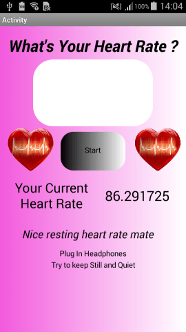
Our 1st year Biomedical Engineering students had very little computer programming experience when they began their first scenario week on Monday, 8th February. So to them, developing a smartphone app that can measure pulse rate seemed almost like an impossible task. However, as they struggled through the week, they learned about building the graphical user interface, switching the phone’s flash on and off, accessing pixel values from the phone’s camera, and performing Fast Fourier Transform to get the frequency of a periodic signal. Gradually, their apps were taking shape. Indeed by Friday all 10 teams had successfully developed their apps, some completed with a customised logo, animation, and even sound effect. It was a tough week, typically 9 to 5 (except Wednesday afternoon off). Many students found it tiring but were very proud of their first ever healthcare apps. Hopefully not their last!
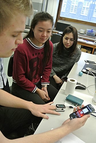
Anxiously waiting for the test result…
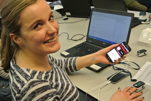
Test result looks good!
Students took turn to demonstrate their apps

This app apparently has the approval of fellow students
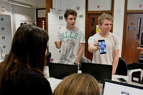
Explaining why this is a great healthcare app!

This app has incorporated an animation in the background!
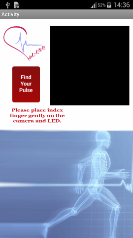
This app has a customised logo (top left corner)!
This app plays simulated heart sounds during the measurement!