X Close

Neurulation Biomechanics

Home

Menu

Archive for the 'Mouse' Category

New mouse model of Terminal Myelocystocele

By Gabriel Galea, on 26 March 2024

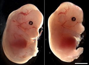
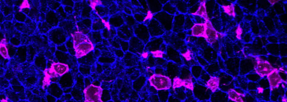
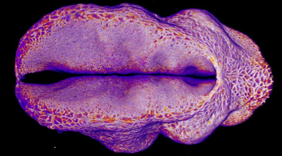
Our recently-published article shows that regional deletion of Fgfr1 in the embryonic trunk produces localised spinal mis-patterning and a terminal myelocystocele-like phenotype in mice. The image below shows a normal mouse fetus (left) and one with a sac-like protrusion at the bottom of the spinal cord (right) which resembles a rare human malformation called Terminal Myelocystocele. Our paper shows that this malformation does not arise because of failed neural tube closure, like more common types of spina bifida do, but happens are the neural tube is fully closed.

Neuroepithelial synchronisation of apical constriction and mitosis

By Gabriel Galea, on 12 December 2022

Our latest paper shows that neuroepithelial cells undergo high-amplitude apical constriction synchronised with cell cycle progression but the timing of their constriction if influenced by tissue geometry. We observe this in mouse and chick embryos, as well as in human iPSC-derived neuroepithelial cells cultured on either flat or curved surfaces. We find this human cell model particularly tractable, except that it requires daily medium changes to keep the cells happy!

Cannabidiol impairs neural tube closure in mouse whole embryo culture

By Gabriel Galea, on 13 April 2022

In our latest paper, we find that exposing mouse embryos to cannabidiol (CBD) in whole embryo culture diminishes their ability to close the neural tube. This happens at concentrations which do not impair the development of other embryonic structures such as somites and does not change cell proliferation or apoptosis, suggesting a selective effect on the neural tube. It’s important not to over-interpret this paper: we exposed embryos to CBD directly not from their mother and we do not know what the mechanism behind the observed effect is. Congratulations to Yosuf who completed most of this work during his iBSC as part of his UCL medical degree.

You can access the paper here.

How does the brain get covered with skin?

By Gabriel Galea, on 12 May 2021

Our latest paper combines physics, embryology and advanced microscopy to work out how the brain gets covered with skin. Read the original paper here, or just watch it happening below!

Closing mouse hindbrain neuropore

Live-imaging showing progression of hindbrain neuropore closure in a mouse embryo.

Uninherited mutations can cause spina bifida

By Gabriel Galea, on 19 February 2021

Our latest research, published in Nature Communications, reveals that spinal cord formation is exquisitely susceptible to mutations which happen during embryo development. We find that mutating one gene, called Vangl2, in just 16% of developing spinal cord cells is enough to cause spina bifida in mice. This is because each mutant cell interferes with the normal function of its neighbours. These mutations are not inherited from either parent, and would not be passed on to the individuals’ children.

We already knew that uninherited (“somatic”) mutations in genes which interact with Vangl2 can be found in 15% of individuals who have spina bifida (previous paper here), but we did not know if these rare mutations were enough to cause such a severe birth defect. We now need to improve diagnostic testing methodology in order to find these mutations without needing to cut out patient tissue.

You can read a lay interpretation of our study here.

Mutant (magenta) spinal cord cells impair their neighbours' function

Hindbrain neuropore closure

By Gabriel Galea, on 3 November 2020

Here’s a link to our new pre-print showing that hindbrain neuropore tissue geometry determines asymmetric cell-mediated closure dynamics. The hindbrain neuropore is a tissue gap over the back of the head which needs to close in order to cover the developing brain with other other cell types. If that does not happen the embryo develops a fatal birth defect called exencephaly (also called anencephaly). Eirini’s work, shown in this pre-print, identifies two different behaviours by which cells around this gap generate mechanical forces needed to close it. Thanks to our collaborations with physicists at Carnegie Mellon, we were able to show that both these behaviours must happen at the same time to describe closure of this gap.

In the image below, the top of the head is on the left, the neck is on the right, and the massive hole between them is the hindbrain neuropore.

Pre-print: New mutations can cause spina bifida

By Gabriel Galea, on 10 September 2020

Check out this new pre-print from our lab. In it we show that mutations which happen during embryonic development, and only affect a small proportion of cells, can be enough to cause spina bifida. It turns out that each mutated cell in our model prevents several other cells from biomechanically contributing to neural tube closure.

Pre-prints are repositories of papers which have not yet been fully peer reviewed and should therefore not be viewed as definitive. We decided to share our findings in this format while they are being formally reviewed because we are excited about the implications of our findings for the diagnosis and prevention of spina bifida as well as for genetic counselling of parents and patients.

Closing mouse spinal neural tube. The green dots are mutant cells.

Closing mouse spinal neural tube. The green dots are mutant cells. This small number of mutant cells is enough to cause spina bifida.