X Close

Neurulation Biomechanics

Home

Menu

Archive for the 'Live-imaging' Category

Neuroepithelial synchronisation of apical constriction and mitosis

By Gabriel Galea, on 12 December 2022

Our latest paper shows that neuroepithelial cells undergo high-amplitude apical constriction synchronised with cell cycle progression but the timing of their constriction if influenced by tissue geometry. We observe this in mouse and chick embryos, as well as in human iPSC-derived neuroepithelial cells cultured on either flat or curved surfaces. We find this human cell model particularly tractable, except that it requires daily medium changes to keep the cells happy!

Measuring the mechanical forces generated by embryos

By Gabriel Galea, on 18 February 2022

Check out our latest pre-print in which we quantify the mechanical forces generated by the closing neural tube in chick embryos. it still feels like science fiction: massive congratulations to Eirini for completing this exciting work.

You can access the pre-print on Research Square, here.

How does the brain get covered with skin?

By Gabriel Galea, on 12 May 2021

Our latest paper combines physics, embryology and advanced microscopy to work out how the brain gets covered with skin. Read the original paper here, or just watch it happening below!

Closing mouse hindbrain neuropore

Live-imaging showing progression of hindbrain neuropore closure in a mouse embryo.

Uninherited mutations can cause spina bifida

By Gabriel Galea, on 19 February 2021

Our latest research, published in Nature Communications, reveals that spinal cord formation is exquisitely susceptible to mutations which happen during embryo development. We find that mutating one gene, called Vangl2, in just 16% of developing spinal cord cells is enough to cause spina bifida in mice. This is because each mutant cell interferes with the normal function of its neighbours. These mutations are not inherited from either parent, and would not be passed on to the individuals’ children.

We already knew that uninherited (“somatic”) mutations in genes which interact with Vangl2 can be found in 15% of individuals who have spina bifida (previous paper here), but we did not know if these rare mutations were enough to cause such a severe birth defect. We now need to improve diagnostic testing methodology in order to find these mutations without needing to cut out patient tissue.

You can read a lay interpretation of our study here.

Mutant (magenta) spinal cord cells impair their neighbours' function

Hindbrain neuropore closure

By Gabriel Galea, on 3 November 2020

Here’s a link to our new pre-print showing that hindbrain neuropore tissue geometry determines asymmetric cell-mediated closure dynamics. The hindbrain neuropore is a tissue gap over the back of the head which needs to close in order to cover the developing brain with other other cell types. If that does not happen the embryo develops a fatal birth defect called exencephaly (also called anencephaly). Eirini’s work, shown in this pre-print, identifies two different behaviours by which cells around this gap generate mechanical forces needed to close it. Thanks to our collaborations with physicists at Carnegie Mellon, we were able to show that both these behaviours must happen at the same time to describe closure of this gap.

In the image below, the top of the head is on the left, the neck is on the right, and the massive hole between them is the hindbrain neuropore.

Chick embryo live imaging

By Gabriel Galea, on 11 September 2020

Eirini spent a very long time looking at chick neuropores closing during live-imaging on our two-photon microscope yesterday. By the end of the session it felt like they were talking to her…

Sometimes you really need to look away from the monitor/eyepiece.

Embryonic cells are stressed

By Gabriel Galea, on 7 September 2020

When a mechanical force is applied to an object, that object deforms and withstands physical stress.

Think of applying a force to stretch a rubber band. It stretches and withstands mechanical tension. If you cut it, that tension is relaxed and the rubber band pings back to its preferred length.

If you understand that, you’re well on your way to understanding a central tenant of biomechanics. Each cell in the embryonic neural tube acts like a little rubber band, stretching its neighbours. To work out how much tension a cell is withstanding we have to cut it and measure how far it pings (we call this “recoil”).

GIF showing laser ablation

So how do you cut a cell? Cells are far too tiny to cut them with anything physical like a scalpel. Instead we use a high-powered laser to very precisely cut a cell border. In the image above you will see the laser cut indicated by a red line as the white borders around cells ping apart. These cells should be pulling the neural tube closed, so by cutting them we can infer whether they were doing their job correctly.

Neurulation Biomechanics Lab

By Gabriel Galea, on 5 September 2020

Welcome to the Neurulation Biomechanics Lab!

We’re a growing group of scientists at the UCL Great Ormond Street Institute of Child Health studying how our brain and spinal cord form while we are tiny embryos.

Specifically, we study how embryos change their shape to cover the brain and spinal cord with skin which protects them during development. If that doesn’t happen, the embryo develops a birth defect known as a Neural Tube Defect. Examples of Neural Tube Defects include Spina Bifida. We study this using genetic models and advanced microscopy, which allows us to watch the neural tube close in model species including mice and chicks.

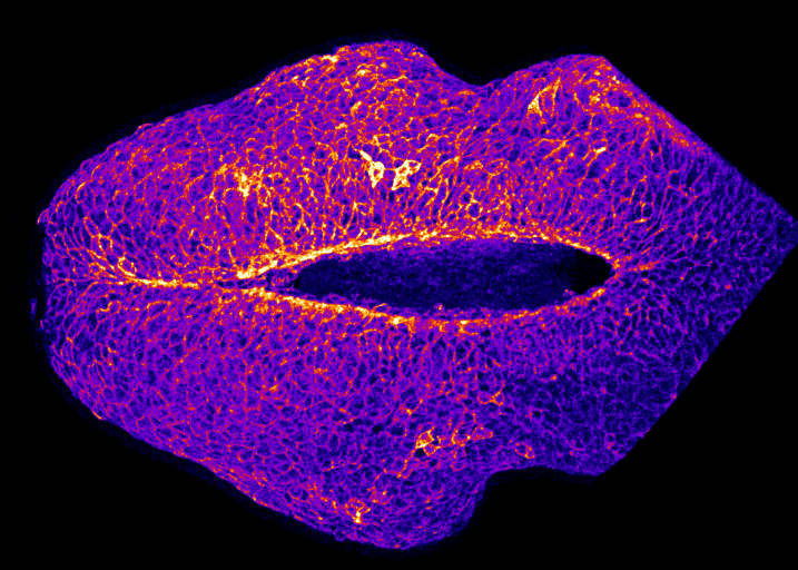
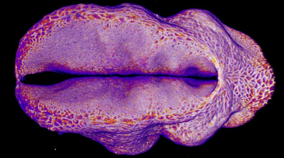
In the image on the right you can see the tail-end of a mouse embryo. Click on it to watch it move. The cyan region is the future skin, which you will see encircles a folding region of red cells. These red cells will form the future spine and you can see how, over around two hours of live-imaging, the embryo makes a lot of progress in covering it. Clearly, the embryo needs to change its shape quite a lot! What you’re seeing is the embryonic process of neurulation.

How do embryonic cells generate, withstand and transmit the mechanical forces required to change the shape of this tissue? This is what we mean by Biomechanics – the mechanics of biological systems.

Do abnormalities in these processes cause Neural Tube Defects? Can we apply what we learn to prevent, or at least explain the causation of, conditions like Spina Bifida? These are many of the questions our lab is interested in and we hope to share our findings here over years to come.