X Close

Neurulation Biomechanics

Home

Menu

Archive for the 'Laser ablation' Category

How does the brain get covered with skin?

By Gabriel Galea, on 12 May 2021

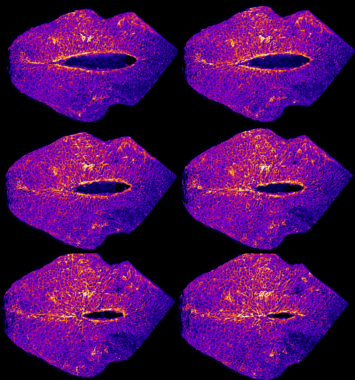
Our latest paper combines physics, embryology and advanced microscopy to work out how the brain gets covered with skin. Read the original paper here, or just watch it happening below!

Closing mouse hindbrain neuropore

Live-imaging showing progression of hindbrain neuropore closure in a mouse embryo.

Hindbrain neuropore closure

By Gabriel Galea, on 3 November 2020

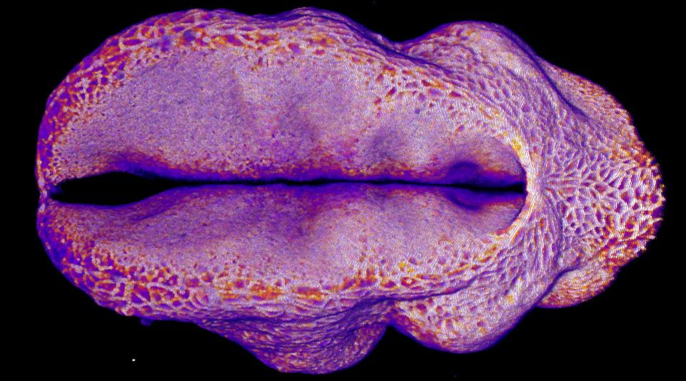
Here’s a link to our new pre-print showing that hindbrain neuropore tissue geometry determines asymmetric cell-mediated closure dynamics. The hindbrain neuropore is a tissue gap over the back of the head which needs to close in order to cover the developing brain with other other cell types. If that does not happen the embryo develops a fatal birth defect called exencephaly (also called anencephaly). Eirini’s work, shown in this pre-print, identifies two different behaviours by which cells around this gap generate mechanical forces needed to close it. Thanks to our collaborations with physicists at Carnegie Mellon, we were able to show that both these behaviours must happen at the same time to describe closure of this gap.

In the image below, the top of the head is on the left, the neck is on the right, and the massive hole between them is the hindbrain neuropore.

Embryonic cells are stressed

By Gabriel Galea, on 7 September 2020

When a mechanical force is applied to an object, that object deforms and withstands physical stress.

Think of applying a force to stretch a rubber band. It stretches and withstands mechanical tension. If you cut it, that tension is relaxed and the rubber band pings back to its preferred length.

If you understand that, you’re well on your way to understanding a central tenant of biomechanics. Each cell in the embryonic neural tube acts like a little rubber band, stretching its neighbours. To work out how much tension a cell is withstanding we have to cut it and measure how far it pings (we call this “recoil”).

GIF showing laser ablation

So how do you cut a cell? Cells are far too tiny to cut them with anything physical like a scalpel. Instead we use a high-powered laser to very precisely cut a cell border. In the image above you will see the laser cut indicated by a red line as the white borders around cells ping apart. These cells should be pulling the neural tube closed, so by cutting them we can infer whether they were doing their job correctly.