Investigating neurovascular coupling in infants with seizures using EEG and DOT
By rmapapg, on 19 October 2017
By Aman Ganglani
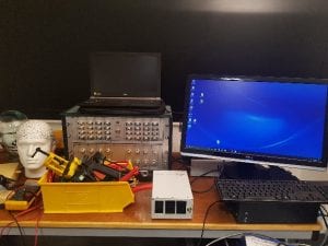
Over the summer I was lucky enough to undertake an eight-week research project in the Biomedical Optics Research Lab (BORL) with the research group focusing on diffuse optical tomography.
Using data acquired from simultaneous electroencephalography (EEG) and diffuse optical tomography (DOT) measurements from the NTS system (developed by Gowerlabs), we looked at the relationship between cerebral blood flow and seizures in neonatal infants with hypoxic ischemic encephalopathy (HIE). This work follows a previous investigation by the group titled ‘Mapping cortical haemodynamics during neonatal seizures using diffuse optical tomography: A case study’ [1]. The data analysed in this study is from the same patient as the previous investigation.
Most of my work was done in Matlab. By employing various statistical tests and by creating my own scripts with the help of the department I further understood the relationship between neonatal seizures and cerebral haemodynamics. Through my own investigation and discussions with the DOT team in formal weekly meetings, I was able uncover patterns and identify further research topics.
A lot of my work was concentrated on the relationship between oxy/deoxy haemoglobin (HbO and HbR respectively) and the spikes on the EEG which indicated seizure like activity. I was able to demonstrate the HbO signal leading the EEG signal through cross correlation tests and visual inspection. I was also able to show evidence of the ‘initial dip’ phenomenon and understood why it remains to be controversial given how it was not seen in every seizure (the ‘initial dip’ phenomenon is observed when there is a dip in HbO prior to a seizure). We also found other time correlations with HbR which warrants further investigation. I ran various other statistical tests such as t-tests to validate my results. Along with this, I also looked at the derivative to investigate if a sudden change in the EEG correlates to a sudden change in haemoglobin levels. Given how I previously found a time lag between the haemoglobin signals and the EEG signal, it came as no surprise that there was no direct correlation without a time lag. I also identified further research topics such as investigating the phase difference between the HbR and HbO signal along with further statistical tests that could be employed on more datasets. All of my code was commented on to specifically allow for other people to continue my work.
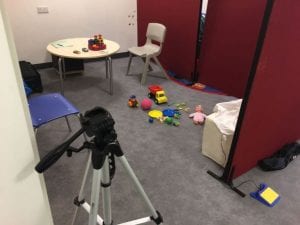
Along with the analysis work I was also able to visit the Evelyn Perinatal Imaging Centre at Rosie hospital in Cambridge. This was where the patients were scanned with EEG and DOT, and I could see how the devices built in the university were tailored to a hospital environment. By then attending meetings with neoLAB. I understood some of the challenges faced by engineers to connect their products with hospital staff. I was also able to do some brief work on image reconstruction which gave me an exciting scope to the future of DOT imaging.
I would like to thank the Engineering department for this amazing opportunity to be a part of this phenomenal project. Everyone at BORL has been incredibly friendly and approachable. A special thanks to Ms Dempsey, Dr Cooper and Dr Hebden for their never-ending support. This opportunity has given me a fantastic insight into the world of research and I look forward to being a part of it.
[1] H.Singh, R.Cooper, C.Lee, L.Dempsey, A.Edwards, S.Brigadoi, D.Airantzis, N.Everdell, A.Michell, D.Holder, J.Hebden, T.Austin. (2014). Mapping cortical haemodynamics during neonatal seizures using diffuse optical tomography: A case study. NeuroImage: Clinical. 5
 Close
Close


