X Close

Events

Home

UCL events news and reviews

Menu

Quantum Fruit – Behind the Scenes at Cheltenham – Day 2/6

By Nicholas Powell, on 14 June 2012

MRI Fruit and Veg

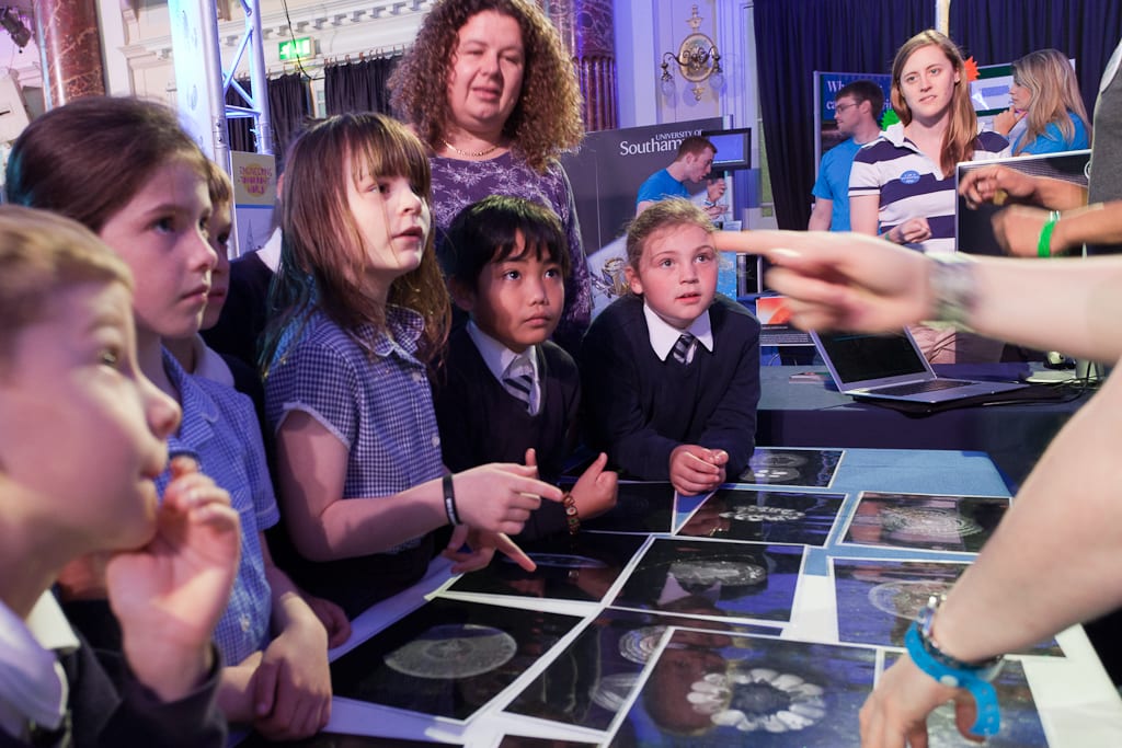
Children guess the MRI-scanned fruit and veg

Behind the Scences at Cheltenham is a daily blog from the UCL CABI team at Cheltenham Science Festival. Every day a member of the team will be talking about their experiences of running a stand.

Day 1 | Day 2 | Day 3 | Day 4 | Day 5 | Day 6

Mark Lythgoe, festival and much-loved CABI director, has been on the hunt for pig testicles all day.

For scanning, you understand.

As briefly mentioned in yesterday’s post, the centre of our stand here in the colourful heart of Cheltenham’s Town Hall is occupied by a tabletop 1 Tesla, 1.5 tonne MRI scanner, prevented from falling through the floor and sinking towards Earth’s core by a strong scaffolding platform.

(more…)

Tears and Water Protons – Behind the Scenes at Cheltenham – Day 1/6

By Thomas A Roberts, on 13 June 2012

CABI at Cheltenham

Katy Ordidge demystifies the physics of MRI

Behind the Scences at Cheltenham is a daily blog from the UCL CABI team at Cheltenham Science Festival. Every day a member of the team will be talking about their experiences of running a stand.

Day 1 | Day 2 | Day 3 | Day 4 | Day 5 | Day 6 |

Two hours into my first shift at Cheltenham and I’d already made one child cry. Not quite the start I’d anticipated.

My colleagues and I from the Centre for Advanced Biomedical Imaging (CABI for short) are at the Cheltenham Science Festival to demonstrate how magnetic resonance imaging (MRI) scanners work and why they are important.

Our “MRI: Can You Scan It?” stand is pitched in the centre of the Discover Zone inside the grand Cheltenham Town Hall. The main draw is a tabletop – in the loosest sense of the word – 1 Tesla Bruker MRI scanner which people can feed with fruit and other small objects to produce live images. Accompanying this are a couple of presentations explaining how MRI works and how it can be applied to imaging your brain.

I was in charge of talking through the latter slideshow and explaining the basics of fMRI: functional magnetic resonance imaging. You’ve probably seen fMRI images in the papers: regions of the brain ‘light up’ in response to a stimulus such as touch, smell or sight. The waterworks incident occurred when I was demonstrating one region of the brain we can probe, using the video below of a dead snake.

(more…)