X Close

Researchers in Museums

Home

Engaging the public with research & collections

Menu

Archive for the 'Uncategorized' Category

Why are animals 3D?

By ucbtch1, on 23 November 2017


Have you heard that our body is mainly composed of 70% water? Although true, the percentage varies from 55% of water in adult women, all the way up to 78% in babies, with the percentage for adult men somewhere in between. This is also true for animals, where some — like the jellyfish — have even 90% of their body composed of water. With this in mind, why don’t animals, including us, look like a soup? How can animals have a defined 3D structure?

Aurelia aurita, moon jellyfish, 5 preserved specimens (C193)

Aurelia aurita, moon jellyfish, 5 preserved specimens (C193)


Animals are made out of cells, the building blocks of our organs and tissues. But cells are basically a bag of water and chemicals; so again, why don’t animals look like giant bags of chemicals? The most obvious reason is that animals have bones that give structure to the rest of the body. But even bones are 31% water, and organs with no bones, such as hearts, still have a unique 3D form. Hearts have defined chambers (see the elephant heart below); they’re not just a mush of cells. The answer lies not in the cells themselves but in what surrounds them.

Elephant dried heart (Z639)

Elephant dried heart (Z639)

Cells are engulfed by the extracellular matrix (ECM) which is mainly composed of proteins. This matrix encompasses the space in-between cells, gives them structural support and acts like a scaffold. It can also act as a pathway for cells to migrate along and it gives out chemical and physical cues that cells respond to. The ECM varies from organ to organ. The brain, for example, is mainly composed of cells with an ECM of only 20% of the total mass. In contrast, cartilage has fewer cells and around 70% of its mass is ECM. Every cell type is surrounded by a specific matrix that will affect its function. Studying this extracellular environment is important to understand how cells develop, how they interact with each other, and how they react to disease.

At the same time, by studying the ECM, researchers can get an idea of how an organ or tissue is structured and how to replicate its intricate architecture. Scientists that work in tissue engineering use a technique which consists of washing away the cells of an organ, literally. By using detergents, the cells are washed away in cycles until just the extracellular matrix is left. In this manner, they can analyse its composition and experiment with the matrix with the end goal of growing an organ in the lab. Therefore, one day we could replace diseased or aged organs with new ones without the need for transplantation. The unique composition of the ECM provides cells with the support they need to survive, and at the same time, gives animals and their organs a defined 3D structure.


Sources:

https://water.usgs.gov/edu/propertyyou.html

Make a Museum about Pokémon!

By Josie Mills, on 21 November 2017

Post-it Note found on the visitor feedback board in the Petrie Museum of Egyptian Archaeology (if this was left by you - do get in touch!)

Post-it Note found on the visitor feedback board in the Petrie Museum of Egyptian Archaeology (if this was left by you – get in touch!)


While working in the Petrie Museum last week I glanced at the board reserved for visitor feedback, noticed this post-it and couldn’t resist taking a photo… As a child of the nineties I have a soft spot for Pokémon and have wholeheartedly embraced revival of the plucky little critters. I’ve particularly enjoyed the nostalgia of sharing the new games with my younger brother, Daniel; if only there were a degree in poké-studies!

Last year the app Pokémon Go made headlines world-wide and the presence of the virtual creatures in museums and archives was widely discussed within the heritage industry. Perhaps unsurprisingly, I’m team positive for Pokémon Go in museums, and the staff at UCL Museums have written some great blog posts on the subject. See this post by Grant Museum Manager Jack Ashby and Research Engager Arendse’s blog.


A wild Pigeotto appears as I am doing my PXRF analysis (Image: J Mills 2016)

A wild Pigeotto appears as I am doing my PXRF analysis (Image: Author’s own photo)


In fact, museums, archaeological ruins, and science labs also feature in the Pokémon games, serving as venues to transform fossils found in the wild into rare Pokémon. These Pokémon, like those mentioned in Arendse’s post, were inspired by real fossils, for example Omanyte from ammonites, and Aerodactyl from pterodactyl. My personal favourite is Relicanth, a fish bearing a close resemblance to the Lazarus species the coeleocanth!


Ammonite

Left: Omanyte (Image: Bulbapedia; Right: Ammonite (Image: British Geological Survey)


Omanyte and Ammonite

Here you can see the similarities between the shell of the Pokémon and the preserved ammonite Mantelliceras, which lived during the Late Cretaceous/Cenomanian period around 100 million years ago. This was a time when much of the Chalk in England formed and Cretaceous ammonite fossils are common in chalky areas of the South Coast. Interestingly fossil ammonites are often displayed ‘upside down’ (effectively with their head in the air!) whereas the Pokémon are orientated to move with their tentacles at the base, more like the original creatures.


Top: Aerodactyl (Image: https://bulbapedia.bulbagarden.net/wiki/Aerodactyl_(Pok%C3%A9mon ); Bottom: Pterodactyl (Image: http://dinosaurpictures.org/Pterodactyl-pictures)

Top: Aerodactyl (Image: Bulbapedia); Bottom: Pterodactyl (Image: Dinosaur Pictures)


Aerodactyl and Pterodactyl

Perhaps the most literal translation of the three are the obvious similarities between the Aerodactyl Pokémon and the extinct Pterodactyl, which lived during the Jurassic period 200-150 million years ago. The fossils on the game are rare and can only be found in certain areas; similarly pterodactyl fossils are also localised in real life, with most excavated from the Solnhofen limestone in Germany.

Top: Relicanth (Image: https://vignette3.wikia.nocookie.net/pokemon/images/a/ad/369Relicanth_AG_anime.png/revision/latest?cb=20141006041759); Bottom: Coelacanth (Image: http://vertebrates.si.edu/fishes/coelacanth/coelacanth_wider.html)

Top: Relicanth (Image: Bulbapedia); Bottom: Coelacanth (Image: Smithsonian)


Relicanth and Coelacanth

Even Relicanth’s name nods to the antiquity and mysterious-ness of the real-life fish the Coelacanth. The Coelacanth is a ‘Lazarus’ species, a term that refers to a taxon that was thought to be extinct but reappears again in the wild. The cryptic nature of the Coelacanth is reflected by the camouflage cartoon pattern of the Pokémon, a graphic allegory of the fish’s complex history!

Although the Pokémon hype has gradually dwindled, perhaps this message from our younger audience (or any nineties kids out there), highlights what we can take from Pokémon: the appeal of learning about animals and artefacts, the surprise of finding new things where you didn’t expect them, and the lure of encountering the rare and interesting.

More practically, specimens that have inspired both fossils and non-fossil Pokémon are on display across the Grant Museum. However, if anybody is interested in funding a museum solely about Pokémon, please get in touch: I know someone who might be suitable for curator *cough*…

How to visualize the insides of an animal?

By ucbtch1, on 26 October 2017

When studying animals, sometimes we need to study them from the inside out —literally. One way to do this is to cut them open and looking at their internal structures, such as with the bisected heads or the microscope slides in the Grant Museum of Zoology. Another way to visualize the inside of an animal is to stain a particular body part while making everything else clear; researchers can do this by using chemicals and colour stains. For example, in the Grant Museum of Zoology, we can find specimens like the tarsier, with its skeleton stained in red, or the zebrafish with red and blue parts.

Adult tarsier stained with Alizarin Red to show calcium (Z2718)

Adult tarsier stained with Alizarin Red to show calcium (Z2718)

Adult zebrafish stained with Alcian Blue and Alizarin Red (V1550)

Adult zebrafish stained with Alcian Blue and Alizarin Red (V1550)


The process of staining these animals begins with the removal of the skin, viscera and fat tissue.  Then, soft tissues like muscle are cleared using a variety of different methods which mostly involve exposing the specimen to different baths of chemicals. Next, the bones are stained with Alizarin Red and the cartilage with Alcian Blue. It’s a long process that can take a couple of days because the stain needs to properly penetrate the tissues, but the results are amazing.

Initially, both Alizarin Red and Alcian Blue were used as textile dyes, but now they also have numerous biological applications. Alizarin Red staining is a method to visualize mineralized tissue because it stains calcium and Alcian Blue stains specific structures mainly found in cartilage. These stains constitute an important part of research because they allow researchers to visualize the intricate structure of tissues and thus understand how they form throughout development.

ADSCs

Image credit: Eleonora Zucchelli

In the lab where I study, researchers work with adipose (fat) derived stem cells which have the capacity to become different kinds of mature cells. These stem cells are grown under specific conditions and by changing these conditions scientists can direct them into becoming mature cells like fat, bone or cartilage — a process called differentiation. But this process can take anywhere from a couple of weeks up to a couple of months! In order to determine if the differentiation is working, researchers stain the stem cells with Alizarin Red and Alcian Blue to identify if they are in fact turning into bone or cartilage. In the images depicted, undifferentiated adipose derived stem cells (ADSCs) on the top appear clear but their differentiated counterparts are stained in blue or red. This means the differentiation is working.

There are many other stains used on animals or cells. The process of clearing and staining can be very complicated depending on the specimen and what one wishes to stain, but the results can be quite fascinating. What animal would you like to see stained from the inside.


Mouse stained with alizarin red (Z3155)

Mouse stained with alizarin red (Z3155)


References:

PUCHTLER, H., Meloan, S. N., & TERRY, M. S. (1969). On the history and mechanism of alizarin and alizarin red S stains for calcium. Journal of Histochemistry & Cytochemistry17(2), 110-124.

McLeod, M. J. (1980). Differential staining of cartilage and bone in whole mouse fetuses by alcian blue and alizarin red S. Teratology22(3), 299-301.


Question of the Week: What’s this Museum For?

By Hannah L Wills, on 19 October 2017

By Hannah Wills


A couple of weeks ago, whilst engaging in the Grant Museum, I started talking to some secondary school students on a group visit to the museum. During their visit, the students had been asked to think about a number of questions, one of which was “what is the purpose of this museum?” When asked by some of the students, I started by telling them a little about the history of the museum, why the collection had been assembled, and how visitors and members of UCL use the museum today. As we continued chatting, I started to think about the question in more detail. How did visitors experience the role of museums in the past? How do museums themselves understand their role in today’s world? What could museums be in the future? It was only during our discussion that I realised quite how big this question was, and it is one I have continued to think about since.

What are UCL museums for?

The Grant Museum, in a similar way to both the Petrie and Art Museums, was founded in 1828 as a teaching collection. Named after Robert Grant, the first professor of zoology and comparative anatomy at UCL, the collection was originally assembled in order to teach students. Today, the museum is the last surviving university zoological museum in London, and is still used as a teaching resource, alongside being a public museum. As well as finding classes of biology and zoology students in the museum, you’re also likely to encounter artists, historians and students from a variety of other disciplines, using the museum as a place to get inspiration and to encounter new ideas. Alongside their roles as spaces for teaching and learning, UCL museums are also places for conversation, comedy, film screenings and interactive workshops — a whole host of activities that might not have taken place when these museums were first created. As student engagers, we are part of this process, bringing our own research, from a variety of disciplines not all naturally associated with the content of each of the museums, into the museum space.


A Murder-Mystery Night at the Grant Museum (Image credit: Grant Museum / Matt Clayton)

A Murder-Mystery Night at the Grant Museum (Image credit: Grant Museum / Matt Clayton)


What was the role of museums in the past?

Taking a look at the seventeenth and eighteenth-century roots of the Ashmolean Museum in Oxford and the British Museum in London, it is possible to see how markedly the role and function of the museum has changed over time. These museums were originally only open to elite visitors. The 1697 statues of the Ashmolean Museum required that ‘Every Person’ wishing to see the museum pay ‘Six Pence… for the Space of One Hour’.[i] In its early days, the British Museum was only open to the public on weekdays at restricted times, effectively excluding anyone except the leisured upper classes from attending.[ii]

Another feature of these early museums was the ubiquity of the sense of touch within the visitor experience, as revealed in contemporary visitor accounts. The role of these early museums was to serve as a place for learning about objects and the world through sensory experience, something that, although present in museum activities including handling workshops, tactile displays, and projects such as ‘Heritage in Hospitals’, is not typically associated with the modern visitor experience. Zacharias Conrad von Uffenbach (1683-1784), a distinguished German collector, recorded his visit to Oxford in 1710, and his handling of a range of museum specimens. Of his interactions with a Turkish goat specimen, Uffenbach wrote, ‘it is very large, yellowish-white, with… crinkled hair… as soft as silk’.[iii] As Constance Classen has argued, the early museum experience resembled that of the private ‘house tour’, where the museum keeper, assuming the role of the ‘gracious host’, was expected to offer objects up to be touched, with the elite visitor showing polite and learned interest by handling the proffered objects.[iv]

Aristocratic visitors handle objects and books in a Dutch cabinet of curiosities, Levinus Vincent, Illustration from the book, Wondertooneel der Nature - a Cabinet of Curiosities or Wunderkammern in Holland. c. 1706-1715 (Image credit: Universities of Strasbourg)

Aristocratic visitors handle objects and books in a Dutch cabinet of curiosities, Levinus Vincent, Illustration from the book, Wondertooneel der Nature – a Cabinet of Curiosities or Wunderkammern in Holland. c. 1706-1715 (Image credit: Universities of Strasbourg)


How do museums think about their function today?

In understanding how museums think about their role in the present, it can be useful to examine the kind of language museums employ when describing visitor experiences. The British Museum regularly publishes exhibition evaluation reports on its website, detailing visitor attendance, identity, motivation and experience. These reports are fascinating, particularly in the way they classify different visitor types and motivations for visiting a museum. Visitor motivations are broken down into four categories: ‘Spiritual’, ‘Emotional’, ‘Intellectual’ and ‘Social’, with each connected to a different type of museum function.[v]

Those who are driven by spiritual motivations are described as seeing the museum as a Church — a place ‘to escape and recharge, food for the soul’. Those motivated by emotion are understood as searching for ‘Ambience, deep sensory and intellectual experience’, the role of the museum being described as akin to that of a spa. For the intellectually motivated, the museum’s role is conceptualised as that of an archive, a place to develop knowledge and conduct a ‘journey of discovery’. For social visitors, the museum is an attraction, an ‘enjoyable place to spend time’ where facilitates, services and welcoming staff improve the experience. Visitors are by no means homogenous, their unique needs and expectations varying between every visit they make, as the Museum’s surveys point out. Nevertheless, the language of these motivations reveals how museum professionals and evaluation experts envisage the role of the modern museum, a place which serves multiple functions in line with what a visitor might expect to gain from the time they spend there.

What will the museum of the future be like?

In an article published in Frieze magazine a couple of years ago, Sam Thorne, director of Nottingham Contemporary, invited a group of curators to share their visions on the future of museums. Responses ranged from the notion of the museum as a ‘necessary sanctuary for the freedom of ideas’, to more dystopian fears of increased corporate funding and the museum as a ‘business’.[vi] These ways of approaching the role of the museum are by no means exclusive; there are countless other ways that museums have been used, can be used, and may be used in the future. My thinking after the conversation I had in the Grant Museum focussed on my own research and experience with museums, but this is a discussion that can and should be had by everyone — those who work in museums, those who go to museums, and those who might never have visited a museum before.


What do you think a museum is for? Tweet us @ResearchEngager or come and find us in the UCL museums and carry on the discussion!


References:

[i] R. F. Ovenell, The Ashmolean Museum 1683-1894 (Oxford: Clarendon Press, 1986), 87.

[ii] Fiona Candlin has written on the class politics of early museums, in “Museums, Modernity and the Class Politics of Touching Objects,” in Touch in Museums: Policy and Practice in Object Handling, ed. Helen Chatterjee, et al. (Oxford: Berg, 2008).

[iii] Zacharias Konrad von Uffenbach, Oxford in 1710: From the Travels of Zacharias Conrad von Uffenbach, trans. W. H. Quarrell and W. J. C. Quarrell (Oxford: Blackwell, 1928), 28.

[iv] Constance Classen, “Touch in the Museum,” in The Book of Touch, ed. Constance Classen (Oxford Berg, 2005), 275.

[v] For this post I took a look at ‘More than mummies A summative report of Egypt: faith after the pharaohs at the British Museum May 2016’, Appendix A: Understanding motivations, 27.

[vi] Sam Thorne, “What is the Future of the Museum?” Frieze 175, (2015), accessed online.

Embryological Wax Models

By ucbtch1, on 14 September 2017

The Grant Museum has a number of embryological wax models on display (Images 1, 2, 4, and 5 amongst others not shown here). These models, while often ignored by visitors, are actually quite remarkable as they showcase the brilliance and mystery of embryological development. They were created to help elucidate essential questions like: how do humans and other animals form? How are a bunch of seemingly insignificant cells, with no shape other than a ball, able to grow so much and in such detail to form intricate patterns like our eyes? How can one cell transform itself into such different tissues, from hard rock bone to the jelly like liver? In order to understand how a human body is formed it is vital to study the very first stages of its creation, i.e. when we are just a bunch of cells.

Image 1. Placental mammal embryo (Z3100)

Image 1. Placental mammal embryo (Z3100)

The very first cells that are formed after fertilization are called stem cells and they start with an unlimited capacity to form any type of cells. With time, they start to differentiate and mature into specialized cells with a limited lifetime. In this process, little by little they lose their unlimited capacity until they can only form cells that are similar in lineage. In this manner, totipotent stem cells can form any body part including extraembryonic tissue like the placenta. On the next level, pluripotent stem cells (embryonic stem cells for example) are capable of forming any body part but have lost the capacity to form extraembryonic tissue. And finally, multipotent stem cells, much more restricted, can only form cells from a specific tissue or organ.

This may appear as a straightforward process, but the development of an animal is a deeply specific, delicate, and sophisticated interplay of signals and coordinated transitions. Think of it as an orchestrated dance of on and off switches leading to specialisation and exponential growth. In fact, it is so complex that we still don’t understand it entirely.

Although not all human, the wax models display the first stages of development of vertebrates and closely related animals. First, one cell divides symmetrically into two, then four, then eight and so on (Image 1 and front models of Image 2). Afterwards, cells start organizing themselves answering to chemical and physical signals and different patterns start to appear (Image 2 models in the back). Eventually, an axis emerges on which cells migrate along which will give rise to the head on one side and the body and limbs on the other (Image 4). 

Slowly but surely, we all go from looking like little worms to fully grown animals (Image 3). It is important to note that in this initial period most embryos have a very similar appearance, at least between vertebrates. These similarities tell us that a lot of the genes that govern this initial growth haven’t changed between species over time. It’s like nature is saying, “well, if it ain’t broke don’t fix it” and so these mechanisms have been conserved in different animals.

Image 2. Branchiostoma sp, Lancelet Embryo (fish-like invertebrate; T114)

Image 2. Branchiostoma sp, Lancelet Embryo (fish-like invertebrate; T114)

These early stages are crucial moments because if one little element of the spatial/temporal organization is out of place, improper organization can lead to lifelong malformations, diseases, or even the termination of the embryo. Hence the importance of understanding how this process works. We know that even in adult life, there are still stem cells proliferating and forming new tissue to a certain degree. Some organs, like the skin, have a lot of stem cells to replace old cells when they die or get injured. But other organs like the brain, have a very limited capacity to grow new cells—one of the reasons why a brain is much more difficult to fix.

Image 3. Development of the external form of the human face (LDUCZ-Z480) and development of external form of human embryo (LDUCZ-Z430)

Image 3. Development of the external form of the human face (LDUCZ-Z480) and development of external form of human embryo (LDUCZ-Z430)

Image 4. Vertebrate embryos. Image taken from https://www.ncbi.nlm.nih.gov/books/NBK9974/

Image 4. Vertebrate embryos. Image taken from https://www.ncbi.nlm.nih.gov/books/NBK9974/

So can we get back all the limitless capacity there once was in the developing embryo? Even though the genetic and molecular mechanisms governing all these changes are still somewhat elusive, researchers are using stem cells and powerful genetic tools to answer this question and decipher every single step of how a human is formed in the womb. Moreover, if we can understand the process, then we can recreate and modify it in the lab, and this is exactly what the field of stem cells and regenerative medicine is trying to do. Imagine having the capacity to grow new organs to be used for transplantation or drug testing. How about growing a brand new functioning leg or arm for amputees? Or studying the mechanisms of diseases like Parkinson’s, leukaemia, diabetes, amongst others. The benefits of harnessing the regenerative potential of cells are far-reaching.

Image 5. Development of the external form of the human face (LDUCZ-Z480).

Image 5. Development of the external form of the human face (LDUCZ-Z480).

The exciting field of stem cells and regenerative medicine has come a long way, more than a century has passed from the first time the term stem cells was used in 1906 up until the creation of genetically modified human embryos in 2017. The embryological wax models represent initial efforts of identifying how changes give rise to specific structures and ultimately how an animal comes into existence. Furthermore, the future still holds exciting breakthroughs, there is still a lot to understand about human development and the wax models are a fantastic resource to portray the morphogenetic changes we all once went through.


Resources:

Gilbert SF. Developmental Biology. 6th edition. Sunderland (MA): Sinauer Associates; 2000. Comparative Embryology. Available from: https://www.ncbi.nlm.nih.gov/books/NBK9974/

http://www.labonline.com.au/content/life-science-clinical-diagnostics-instruments/article/why-do-all-animal-embryos-look-the-same–236915402

Question of the Week: Why do brains have wrinkles?

By ucbtch1, on 27 April 2017

The brains displayed at the entrance of the Grant Museum are mostly mammal’s brains but we can observe differences in sizes and in how smooth or wrinkly they are. The folds of a brain are called gyri and the grooves are called sulci. These morphological features are produced by the folding of the cortex, the part of our brain responsible for higher cognitive processes like memories, language and consciousness. During development, all brains start off with a smooth surface and as they grow, gyrification (the development of the gyri and sulci) occurs. It is interesting to note that the major folds are very consistent amongst individuals, meaning that development is similar sometimes even amongst species.

c_ucl_gmz_matt_clayton020

The brain collection on display at the Grant Museum of Zoology (Image credit Grant Museum of Zoology).


It has been assumed that the wrinkles in brains correlate with an animal’s intelligence. The reasoning behind this is that a bigger brain, and hence more neurons, need more space. The folds allow the cortex to increase its area while being packed in a confined space like our cranium. There are several factors and hypothesis of how gyrification occurs. Recently, researchers at Harvard developed a 3D gel model based on MRI (magnetic resonance imaging) images to understand how this process occurs. They found out that it all boils down to the mechanical properties of the cortex. While neuronal cells grow and divide, the increasingly bigger brain leads to a compression of the cortex and to the formation of the folds. The researchers were able to mimic the folds of the cortex and were stunned at how similar their gel model looked to a real human brain.

tumblr_o21mv85Nt01t5fphqo1_1280

Gel model of a foetal brain (Image credit: Mahadevan Lab/Harvard SEAS).


Even though most of the brains on display in the Grant museum have gyri and sulci, in nature, most animals have smooth brains. In general, larger brains have folds while smaller brains do not, even small mammals like rats or mice have smooth brains. In humans, a lissencephalic brain is one without gyri and sulci and is a result of a rare disorder that is characterised by mental abnormalities. From the collection of brains in the Grant Museum, there is only one lissencephalic brain—next time you visit the museum see if you can spot it. Additionally, try to find the brain coral. Because of its intricate maze–like pattern, Diploria labyrinthiformis has very similar ridges and grooves as a brain, and so is referred to as brain coral. Overall, I find looking at brains and their grooves fascinating, each species with their own pattern and each groove in a specific place. Makes me wonder how brain coral gets its patterns.

brain coral 3

Diploria labyrinthiformis also known as brain coral(Grant Museum C1084).


References:

Roth, G. and Dicke, U., 2005. Evolution of the brain and intelligence. Trends in cognitive sciences9(5), pp.250-257.

Ronan, L. and Fletcher, P.C., 2015. From genes to folds: a review of cortical gyrification theory. Brain Structure and Function220(5), pp.2475-2483.

Manger, P.R., Prowse, M., Haagensen, M. and Hemingway, J., 2012. Quantitative analysis of neocortical gyrencephaly in African elephants (Loxodonta africana) and six species of cetaceans: comparison with other mammals. Journal of Comparative Neurology520(11), pp.2430-2439.


3,500-year-old bread and beer from the New Kingdom, Egypt

By tcrngon, on 9 February 2017

The Petrie Museum at University College London contains a vast and varied collection of archaeological artefacts from Egypt, ranging from Palaeolithic stone tools to the first naturalistic mummy portraits from Roman Egypt. Many are also the organic materials at the Petrie Museum, which preserved archaeologically thanks to the dry and arid climate conditions in Egypt. Amongst these, the museum houses some remarkable examples of foods and drinks, such as several bread loaves, fruits and even beer residue recovered during excavations from different archaeological sites in Egypt.

In order to create a direct link with my PhD project on Neolithic cereal foods from Çatalhöyük (Turkey) and to show visitors how cereal foods get preserved archaeologically and how important these were in past societies, I put together a display with the help of Dr. Alice Stevenson (former curator of the Petrie Museum), with some of the best examples kept at the museum (Fig. 1). We chose a loaf of bread from Hatshepsut’s tomb in Deir el Bahri (ca. 1458 BC), beer residue from inside a ceramic vessel and emmer wheat spikelets. Emmer, an ancient crop originated in the Near East, was domesticated 12,000 years ago in Syria as new archaeobotanical research has recently shown (Arranz-Oteagui et al. 2016); and it was also, together with barley, the staple crop in ancient Egypt used by the community on a daily basis.

Bread aIMAG4787nd beer were extremely important in past societies, maybe even more important that they are for us nowadays. Bread, and also beer in ancient Egypt were basic dietary items which were consumed everyday with every meal of the day. First studies on Egyptian bread were carried out by Delwen Samuel (1994), as part of her PhD research at University of Cambridge. During and after her doctoral research, she studied approximately 20 loaves of bread from ancient Egypt, among which there was the loaf of bread on display today at the Petrie Museum. Not only the study of bread is important because we find out about the ingredients which were involved in its preparation, but also because we can learn about the way in which cereals were processed, prepared and how these breads were baked or cooked. Samuel (1994; 2000) explains how in the preparation of these breads, there was a very complicated process involved, from the dehusking of the emmer wheat to the baking of bread:

emmer needs an extensive treatment; when threshed, it breaks into packets called spikelets, each of which is a thick envelope of chaff tightly surrounding two kernels. Vigorous but careful processing is needed to break the chaff apart without damaging the grain kernels, before winnowing and sieving clean the chaff from the kernels”. And she continues: “Whole spikelets were moistened with a little water and pounded with wooden pestles in limestone mortars (…) the damp mixture of freed grain kernels and broken chaff then had to be dried, probably by spreading the mass in the sunshine. This was followed by a series of winnowing steps, which removed the fine chaff, and by sieving, which removed the heavier pieces”.

After this arduous proceIMAG4782ss the grain was finely ready to be ground into flour using saddle querns like those that we can see on display at the Petrie Museum (Fig. 2). Doughs were then baked in bread moulds directly on the embers of the hearths during the Old Kingdom; bread was later baked inside pottery cylinders, on the inner walls, creating a tannur environment. It will not be until IMAG4793the New Kingdom that Egyptians build real bread ovens (tannurs).

Using Scanning Electronic Microscopy (SEM) to analyse the microstructure of these breads, Samuel found out that, although mainly made of emmer flour, some of them also contained barley flour and other foods such as figs and dates. Also, under the SEM, it was possible to see that some yeast granules had preserved the desiccation process and were visible. Although yeast is naturally found in the environment, we do not know when or how, was first used for bread baking and so far Samuel’s evidence for the use of yeast for bread baking by ca. 1400 is still the earliest example of its use. Some archaeologists believe its first discovery was probably accidental and that it was probably connected to the preparation of alcoholic drinks or brewing. For Egypt, this could very well be the case, since beer making is widely documented in ancient Egypt. Many are the decorative depictions about brewing: for example, a group of Old Kingdom statuettes is on display at the Cairo Museum, including a female brewer from the mastaba of Meresankh at Giza (JE66624) and a man working with a pottery vessel from the mastaba of Ptahshepses at Saqqara (Cairo CGII~; Saleh and Sourouzian 1987: nos. 52 and 53 respectively). A variIMAG4867ety of texts also mention bread and beer production. The majority of these are scribal exercises, concerned with converting quantities of grain into loaves and beer of specific strength or quantity. Examples of these exercises can be found in the Rhind Mathematical Papyrus (Peet I 923: 112- 22).IMAG0341

Through the study of desiccated beer residue from vessels contents we can learn about the early brewing activities. According to Samuel (1996), Egyptians might have been using up to four different techniques for brewing, which differ from our modern traditional techniques. Samuel concluded that most likely used sprouted/malted grain for brewing but also unsprouted grain which could have been processed in other ways in order to be brewed, such as moisturising non-germinated grain and then heating it. Certainly, this could explain the many different names for the term “beer” in Egypt and thus the possibility of having different types of beer, each of them with different taste and characteristics.

Sources:

Arranz-Otaegui, A., Ibanez, JJ., and Zapata-Pena, L. 2016. Hunter-gatherer plant use in southwest Asia: the path to agriculture. In Hardy, K. and Kubiak-Martens, L. (eds). Wild Harvest. Plants in the hominin and pre-agrarian human worlds. Oxford: Oxbow books. Pp. 91-110

Samuel, D. 1994. An Archaeological study of Baking and Bread in New Kingdom Egypt. PhD, University of Cambridge. https://www.repository.cam.ac.uk/handle/1810/245007

Samuel, C. 1996. Archaeology of ancient Egyptian beerJournal of the American Society of Brewing Chemists 54:3-12.

Samuel, D. 2000. Brewing and baking in Ancient Egyptian materials and technology. Edited by P. T. Nicholson and I. Shaw, pp. 537-576. Cambridge: Cambridge University Press.

Introducing our new engagers

By Stacy Hackner, on 23 May 2016

We are very lucky to have hired a new cohort of engagers starting this summer! They are all first-year PhD students with specialties in archaeology & heritage, molecular biology, crime science, English literature, bioscience, and history of science. We’re excited to have them on board our team – make sure to visit the Grant and Petrie this summer to hear about their inspiring research. Check out the Bios page to find out more about their work.

Question of the Week:

What is an axolotl and why is it so unique?

By ucbtch1, on 4 May 2016

By Citlali Helenes Gonzalez

From the many specimens that are on exhibit at the Grant Museum of Zoology, it is hard to choose one that stands out. For me, it has to be the axolotl, a small little creature that passes by unnoticeable. It is not the biggest or the strangest but it has a unique feature that we humans desire to achieve: tissue regeneration. Although newts, salamanders and starfish can also regenerate tissue, the axolotl is probably the most interesting of these animals because of the extent of its capacity.

20160210_134444When we humans have an injury, our body starts forming scar tissue over the injury. With just a few exceptions, like the skin that is regenerating constantly and the liver that does something similar to regeneration, the rest of our body has very limited regeneration capacity.

The axolotl, on the other hand, after an injury, the tissue at the wounding site starts to regrow new healthy tissue instead of scar tissue. It does so to the extent that it can regrow a whole limb, jaw, tail, spinal cord and even some parts of its brain. Scientists have even transplanted organs from one axolotl to another with no rejection issues. And if all of this is not enough for you to be amazed, the axolotl is over 1000 times more resistant to cancer than mammals. This is why it has been used as a model animal for the study of regeneration and development.

Studying the axolotl has huge implications for medical research because if we can learn how the mechanisms of regeneration in this little animal work, then maybe we can simulate it in humans. And in the long run, if we learn how to regenerate and repair tissue, this could mean no more need for transplants, no more prosthesis of arms or legs, helping burn victims just to name a few benefits.

Unfortunately, this little animal native to Mexico has been listed as an endangered species due to the destruction of its natural habitat in the lakes and canals of Mexico City. But scientists across the globe are still studying them and looking closer into their genome to try to unlock the secrets of tissue regeneration. So next time you stop by the Grant Museum take a closer look at this fascinating animal that even though its small size has a lot to offer to human medical research.


Further reading:

Roy, S. and Lévesque, M., 2006. Limb regeneration in axolotl: is it superhealing? The Scientific World Journal6, pp.12-25.

http://blogs.scientificamerican.com/guest-blog/regeneration-the-axolotl-story/


“Strange Creatures” – Reflections – Part One

By uclznsr, on 25 June 2015

Strange Creatures: The Art of Unknown Animals — Reflections: Part One

As the Grant Museum’s blockbuster exhibition, ‘Strange Creatures: The Art of Unknown Animals,’ draws to a close this Saturday, I reflect, in the first of two blogposts, on the provocations and implications of this thought-provoking, juxtapositional display of scientific and creative artefacts. 

Glass, despite its transparency, is a singularly opaque and contradictory substance. Neither entirely liquid nor completely solid, glass, at super-heated temperatures can take on any form. However, at room temperature, despite its unyielding firmness, it is considered too chemically disordered to be considered a solid, as such. In technical terms, it is called, somewhat paradoxically, an ’amorphous solid’.

A further paradox: Glass is both naturally occurring and manufactured by humans. Midnight-black obsidian is created by the rapid cooling of silica, first made deliquescent by exposure to volcanic lava; the manufacture of human-made glass follows the same basic pattern. It is both geologically ancient and surprisingly modern; prehistoric humans utilised and greatly valued obsidian for its sharp edges, while, human-produced glass dates only as far back as the third millennium BC.

A consideration of the history of its use and manufacture in human society presents us with further contradictions. Used first for decorative purposes by Mesopotamians and in Ancient Egypt, in Ancient Rome, glass became a practical material, used for bottles, vessels, pitchers, cups, plates, goblets, and other household objects. Later on in the human story, glass would be a catalyst for scientific discovery and scholarship – glass lenses not only enabled ageing scholars to work past the onset of myopia, but acted as an enhancement to human vision. That illumination and enlightenment should be the dominant metaphor of scientific discovery as well as a central theme in Western religious discourse is no surprise then, given that glass is the material threshold that opens out onto both these worlds. The stained glass windows of European churches and cathedrals cast brilliant, coloured light upon biblical stories and the lives of saints as well as the people that stir within them, while glass lenses allow us to see far into the heavens and explore the microscopic mysteries of biological life.

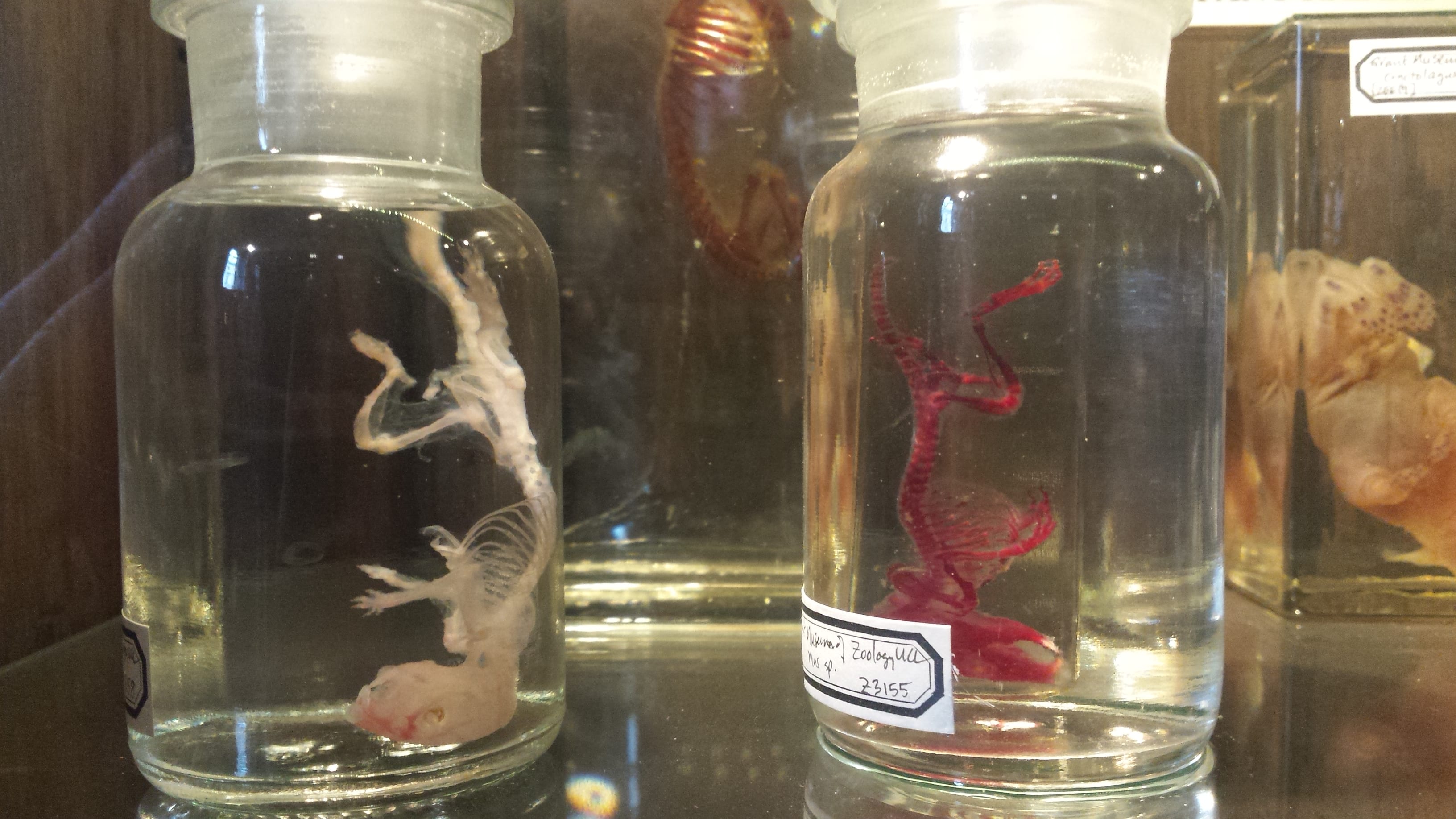
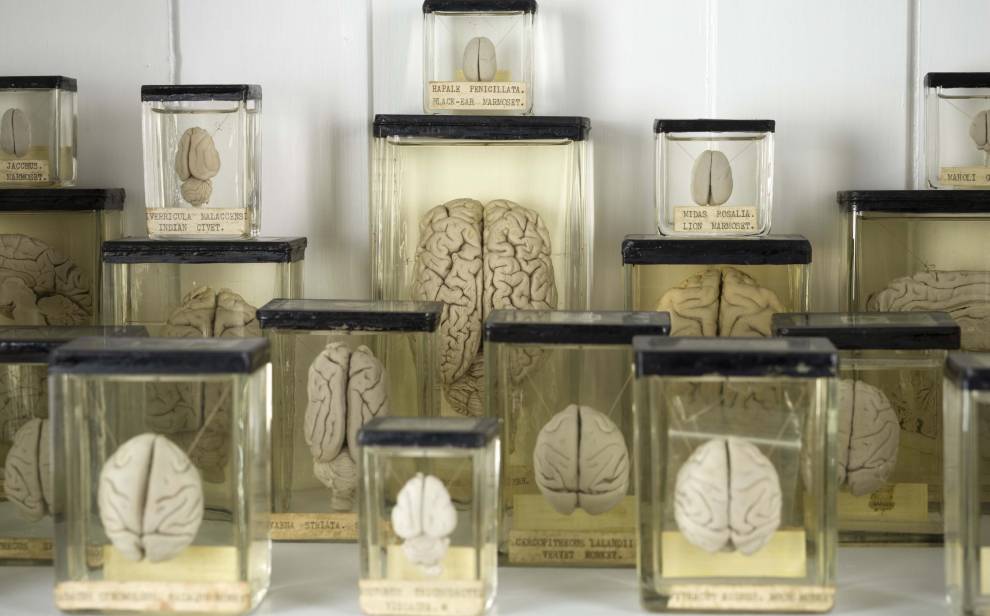
Enter the Grant Museum of Zoology and Comparative Anatomy and one begins to see how the paradoxes of this material pervade biological science and, perhaps more significantly, the way in which we view it. Glass cabinets store a panoply of jars, in which colourless bags of flesh float silently in a nameless liquid, suspended in time. Transparent panes function as windows through which the spectacle of science can be viewed as well as the boundary by which it is ensured we remain spectators and not fumbling participants. The method of liquid preservation preferred by the nineteenth-century biologists and anatomists that used the collection in the Grant Museum is made possible by the unique properties of glass which allows them to store specimens in a sterile vessel as well as view the specimen contained within. This not only provides a double, tangible, scientific benefit – storage and visibility – but also makes possible the extravagant and morbid theatricality of the museum, something often considered antithetical to the staid objectivity of scientific truth.

By the entrance to the Grant Museum, adjacent to a collection of preserved brains, there sits a group of organisms encased in glass whose vitreous appearance seems distinctly unreal or unnatural. Attend to the object labels, and it becomes clear that, in fact, this unreality is no mere appearance, for these specimens are not merely sitting behind glass panes and stored in glass jars, but are themselves made from glass.


Synapta Maculata – Courtesy of Grant Museum


The objects in question are an otherworldly assembly of Blaschka models, replica versions of marine animals such as jellyfish, sea-slugs, and anemones. Their singularity as objects derives from the delicate, alien beauty of the animals of which they are simulacra, as much as it does from the material out of which they are made. The Blaschka family, renowned 19th-century Bohemian glass blowers, were in high demand for their ability to create apparently accurate models of marine invertebrates, the translucent, diaphanous flesh of which was difficult to preserve at the time. More striking still than the evocative, glassy fragility of these model animals is their seeming uncanny indistinguishability from the organisms of which they are merely representations.

The paradox of glass, then, is embodied in the very objects themselves which hover in the uncertain, amorphous zone between nature and art, by presenting themselves as the handicraft of evolutionary creation and and not the invention of and monuments to human artifice. For many visitors, it is difficult, if not impossible, to differentiate “real” specimens from these glass models without the aid of an object label or the keen observational skill of a zoologist. In another cabinet, a glass sea anemone, another Blaschka model, sits inconspicuously alongside a host of preserved anemones, jellyfish, and other invertebrates – very few people notice anything untoward or fake. It is only when visitors’ attention is drawn to the fact that they are, in fact, ‘false’ approximations of animals that these objects engender the awe they deserve. The moment their unreality or falseness is revealed is a sort of “lightbulb moment” for visitors, a drawing back of the veil to expose the secrets of their illusory nature. There is a titillation, as in the theatre or at a magic show, when we are confronted by our own suspension of disbelief, when that which presents itself as real reveals itself to be staged, drawing the spectator out of the comfortable – yet knowingly false – sense of belief that makes the spectacle itself possible.

Limax arborum – Courtesy of Grant Museum

And yet, for the multitude of museum-goers that fail to notice the object labels or who simply find the spectacle of these models sufficient for otherwise-detained powers of scrutiny, these simulacra are, in effect, real. In this sense, they work as well as genuine organic specimens, the function of which is simply to present itself to the eye of the viewer — to produce a spectacle of science. Should this state of affairs concern us? The supposedly objective truths of science recede into the shadows in view of the much brighter light of the artificial, man-made, and subjective truths of ‘art’ and the theatricality of the museum. What are the implications of this? And is the duty of the museum as such to make clear what is real and what is not?

Many specimens and objects in the Grant Museum pose similar questions: To what extent can deliberately posed taxidermic specimens offer us anything approaching an objective representation of an animal? Do plaster casts of ancient fossils adequately represent their real correlatives in other museums? Do models, originally made for teaching, belong in a museum that presents itself a museum of Zoology and Comparative Anatomy (and not a museum of the practice of these now historical, and in the case of the latter, defunct, disciplines)? And what, if anything, is the significance of these scientific models being made from glass, the very material that in microscopes lenses and slides, petri dishes, and storage jars, makes possible the practice and spectacle of contemporary biological science in the first place?

The philosophical, practical, and aesthetic puzzles the Blaschka models raise are a central component of the Grant Museum’s current – and hugely successful – exhibition, Strange Creatures: The Art of Unknown Animals. The exhibition is intended to explore the history and practice of representing previously unknown or exotic animals and the implications this practice has for art, science, museums, and the practice of representation as such. A number of Blaschka models are included in the exhibition, as are a number of fascinating work from UCL’s other collections.

16thC Animals: Allegory, Art, and Authenticity 

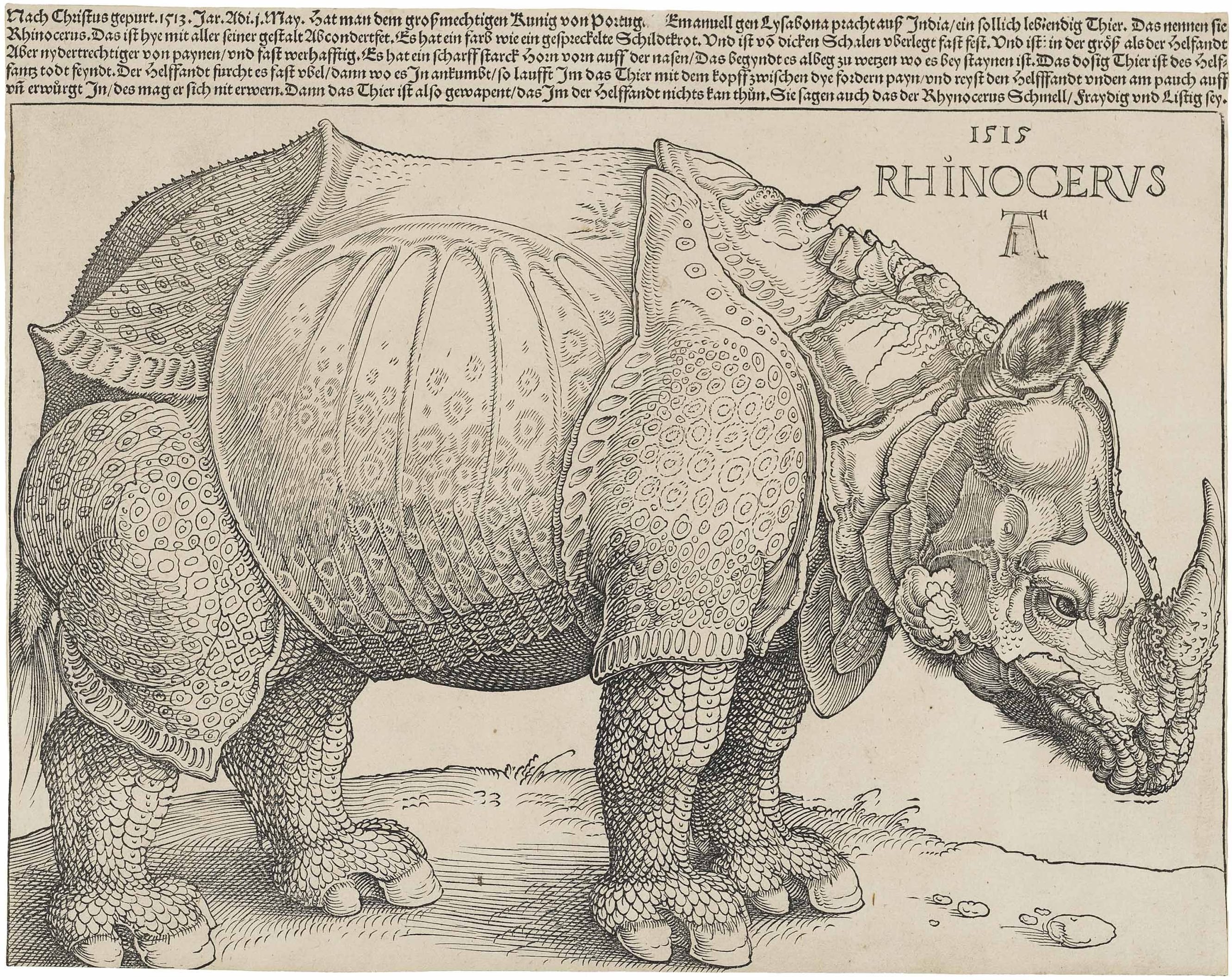
The influence of the 16th-century German artist, Albrecht Dürer, looms large in this exhibition. Two works in this exhibition, both from the 16th century are derived or copied from his work. One, an engraving by an anonymous artist based on Dürer’s St Jerome in his Study includes an anatomically errant lion. The second, an engraving of a rhinoceros, based on a hugely influential woodcut by Dürer of the same animal from 1515, shows this animal to be heavily armoured, covered in scales, and in possession of an additional horn at the top of its spine. Dürer’s original work was not based on any real life encounter with the animal, but on an account and a sketch produced by those who witnessed an Indian rhinoceros at the Lisbon court of King Manuel I.

Dürer's Rhinoceros - 1515

Dürer’s Rhinoceros – 1515

Such was its influence on 16th century image-making that it produced an entire genre of rhinoceros representations and it is in this genre that the engraving in the Grant Museum exists. It would not be until the 18th century that Durer’s rhinoceros would be replaced by more anatomically accurate versions, supplanting the cultural dominance of the partially mythical armoured version that dominated the European imaginary. However, this particular representation and its progeny continued to occupy an exalted place in the West’s vision of the East and its exotic animals, apparently appearing in textbooks until the 1930s.

It is the former Durer-derived work, however, the engraving of St Jerome working in his study, that arguably offers the visitor a more engaging opportunity to meditate on the relation between art and nature. If Durer’s rhinoceros embodies the way in which artistic and scientific representations of animals – accurate or not – can exert an influence for hundreds of years on the scientific and cultural imagination, the copied reverse engraving of St Jerome in his Study dramatises a complex interaction between the representation of the natural world, allegory, and the European and Christian mythos.

St Jerome in his Study - 1514

St Jerome in his Study – 1514

The accuracy or provenance of the representation of the lion represented in this work, which appears strangely misshapen to the contemporary eye, is subordinate in this work to the numerous objects of allegorical import that surround Jerome as he bends dutifully over his bible. A cross sitting on his desk, an image of sacrifice and resurrection, is located at the midpoint between Jerome’s lowered head and a skull, a memento mori, which, sitting in the windowsill, is a reminder of finiteness of life (“remember [that you have] to die”). There is an uncanny resonance here between the deliberate allegory of this skull and the actual crania that inhabit almost every corner of the museum. In this light, the Grant Museum takes on the feeling of an overwhelming, repetitive chronicle of death and a chilling reminder of our own mortality, even as we walk amidst this multitude of animals and consider the abundant and varied nature of life. Elsewhere in this engraving, an hourglass symbolises the inexorable passage of time and a gourd hanging from the ceiling recalls a now obscure scholarly debate between Jerome and St Augustine over the translation of the Greek term kikayon (Augustine interprets this as ‘gourd’ whereas Jerome translates it as ‘ivy’), the reference to which is assumed to reflect Jerome’s dedicated, scholarly character.

The lion is itself suffused with allegorical significance. Jerome is said to have pulled a thorn from the lion’s paw and, in doing so, taming it and in this work it lies peacefully and obediently by his desk, as does a dog – itself a symbol of loyalty. Thus, while the rhinoceros offers us an exemplar of the way in which inaccurate representation can come to construct (cultural) reality, St Jerome shows us how scientific accuracy in an artwork can be less important than its allegorical, symbolic, or referential significance. It becomes difficult not to view the skeletons of lions and dogs, as well as the multitude of animal memento mori in the Grant Museum, less as objects of science, but as frames over which we drape allegorical and unconscious meaning.

Michel Foucault, in an essay on authorship and authenticity, reminds us that it is with St Jerome that the scholarly practice of seeking the authorial provenance of a text originates. Jerome, we are told, pioneered certain techniques for confirming the authenticity of a work by identifying certain traits within it that could tie to a particular author. Such desire for confirmation and authenticity is made difficult in these engravings, not only because they are themselves copies of older works, but because their ostensibly inaccurate representations of animals exert such an influence on our culture that their scientific “authenticity” is no longer relevant. The importance of St Jerome’s task in seeking out authorial authenticity was to confirm the value of the text and to affirm its “truth”. To dismiss the value and truth of Dürer’s Rhinoceros or St Jerome because of an assumed inauthenticity and inaccuracy is to miss entirely the point of this exhibition: art, science, indeed representation, incorrect or otherwise, participate in the authorship of our reality.

Cuddly Toys, Plastic Dinosaurs, and Commercial Interests

The exhibition, however, is not entirely taken up with implicit invitations to consider human mortality or meditate on the origin of Western exegetical practice. In another part of the museum, a teddy bear looks out from one case, a representation of a recently discovered mammal called the Olinguito. Here, we are invited to consider the way in which media representations and marketing ploys can distort the import of scientific knowledge. To be sure, the discovery of the Olinguito was hugely significant, the first new mammal to be found in many years. And yet, is its discovery more important than any other advances in biology? The exhibition shows how the Olinguito discovery was reported widely upon in major newspapers, to the benefit of news outlets in search of copy and marketable stories, but perhaps to the detriment of the representation of the complexity and heterogeneity of scientific and biological research.

Toys, the seemingly innocent playthings of children, feature in another corner of the museum. Here, multiform plastic dinosaurs rendered in primary colours remind us of the pervasiveness of inaccurate representations of animals and their effect on our implicit understanding of them. Opposite this little testament to the indifference of toy-makers and cartoonists to scientific accuracy, a small screen displays an explanation of the latest research into the anatomy of the dinosaur which is arguably the most frequent victim of distortion, inaccuracy, and consequent mythologisation: the Tyrannosaurus Rex. We have, it seems, been representing — and, indeed, imagining —  this mythological figure all wrong. According to new research, we are to understand that this animal was in fact more svelte than we previously imagined, and this fact is rendered in suggestive 3-D imaging on the screen before us. The gap between this hi-tech scientific rendering and the less-than-scientific children’s toys is clear: it suggests to us that humans, whether artists, toy designers, film-makers, and cartoonists are not to be trusted with representing the strange or exotic —  for they will reliably get it wrong. And more insidiously, this inaccuracy occurs sometimes in the service of commercial interest.

The juxtaposition of the this newly rendered 3D dinosaur model – the red cables of its musculature stretching, contracting, and expanding as its legs propel it forward – with Dürer’s rhino tells us something different, however. And it is a chastening point because for all our contemporary confidence that this digital rendering of T-Rex is accurate, it is clear that a similar feeling possessed those who felt themselves to be improving on Dürer’s rhinoceros.

We may never improve on this model, or we may find evidence that will refute its accuracy or simply modify it. However, such is the peril of representing the unknown, of re-creating something that is, in real terms, entirely inaccessible to human perception, that it is – from a philosophical point of view, if not a scientific one – a doomed project.

Next Week: More Reflections

In the next part of this blog, I will reflect further on the perils of representation in science and art, and finally arrive at what is the centrepiece to this exhibition: Stubbs’ Kangaroo or The Kangaroo from New Holland (1772). This painting is hugely historically significant, not only for the stories it tells about Captain Cook, the practice of painting newly discovered animals, and the history of biology, but also because of what it does not say about Australia’s indigenous culture and the contrasting traditions of representation that existed thousands of years prior to the arrival of Europeans.

Strange Creatures: The Art of Unknown Animals runs until the 26 of June