X Close

Researchers in Museums

Home

Engaging the public with research & collections

Menu

Archive for the 'Museum Collections' Category

Why did Ancient Egyptians Love Cats?

By Josie Mills, on 17 April 2018

You really wouldn’t want to get into a cat versus dog argument with me (cats are superior obviously) and as it turns out the Ancient Egyptians agree! Ancient Egyptian iconography is packed with representations of cats — from tomb paintings to statues, their feline friends were everywhere. But did they always love cats? And why did they love them so much?

It’s thought that humans and cats began interacting in Ancient Egypt after 4000 BCE as this is when cats start to appear in visual representations like hieroglyphs and tomb paintings. It’s unlikely that these cats were fully domesticated and were probably one of the two species of wild cat that existed in Egypt at the time: the Jungle Cat and the African Wild Cat. Interestingly, although there was more than one type of cat, Egyptians only had a single word for feline, the onomatopoeic ‘miu’ or ‘miit’, meaning literally ‘he or she who mews’.


A fragment of the wall painting from the tomb of Nebamun, which is dated to 1350 BCE. The scene shows Nebamun fishing in the marshes with his wife and daughter. Just to the left of his right knee is a cat amongst the wildfowl. (Courtesy of the British Museum © The Trustees of the British Museum. Museum number EA37977)


Between 4000 – 2000 BC humans and cats gradually began to live in closer company. Archaeologists believe that the main driving force behind initial cat domestication was their usefulness as pest control. Ancient Egyptian economy was largely based on farming with grain and its distribution was important to many Egyptians livelihoods. Grain was held in buildings called granaries and people realised that granaries visited by roaming cats had fewer problems with vermin. These cats, who had initially just stopped off to snack on mice, were encouraged to stick around and treated with kindness — finally slinking their way into the domestic home around 2000 BC.

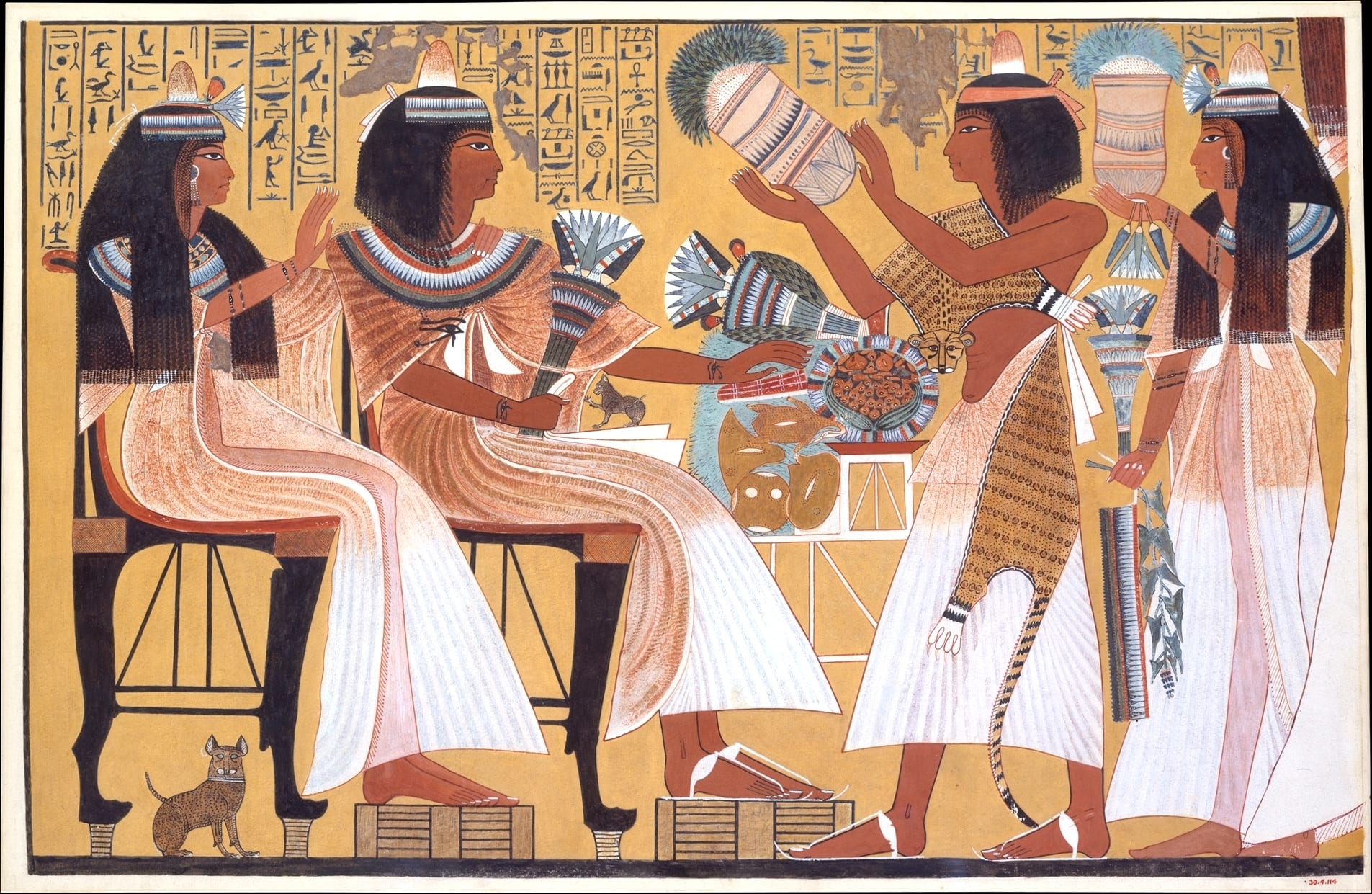
However, cats didn’t just chow down on small vermin like rodents; they were also known to kill poisonous snakes. Snakes were a real issue in Ancient Egypt and the presence of cats reduced the threat of poisoning. Through this behaviour, cats were perceived to have a protective nature which, combined with their ability to have lots of kittens, made them a symbol of the home, women, and fertility. Tomb paintings dated to the New Kingdom often feature cats as dedicated companions of women, usually seated under their chairs.


This image shows a wall painting from the tomb of Ipuy, at Deir el-Medina. Ipuy has a small kitten sitting on his lap whilst a cat sits under his wife’s chair (Image credit: https://www.nilemagazine.com.au/march-2015-archive/2015/3/22/ancient-egypts-best-dressed-cats)


Their representation in popular culture and usefulness around the home and workplace gave cats a prominent position in Egyptian society. Some people were even named after cats, Miut and Miit, Ta-mitt (female cat) and Pa-mitt (tom cat). Killing a cat was punishable by death, even if it was an accident, and when a family cat died it was common for its owners to shave their eyebrows as part of the mourning process. See I wasn’t kitten when I told you cats were important!

Cats also had a significant impact on religion in Ancient Egypt, despite being a relatively late addition to the Pantheon (c. 2000 – 1000 BC). The earliest representation of a cat or lion in Egyptian religion  was the fur-midable Mafdet, a cat-like deity associated with justice and execution. Interestingly Mafdet probably translates as ‘runner’, and it’s possible she embodied a cheetah or jaguar.

Mafdet was followed by Sekhmet, meaning strength and ferocity, a lion-headed goddess. She played a key part in the Egyptian creation myth when Hathor, daughter of Ra, was transformed into Sekhmet to remind humans of the God’s power (seriously gruesome events ensued). She has a reputation as a ferocious deity but also a stalwart protector of the innocent.

Bastet is probably the most famous cat-headed goddess. Much more moderate than her predecessors, she was associated with fertility, womanhood, and the home. Bastet was a very popular goddess through to the Ptolemaic and Roman Periods in Egypt; she even had a cult centre of worship called Bubastis.

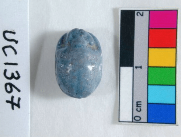
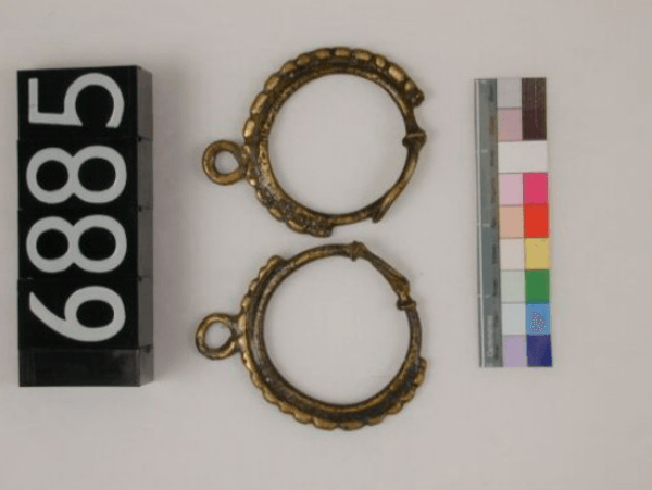
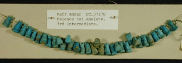
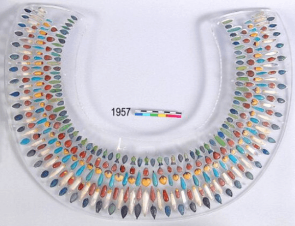
The Cult of the Cat was not restricted to Bubastis and spread across Ancient Egypt with large temples dedicated to the cat goddesses, which house and cared for hundreds of cats. Cats were even mummified in a similar way to humans and placed in temples after their death. The Petrie Museum has its very own mummified cat (sort of), which is part of the Langton Collection, a substantial bequest of artefacts that are all cat related. They were originally brought together by Mr and Mrs Langton, who excavated and worked in Egypt in the early twentieth century, who wanted to highlight the importance of the Cult of the Cat!


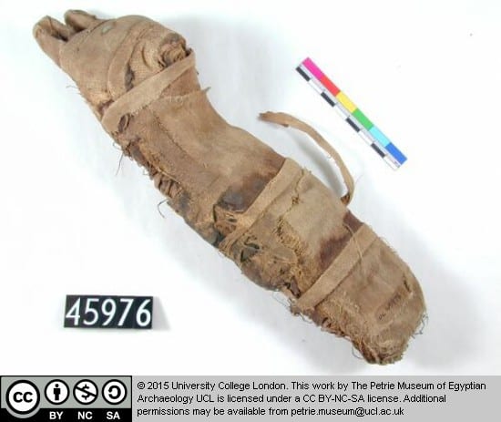
Mummified remains inside linen bandages shaped to look like a cat. An x-ray of this artefact revealed that it only contained two leg bones! Dated to the Late/Roman Period (Petrie Museum, 45976)


The popularity of cats in a religious context peaked during the Ptolemaic period (332 – 30 BC), when political unrest was rife across Egypt. One of the reasons that I know about this period (when I should really be concentrating on Neanderthals) is through playing the video game Assassin’s Creed Origins, which is set during the reign of the Ptolemies. The game is incredibly accurate and a recent update allows you to play in discovery mode, effectively turning Ancient Egypt into a virtual museum. One of my favourite features of the game are the little cats that weave around your feet as you explore towns and villages. These cats have sandy, light red brown or striped coats inspired by cats painted in tombs. Hilariously, and much to my initial frustration, cats can choose whether to interact with you or not! In my opinion greatly adding to the realistic nature of the game…


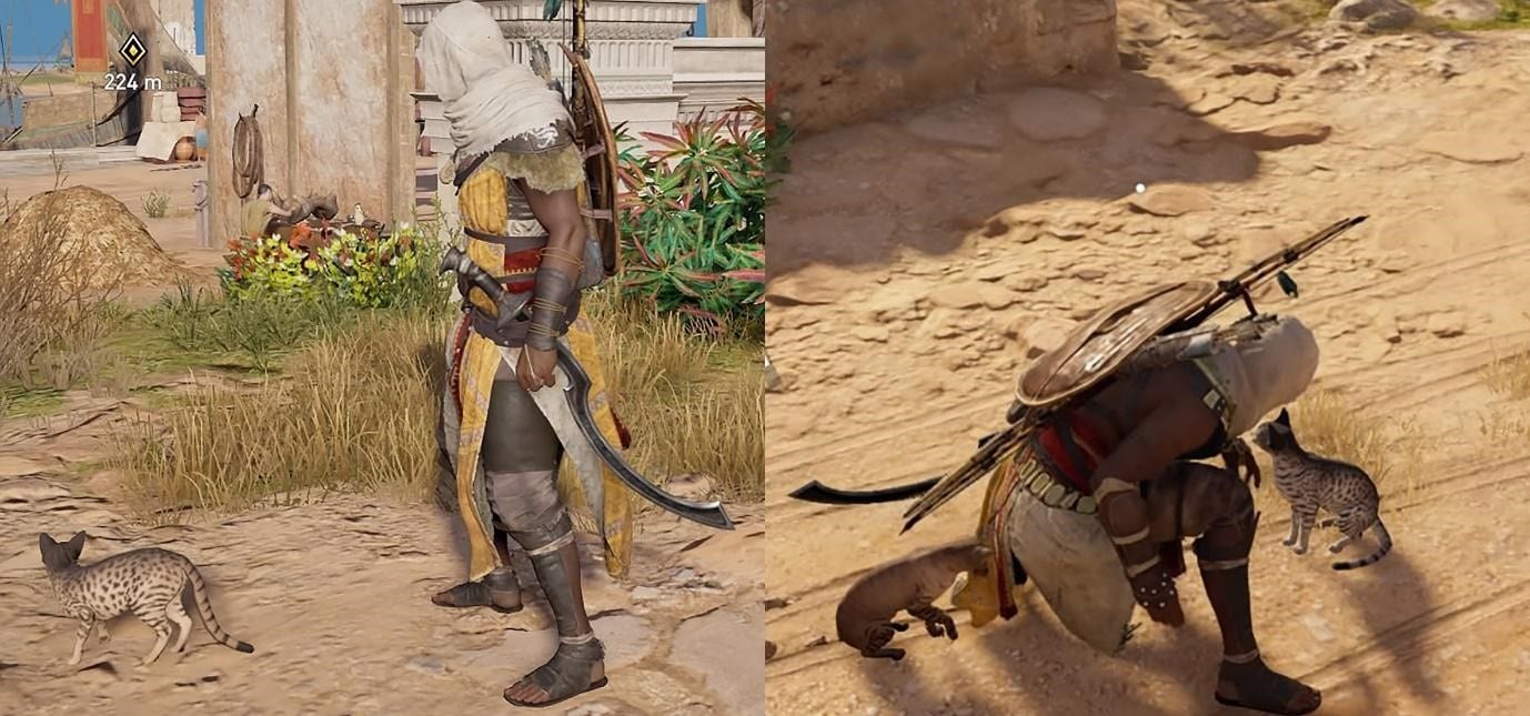
On the left a cat petting fail, on the right a cat petting success! Screenshots taken from Assassin’s Creed Origins made by Ubisoft


There’s ample evidence that Ancient Egyptians loved cats and the prominent role they played in day-to-day life and religious worship. Five thousand years later I’m not sure how much has changed. Incidentally if you’d like to read more about cats in a medieval context (of course you would!) check out my fellow engager Arendse’s blog post.

References

Challis, D. 2015. Miw: the Langton Cat Collection. In: Stevenson, A (ed.) Petrie Museum of Egyptian Archaeology Characters and Collections. UCL Press: London 72-74

Malek, J. 1993. The Cat in Ancient Egypt. British Museum Press: London

https://en.wikipedia.org/wiki/Cats_in_ancient_Egypt


Jewels of an Ancient Civilization

By Julia R Deathridge, on 1 March 2018

Whenever I’m in the Petrie Museum I’m always drawn to the jewellery. This is because a) much like a magpie my attention is easily attracted to shiny pretty objects, and b) I would actually wear a lot of the pieces on display, probably to some future fancy event that I’ll one day attend post PhD life. So I decided to do a little research on the history of jewellery in ancient Egypt and pick out my favourite pieces from the collection.

Gold wide collar necklace, dynasty 18. From the tomb of the three minor wives of Thutmose III. CC BY-NC 2.0 © Peter Roan

The rise of extravagant jewellery

As far back as the Stone Age, our ancestors have been decorating themselves in jewellery. Originally these were just simple pieces crafted from easily available resources such as seashells, bone and animal skins. However, the ancient Egyptians had other ideas, and they would go on to create trends and styles of jewellery that would live on to this day.

The discovery of gold in ancient Egypt, along with the use of precious gems, resulted in the creation of highly lavish jewellery pieces that epitomised the luxury culture of nobles and royals. As technology advanced and materials became more readily available, the popularity and extravagance of jewellery also increased, making it one of the most desirable trade items of the ancient world.

Jewellery and religion

Jewellery was extremely popular in ancient Egypt. Everyone wore it, whether they were male, female, rich or poor. But jewellery was not just about adorning oneself with pretty gems; it also acted as symbol of status and was steeped in religious beliefs.

Small charms, known as amulets, were of particular religious importance to ancient Egyptians. They believed that these charms had magical powers of protection and healing, and would bestow good fortune to the wearer. Much like charm bracelets today, these charms were commonly worn as part of a necklace or bracelet, and the shape or symbol of the amulet would specify a particular meaning or power.

Violet faience scarab bead (Petrie Museum: UC1367)

Jewellery offered magical powers to the dead as well as the living, and ancient Egyptians were often buried wearing their prized jewels. One of the most common amulets to be buried with was the scarab, as it symbolised rebirth and would ensure reincarnation to the next level.

 Materials and metals

The materials that a jewellery piece was made out of acted as an indicator for social class. Nobles would wear jewellery made up of gold and precious gems, and others would wear jewellery made from copper, colourful stones and rocks.

Gold was the most commonly used precious metal, due to its availability in Egypt at the time and its softness, which made it the perfect material for establishing elaborate intricate designs. Moreover, the non-tarnishing properties of gold added to the magical prowess of the metal, leading ancient Egyptians to believe that it was the ‘flesh of the gods’.

Another regularly used material was the semi-precious stone Lapis Lazuli. The deep blue colour of Lapis Lazuli symbolised honour, royalty, wisdom and truth. Other prized stones included obsidian, garnet, rock crystal and carnelian, pearls and emeralds. However, artificial more affordable versions of these precious gems were also crafted, and commonly worn by the lower classes. Much like the fake diamonds and pearls of today, these artificial gemstones were practically indistinguishable from the real thing.

I want that jewellery!

So now we’ve had a little history. Lets get on to the important stuff – which pieces of jewellery I would most like to wear!

First, lets start with the earrings. It wasn’t actually until King Tutankhamen that earrings became a popular jewellery item among ancient Egyptians. The style and use of earrings is likely to have been brought over from western Africa. My favourite earrings are these beautiful hoops, which would not look out of place on stall in a Brick Lane market!


Another piece that would nicely fit into my jewellery collection is a string of faience cat amulets. Firstly, it will go brilliantly with all my other cat jewellery. Secondly, cats were highly regarded in ancient Egypt and these cat amulets would likely to have been of great importance to the owner.


Faience, turquoise glaze, sting of cat amulets (Petrie Museum: UC37170)


Finally, the ultimate extravagant piece from the collection that I would love to own, is this wide collar necklace, which was likely to have been worn by Akhenaten, Tutankhamen’s father. Each bead was excavated separately and the design of the necklace was reconstructed for the Petrie collection. Additionally, conservation revealed a turquoise bead (11th from the right) to have a cartouche of Tutankhamun. When you’re next in the Petrie, see if you can spot it!


Reconstructed bead necklace. Armana period (Petrie Museum: UC1957)


Question of the Week: Why Do Wombats Poop Cubes?

By Arendse I Lund, on 14 February 2018


A wombat waddling along (Image: © Jack Ashby)

With pudgy little legs and a determined waddle, wombats are amongst Australia’s cutest marsupials. I mean, have you ever seen a wombatlet (not the technical term, unfortunately) sneeze? There’s lots to love about wombats—including their cube-shaped poop.

Wombat faeces—not a snack treat (Image: Bjørn Christian Tørrissen)

This odd wombat feature has sparked a lot of gleeful speculation. The prevailing thought is that these six-sided excrements are caused by a combination of the digestion time, the shape of the large intestine, and the dryness of the resulting fecal matter.

Wombats have a slow digestive system—it takes up to 2.5 weeks for food eaten to make its way down the alimentary canal, through the stomach, small intestine, and finally out the anus as fecal matter. (On the scale of animal defecation time, wombats aren’t even in the running. One snake was recorded as “holding it” for 420 days.)

A common wombat, or Vombatus ursinus, skull with large teeth for masticating grasses and roots, and a skeleton with large front claws for digging (Images: Grant Museum of Zoology, Z68 and Z67)

After being processed by the stomach, the digested matter transverses the large intestine, which is a long tube-like organ with ridged sides. These ridges may help to break the matter into compact sections. Since the final part of the intestine is much smoother, these cubed sections retain their shape all the way to the anus.

A wombat’s long digestive time means that this matter becomes condensed and, ultimately, dry as the nutrients are extracted. Wombats have some of the driest faeces amongst mammals and, it turns out, it’s a handy evolutionary trait. Wombats use their droppings to mark territory; with a propensity to defecate on logs and other elevated objects, cubes won’t roll off, unlike cylindrical droppings. As wombats drop between 80 and 100 scats a day, it would be a pain if they, well, scattered.


According to Jack Ashby, Manager of the Grant Museum of Zoology, “Another thing to note about wombat poo is that since wombats have backwards-facing pouches, larger wombatlets end up spending a lot of time with their faces in poo. It has been suggested that this is an important way that they gain helpful gut bacteria that they need to digest the wombat diet of tough Australian grasses.”

If you want to see fake wombat faeces in action, Robyn Lawrence created a video demonstrating a wombat’s digestive system. She uses Jell-O to illustrate the forming and squeezing of the food into cube shapes, which then passes unchanged through the colon and out the fake anus.

So no, the wombat rectum isn’t square.

———

Further Reading:

Menkhorst, P. A Field Guide to the Mammals of Australia. South Melbourne: Oxford University Press, 2001.

Triggs, Barbara. The Wombat: Common Wombats in Australia. University of New South Wales Press, 2002.

I spy with my little eye… Micrarium Top 5

By tcrnkl0, on 9 January 2018

Want a tour through the Grant Museum’s iconic display of the tiny creatures that populate our world? Well unfortunately, it’s much too small for that! However, here I’ll tell you about five of my favourite slides to be on the lookout for when you visit.

The Micrarium. Photo by author.

The Micrarium’s floor-to-ceiling lightboxes illuminate 2323 microscope slides featuring insects, sea creatures, and more, with another 252 lantern slides underneath. While this sounds like a lot of slides, it’s only around 10% of what the museum holds. Natural history museums often find it difficult to display their slide collections, but the diminutive creatures often featured on them make up most of our planet’s biodiversity.

I start most of my conversations with visitors during Student Engager shifts here – the Micrarium provides a clear illustration of my PhD research about how challenging aspects of diversity (of all kinds) are integrated into existing collections. It’s also an ideal place within the museum to try to pause people in the flow of their visit – it’s hard to resist stopping to snap a selfie or two.

Selfie by author.

The soft glow of the Micrarium’s backlit walls often draws people into the space without realising the enormity (or tininess!) of what they’re looking at. Over time, I’ve cultivated a number of favourites that I point out  in order to share the variety, strangeness, and poetry of the individual slides.

Small and mighty

‘Stomatopoda “Erichtheus” larva’. Photo by author.

I was attracted to this slide because at first I thought it looked like a little flying squirrel. In actuality, it’s the larvae of a mantis shrimp.

The mantis shrimp is an incredible animal. To start, they have the most complex eyes of any animal, seeing a spectrum of colour ten times richer than our own. Its two ‘raptorial’ appendages can strike prey with an amount of force and speed, causing the water around them to boil and producing shockwaves and light that stun, smash and generally decimate their prey.

For more, check out this comic by The Oatmeal that illustrates just how impressive mantis shrimp are.

‘and toe of frog, wool of bat, and tongue of dog’

‘Eye of beetle’. Photo by author.

This is one of my favourite labels in the collection – was a zoologist also dabbling in witchcraft ingredients?  Probably not. But, I’d love to know what the slide was originally used for.

The slide itself also looks unusual due to its decorative paper wrapping. These wrappings were common to slides from the mid-19th century, which were produced and sold by slide preparers for others to study.

Many of the slides in the Micrarium were for teaching students who could check out slides like library books. So, perhaps it illustrated some general principles about beetle eyes rather than being used for specialist research.

Cat and Mouse

Fetal cat head (L). Embryonic mouse head (R). Photo by author.

One of the secrets of the Micrarium is that there are bits of larger animals hidden among all of the tiny ones. I like how the mice look surprisingly cheerful, all things considered. Bonus: see if you can also find the fetal cat paws!

Seeing stars

‘OPHIUROIDEA Amphiura elegens’. Photo by author.

This is a young brittle star, which in the largest species can have arms extending out to 60cm. Brittle stars are a distinct group from starfish; most tend to live in much deeper depths than starfish venture. They also move much faster than starfish, and their scientific name ‘Ophiuroidea’, refers to the slithery, snake-like way their arms move.

This slide can be found at child height, and it’s nice to show kids something they’re likely to recognise.

And finally:

Have you seen the bees’ tongue?

‘Apis (Latin for bee) tongue’ Photo by author.

Showing visitors this slide of the bee’s tongue almost always elicits surprise and fascination. Surprise at the seemingly strange choice to look at just the tongue of something so small and fascination at how complex it is.

We don’t normally think of insects having something so animal-sounding as a tongue (more like stabby spear bits to sting or bite us with!). But, bee tongues are sensitive and impressive tools: scientists have observed bee tongues rapidly evolving alongside climate change.

Good luck finding these…or your own Top 5! Share any of your favourites in the comments.

The Grant Museum blog did a similar post five years ago when the Micrarium opened. These don’t overalp with my Top 5 (which is easy to avoid when there are 2323 slides), so you should also check that out.

Why are animals 3D?

By ucbtch1, on 23 November 2017


Have you heard that our body is mainly composed of 70% water? Although true, the percentage varies from 55% of water in adult women, all the way up to 78% in babies, with the percentage for adult men somewhere in between. This is also true for animals, where some — like the jellyfish — have even 90% of their body composed of water. With this in mind, why don’t animals, including us, look like a soup? How can animals have a defined 3D structure?

Aurelia aurita, moon jellyfish, 5 preserved specimens (C193)

Aurelia aurita, moon jellyfish, 5 preserved specimens (C193)


Animals are made out of cells, the building blocks of our organs and tissues. But cells are basically a bag of water and chemicals; so again, why don’t animals look like giant bags of chemicals? The most obvious reason is that animals have bones that give structure to the rest of the body. But even bones are 31% water, and organs with no bones, such as hearts, still have a unique 3D form. Hearts have defined chambers (see the elephant heart below); they’re not just a mush of cells. The answer lies not in the cells themselves but in what surrounds them.

Elephant dried heart (Z639)

Elephant dried heart (Z639)

Cells are engulfed by the extracellular matrix (ECM) which is mainly composed of proteins. This matrix encompasses the space in-between cells, gives them structural support and acts like a scaffold. It can also act as a pathway for cells to migrate along and it gives out chemical and physical cues that cells respond to. The ECM varies from organ to organ. The brain, for example, is mainly composed of cells with an ECM of only 20% of the total mass. In contrast, cartilage has fewer cells and around 70% of its mass is ECM. Every cell type is surrounded by a specific matrix that will affect its function. Studying this extracellular environment is important to understand how cells develop, how they interact with each other, and how they react to disease.

At the same time, by studying the ECM, researchers can get an idea of how an organ or tissue is structured and how to replicate its intricate architecture. Scientists that work in tissue engineering use a technique which consists of washing away the cells of an organ, literally. By using detergents, the cells are washed away in cycles until just the extracellular matrix is left. In this manner, they can analyse its composition and experiment with the matrix with the end goal of growing an organ in the lab. Therefore, one day we could replace diseased or aged organs with new ones without the need for transplantation. The unique composition of the ECM provides cells with the support they need to survive, and at the same time, gives animals and their organs a defined 3D structure.


Sources:

https://water.usgs.gov/edu/propertyyou.html

How to visualize the insides of an animal?

By ucbtch1, on 26 October 2017

When studying animals, sometimes we need to study them from the inside out —literally. One way to do this is to cut them open and looking at their internal structures, such as with the bisected heads or the microscope slides in the Grant Museum of Zoology. Another way to visualize the inside of an animal is to stain a particular body part while making everything else clear; researchers can do this by using chemicals and colour stains. For example, in the Grant Museum of Zoology, we can find specimens like the tarsier, with its skeleton stained in red, or the zebrafish with red and blue parts.

Adult tarsier stained with Alizarin Red to show calcium (Z2718)

Adult tarsier stained with Alizarin Red to show calcium (Z2718)

Adult zebrafish stained with Alcian Blue and Alizarin Red (V1550)

Adult zebrafish stained with Alcian Blue and Alizarin Red (V1550)


The process of staining these animals begins with the removal of the skin, viscera and fat tissue.  Then, soft tissues like muscle are cleared using a variety of different methods which mostly involve exposing the specimen to different baths of chemicals. Next, the bones are stained with Alizarin Red and the cartilage with Alcian Blue. It’s a long process that can take a couple of days because the stain needs to properly penetrate the tissues, but the results are amazing.

Initially, both Alizarin Red and Alcian Blue were used as textile dyes, but now they also have numerous biological applications. Alizarin Red staining is a method to visualize mineralized tissue because it stains calcium and Alcian Blue stains specific structures mainly found in cartilage. These stains constitute an important part of research because they allow researchers to visualize the intricate structure of tissues and thus understand how they form throughout development.

ADSCs

Image credit: Eleonora Zucchelli

In the lab where I study, researchers work with adipose (fat) derived stem cells which have the capacity to become different kinds of mature cells. These stem cells are grown under specific conditions and by changing these conditions scientists can direct them into becoming mature cells like fat, bone or cartilage — a process called differentiation. But this process can take anywhere from a couple of weeks up to a couple of months! In order to determine if the differentiation is working, researchers stain the stem cells with Alizarin Red and Alcian Blue to identify if they are in fact turning into bone or cartilage. In the images depicted, undifferentiated adipose derived stem cells (ADSCs) on the top appear clear but their differentiated counterparts are stained in blue or red. This means the differentiation is working.

There are many other stains used on animals or cells. The process of clearing and staining can be very complicated depending on the specimen and what one wishes to stain, but the results can be quite fascinating. What animal would you like to see stained from the inside.


Mouse stained with alizarin red (Z3155)

Mouse stained with alizarin red (Z3155)


References:

PUCHTLER, H., Meloan, S. N., & TERRY, M. S. (1969). On the history and mechanism of alizarin and alizarin red S stains for calcium. Journal of Histochemistry & Cytochemistry17(2), 110-124.

McLeod, M. J. (1980). Differential staining of cartilage and bone in whole mouse fetuses by alcian blue and alizarin red S. Teratology22(3), 299-301.


Question of the Week: What’s this Museum For?

By Hannah L Wills, on 19 October 2017

By Hannah Wills


A couple of weeks ago, whilst engaging in the Grant Museum, I started talking to some secondary school students on a group visit to the museum. During their visit, the students had been asked to think about a number of questions, one of which was “what is the purpose of this museum?” When asked by some of the students, I started by telling them a little about the history of the museum, why the collection had been assembled, and how visitors and members of UCL use the museum today. As we continued chatting, I started to think about the question in more detail. How did visitors experience the role of museums in the past? How do museums themselves understand their role in today’s world? What could museums be in the future? It was only during our discussion that I realised quite how big this question was, and it is one I have continued to think about since.

What are UCL museums for?

The Grant Museum, in a similar way to both the Petrie and Art Museums, was founded in 1828 as a teaching collection. Named after Robert Grant, the first professor of zoology and comparative anatomy at UCL, the collection was originally assembled in order to teach students. Today, the museum is the last surviving university zoological museum in London, and is still used as a teaching resource, alongside being a public museum. As well as finding classes of biology and zoology students in the museum, you’re also likely to encounter artists, historians and students from a variety of other disciplines, using the museum as a place to get inspiration and to encounter new ideas. Alongside their roles as spaces for teaching and learning, UCL museums are also places for conversation, comedy, film screenings and interactive workshops — a whole host of activities that might not have taken place when these museums were first created. As student engagers, we are part of this process, bringing our own research, from a variety of disciplines not all naturally associated with the content of each of the museums, into the museum space.


A Murder-Mystery Night at the Grant Museum (Image credit: Grant Museum / Matt Clayton)

A Murder-Mystery Night at the Grant Museum (Image credit: Grant Museum / Matt Clayton)


What was the role of museums in the past?

Taking a look at the seventeenth and eighteenth-century roots of the Ashmolean Museum in Oxford and the British Museum in London, it is possible to see how markedly the role and function of the museum has changed over time. These museums were originally only open to elite visitors. The 1697 statues of the Ashmolean Museum required that ‘Every Person’ wishing to see the museum pay ‘Six Pence… for the Space of One Hour’.[i] In its early days, the British Museum was only open to the public on weekdays at restricted times, effectively excluding anyone except the leisured upper classes from attending.[ii]

Another feature of these early museums was the ubiquity of the sense of touch within the visitor experience, as revealed in contemporary visitor accounts. The role of these early museums was to serve as a place for learning about objects and the world through sensory experience, something that, although present in museum activities including handling workshops, tactile displays, and projects such as ‘Heritage in Hospitals’, is not typically associated with the modern visitor experience. Zacharias Conrad von Uffenbach (1683-1784), a distinguished German collector, recorded his visit to Oxford in 1710, and his handling of a range of museum specimens. Of his interactions with a Turkish goat specimen, Uffenbach wrote, ‘it is very large, yellowish-white, with… crinkled hair… as soft as silk’.[iii] As Constance Classen has argued, the early museum experience resembled that of the private ‘house tour’, where the museum keeper, assuming the role of the ‘gracious host’, was expected to offer objects up to be touched, with the elite visitor showing polite and learned interest by handling the proffered objects.[iv]

Aristocratic visitors handle objects and books in a Dutch cabinet of curiosities, Levinus Vincent, Illustration from the book, Wondertooneel der Nature - a Cabinet of Curiosities or Wunderkammern in Holland. c. 1706-1715 (Image credit: Universities of Strasbourg)

Aristocratic visitors handle objects and books in a Dutch cabinet of curiosities, Levinus Vincent, Illustration from the book, Wondertooneel der Nature – a Cabinet of Curiosities or Wunderkammern in Holland. c. 1706-1715 (Image credit: Universities of Strasbourg)


How do museums think about their function today?

In understanding how museums think about their role in the present, it can be useful to examine the kind of language museums employ when describing visitor experiences. The British Museum regularly publishes exhibition evaluation reports on its website, detailing visitor attendance, identity, motivation and experience. These reports are fascinating, particularly in the way they classify different visitor types and motivations for visiting a museum. Visitor motivations are broken down into four categories: ‘Spiritual’, ‘Emotional’, ‘Intellectual’ and ‘Social’, with each connected to a different type of museum function.[v]

Those who are driven by spiritual motivations are described as seeing the museum as a Church — a place ‘to escape and recharge, food for the soul’. Those motivated by emotion are understood as searching for ‘Ambience, deep sensory and intellectual experience’, the role of the museum being described as akin to that of a spa. For the intellectually motivated, the museum’s role is conceptualised as that of an archive, a place to develop knowledge and conduct a ‘journey of discovery’. For social visitors, the museum is an attraction, an ‘enjoyable place to spend time’ where facilitates, services and welcoming staff improve the experience. Visitors are by no means homogenous, their unique needs and expectations varying between every visit they make, as the Museum’s surveys point out. Nevertheless, the language of these motivations reveals how museum professionals and evaluation experts envisage the role of the modern museum, a place which serves multiple functions in line with what a visitor might expect to gain from the time they spend there.

What will the museum of the future be like?

In an article published in Frieze magazine a couple of years ago, Sam Thorne, director of Nottingham Contemporary, invited a group of curators to share their visions on the future of museums. Responses ranged from the notion of the museum as a ‘necessary sanctuary for the freedom of ideas’, to more dystopian fears of increased corporate funding and the museum as a ‘business’.[vi] These ways of approaching the role of the museum are by no means exclusive; there are countless other ways that museums have been used, can be used, and may be used in the future. My thinking after the conversation I had in the Grant Museum focussed on my own research and experience with museums, but this is a discussion that can and should be had by everyone — those who work in museums, those who go to museums, and those who might never have visited a museum before.


What do you think a museum is for? Tweet us @ResearchEngager or come and find us in the UCL museums and carry on the discussion!


References:

[i] R. F. Ovenell, The Ashmolean Museum 1683-1894 (Oxford: Clarendon Press, 1986), 87.

[ii] Fiona Candlin has written on the class politics of early museums, in “Museums, Modernity and the Class Politics of Touching Objects,” in Touch in Museums: Policy and Practice in Object Handling, ed. Helen Chatterjee, et al. (Oxford: Berg, 2008).

[iii] Zacharias Konrad von Uffenbach, Oxford in 1710: From the Travels of Zacharias Conrad von Uffenbach, trans. W. H. Quarrell and W. J. C. Quarrell (Oxford: Blackwell, 1928), 28.

[iv] Constance Classen, “Touch in the Museum,” in The Book of Touch, ed. Constance Classen (Oxford Berg, 2005), 275.

[v] For this post I took a look at ‘More than mummies A summative report of Egypt: faith after the pharaohs at the British Museum May 2016’, Appendix A: Understanding motivations, 27.

[vi] Sam Thorne, “What is the Future of the Museum?” Frieze 175, (2015), accessed online.

Label Detective: Are Bacteria ‘Ordinary Animals?’

By tcrnkl0, on 17 October 2017

A few weeks ago, the Grant Museum opened a new exhibit, The Museum of Ordinary Animals: boring beasts that changed the world. As a detective of the mundane myself, I am a huge fan. But I’m particularly curious about the ordinary animals we can’t see.

Rather than focusing on a specific artefact label, I answer the title question by visiting two places in the Museum of Ordinary Animals exhibition that help raise questions about how things are organised and labeled in zoology more broadly.

Case notes: Bacteria are everywhere. As I mentioned in my previous post, we have 160 major species of bacteria in our bodies alone, living and working together with our organ systems to do things like digest nutrients. This is also happens with other animals — consider the ordinary cow, eating grass. Scientist Scott F. Gilbert tells us that in reality, cows cannot eat grass. The cow’s genome doesn’t have the right proteins to digest grass. Instead, the cow chews grass and the bacteria living in its cut digest it. In that way, the bacteria ‘make the cow possible’.

IMG_1102

The Ordinary Cow, brought to you to by bacteria. Credit: Photo by author

Scientifically speaking, bacteria aren’t actually ‘animals’; they form their own domain of unicellular life. But, as with the cow, bacteria and animals are highly connected. Increasingly, scientists say that the study of bacteria is ‘fundamentally altering our understanding of animal biology’ and theories about the origin and evolution of animals.

But, before we get into that, let’s go back to Charles Darwin (1809-1882). Darwin studied how different species of animals, like the pigeon, are related to each other, and how mapping their sexual reproduction shows how these species diversify and increase in complexity over time. This gets depicted as a tree, with the ancestors at the trunk and species diversifying over time into branches.

Picture1

Darwin’s Ordinary Tree of Pigeons. Photos by author

When scientists began to use electron microscopes in the mid-20th century, our ideas about what made up the ‘tree of life’ expanded. We could not only observe plants, animals, and fungi, but also protists (complex small things) and monera (not-so-complex small things). This was called the five kingdom model. Although many people still vaguely recollect this model from school, improved techniques in genetic research starting in the 1970s has transformed our picture of the ‘tree of life’.

It turns out we had given way too much importance to all the ordinary things we could see, when in fact most of the tree of life is microbes. The newer tree looks like this:

Credit: Wikipedia Commons

Credit: Wikipedia Commons

Now there are just three overarching domains of life: Bacteria, Eucarya (plants, animals, and fungi are just tiny twigs on this branch), and Archaea (another domain of unicellular life, but we’ll leave those for another day).

There’s a third transformation of the ‘tree of life’, and this one is my favourite. Since the 1990s, DNA technology and genomics have given us an even greater ability to ‘see’ the diversity of microbial life and how it relates to each other. The newest models of the tree look more like this:

Credit: Wikipedia Commons

Credit: Wikipedia Commons

This is a lot messier. Why? Unlike the very tiny branches of life (plants and animals) that we focused a lot of attention on early on in the study of evolution, most of life on earth doesn’t reproduce sexually. Instead, most microbes transfer genes ‘horizontally’ (non-sexually) across organisms, rather than ‘down’ a (sexual) genetic line. This creates links between the ‘branches’ of the tree, starting to make it look like….not a tree at all. As scientist Margaret McFall-Ngai puts it: ‘we now know that genetic material from bacteria sometimes ends up in the bodies of beetles, that of fungi in aphids, and that of humans in malaria protozoa. For bacteria, at least, such transfers are not the stuff of science fiction but of everyday evolution’.

Status: Are bacteria Ordinary Animals? We can conclusively say that bacteria are not animals. But, they are extremely ordinary, even if we can’t see them with the naked eye. In truth, they’re way more ordinary than we are.


Notes

As with the previous Label Detective entry, this post was deeply inspired by the book Arts of Living on a Damaged Planet, an anthology of essays by zoologists, anthropologists, and other scholars who explore how environmental crisis has highlights the complex and surprising ways that life on earth is tied together. Scott F. Gilbert and Margaret McFall-Ngai, both cited above, contribute chapters.

Embryological Wax Models

By ucbtch1, on 14 September 2017

The Grant Museum has a number of embryological wax models on display (Images 1, 2, 4, and 5 amongst others not shown here). These models, while often ignored by visitors, are actually quite remarkable as they showcase the brilliance and mystery of embryological development. They were created to help elucidate essential questions like: how do humans and other animals form? How are a bunch of seemingly insignificant cells, with no shape other than a ball, able to grow so much and in such detail to form intricate patterns like our eyes? How can one cell transform itself into such different tissues, from hard rock bone to the jelly like liver? In order to understand how a human body is formed it is vital to study the very first stages of its creation, i.e. when we are just a bunch of cells.

Image 1. Placental mammal embryo (Z3100)

Image 1. Placental mammal embryo (Z3100)

The very first cells that are formed after fertilization are called stem cells and they start with an unlimited capacity to form any type of cells. With time, they start to differentiate and mature into specialized cells with a limited lifetime. In this process, little by little they lose their unlimited capacity until they can only form cells that are similar in lineage. In this manner, totipotent stem cells can form any body part including extraembryonic tissue like the placenta. On the next level, pluripotent stem cells (embryonic stem cells for example) are capable of forming any body part but have lost the capacity to form extraembryonic tissue. And finally, multipotent stem cells, much more restricted, can only form cells from a specific tissue or organ.

This may appear as a straightforward process, but the development of an animal is a deeply specific, delicate, and sophisticated interplay of signals and coordinated transitions. Think of it as an orchestrated dance of on and off switches leading to specialisation and exponential growth. In fact, it is so complex that we still don’t understand it entirely.

Although not all human, the wax models display the first stages of development of vertebrates and closely related animals. First, one cell divides symmetrically into two, then four, then eight and so on (Image 1 and front models of Image 2). Afterwards, cells start organizing themselves answering to chemical and physical signals and different patterns start to appear (Image 2 models in the back). Eventually, an axis emerges on which cells migrate along which will give rise to the head on one side and the body and limbs on the other (Image 4). 

Slowly but surely, we all go from looking like little worms to fully grown animals (Image 3). It is important to note that in this initial period most embryos have a very similar appearance, at least between vertebrates. These similarities tell us that a lot of the genes that govern this initial growth haven’t changed between species over time. It’s like nature is saying, “well, if it ain’t broke don’t fix it” and so these mechanisms have been conserved in different animals.

Image 2. Branchiostoma sp, Lancelet Embryo (fish-like invertebrate; T114)

Image 2. Branchiostoma sp, Lancelet Embryo (fish-like invertebrate; T114)

These early stages are crucial moments because if one little element of the spatial/temporal organization is out of place, improper organization can lead to lifelong malformations, diseases, or even the termination of the embryo. Hence the importance of understanding how this process works. We know that even in adult life, there are still stem cells proliferating and forming new tissue to a certain degree. Some organs, like the skin, have a lot of stem cells to replace old cells when they die or get injured. But other organs like the brain, have a very limited capacity to grow new cells—one of the reasons why a brain is much more difficult to fix.

Image 3. Development of the external form of the human face (LDUCZ-Z480) and development of external form of human embryo (LDUCZ-Z430)

Image 3. Development of the external form of the human face (LDUCZ-Z480) and development of external form of human embryo (LDUCZ-Z430)

Image 4. Vertebrate embryos. Image taken from https://www.ncbi.nlm.nih.gov/books/NBK9974/

Image 4. Vertebrate embryos. Image taken from https://www.ncbi.nlm.nih.gov/books/NBK9974/

So can we get back all the limitless capacity there once was in the developing embryo? Even though the genetic and molecular mechanisms governing all these changes are still somewhat elusive, researchers are using stem cells and powerful genetic tools to answer this question and decipher every single step of how a human is formed in the womb. Moreover, if we can understand the process, then we can recreate and modify it in the lab, and this is exactly what the field of stem cells and regenerative medicine is trying to do. Imagine having the capacity to grow new organs to be used for transplantation or drug testing. How about growing a brand new functioning leg or arm for amputees? Or studying the mechanisms of diseases like Parkinson’s, leukaemia, diabetes, amongst others. The benefits of harnessing the regenerative potential of cells are far-reaching.

Image 5. Development of the external form of the human face (LDUCZ-Z480).

Image 5. Development of the external form of the human face (LDUCZ-Z480).

The exciting field of stem cells and regenerative medicine has come a long way, more than a century has passed from the first time the term stem cells was used in 1906 up until the creation of genetically modified human embryos in 2017. The embryological wax models represent initial efforts of identifying how changes give rise to specific structures and ultimately how an animal comes into existence. Furthermore, the future still holds exciting breakthroughs, there is still a lot to understand about human development and the wax models are a fantastic resource to portray the morphogenetic changes we all once went through.


Resources:

Gilbert SF. Developmental Biology. 6th edition. Sunderland (MA): Sinauer Associates; 2000. Comparative Embryology. Available from: https://www.ncbi.nlm.nih.gov/books/NBK9974/

http://www.labonline.com.au/content/life-science-clinical-diagnostics-instruments/article/why-do-all-animal-embryos-look-the-same–236915402

Who wants to adopt a parasite? Or, terror and disgust in the Grant Museum. 

By uclznsr, on 12 July 2017

Of all the creatures, critters, beasts, birds, and baleens in UCL’s Grant Museum, few organisms are as ignored and maligned as the parasites. Visitors tend to skirt their north-easterly facing cabinet, either because they have begun their journey around the opposite side of the museum’s horseshoe layout or to bathe in the light of the museum’s “micrarium”; either way, they are not stopping to dwell for long in front of the roundworms, flatworms, or flukes. Likewise, these “helminths” are amongst the least popular candidates for adoption, which allows visitors to have their name displayed beside their specimen of choice in exchange for a contribution to the museum’s conservation, renovation, and documentation projects. There is a gruesome irony here, since tapeworms can also dwell in the gastrointestinal tract of a human museum visitor for two decades completely unnoticed – and grow up to 55 feet long.

This fact might go some way to explaining why visitors seem to prefer more orthodox – and straightforwardly threatening – specimens. They seem to have little problem with, are even fascinated by, lions, sharks, jellyfish, scorpions, and other animals whose attacks can be painfully and violently fatal to humans. But parasites, whose methods are comparatively insidious, seem implicitly to repel. Visitors to the Grant Museum seem to prefer, therefore, threats which are visible and whose assault we can see coming, rather than incursions upon our safety from an invisible and undetectable enemy. They are not alone in this, of course: the fear of that which cannot be seen, or refuses to be revealed, is not merely an expedient workaround for low-budget horror films, but permeates folk-tales, fairy-tales, and mythology, across the world.

Nevertheless, our revulsion of parasites in particular – and not merely of what is invisible – must itself have a less visible cause, because parasites themselves are not always hidden. Organisms like the tick and the leech feed off and derive nutrients at the expense of their host, and are certainly not invisible as they do so. An infamous scene from the film adaptation of Stephen King’s Stand by Me acts as a testament to the terror the latter species seems to evoke.

Perhaps, then, it is less what is invisible or visible and more a political economic fear – a question of ownership. After all, the parasite yields profit from our bodies, without offering anything in exchange, and often without having the good grace to let us know. Can we even claim to own our own bodies, if they are so easily exploited by these organisms? One man, Dimitri Tsafendas, was certain that a parasite had taken ownership not only of his body but his mind when in 1966 he assassinated the prime minister of South Africa, Henrik Verwoerd. Tsafendas was convinced a tapeworm he had as a boy was still present in his system, and was dictating his every action.

This killing represents an uncanny reversal. Verwoerd is known today for his role as the “architect of apartheid” and the discourses and justifications of racism have often drawn upon the notion of “the other” as a parasite, whose incursion on the body politic represents the threat of impurity, disease, and the loss of national ownership. Verwoerd himself became the victim of such a delusion, albeit on the personal, individual scale. Tsafendas, despite his claims, was deemed criminally insane.

Ornithodoros sanguinis-cameli, J135, The Grant Museum of Zoology

Ticks, J135, The Grant Museum of Zoology

Other theories also allow us to speculate why the parasite exerts such a hold upon the human psyche. The French psychoanalyst Didier Anzieu argues in his The Skin Ego (Le Moi Peau) that the biological protection offered by the bodily ‘wrapping’ of our skin is doubled by a psychic defense mechanism which guards against penetrations of and assaults upon our identity sense of self-unity. To feel revulsion or horror at the sight of a tick burrowing itself into one’s arm, therefore, is not merely because it represents a threat to our biological well-being; our disproportionate terror is a result of the sense of being attacked on a deeper, existential level, an infiltration of the very boundaries that allow us to constitute ourselves as sovereign, unified psychic beings.

This theory complements another psychoanalytic theory, Julia Kristeva’s notion of abjection. She contends in The Powers of Horror that our revulsion at the abject is in fact a way of shoring up the very self-identity, unity, and sense of psychic wholeness that it appears to threaten. This is achieved by “abjection”, the sense of relief and pleasure at ejecting from the body that which is perceived to be impure of dangerous. In this way, she argues, we not only resist assimilating what appears to us as an assault on our being, in doing so ‘I expel myself, I spit myself out, I abject myself within the same motion through which “I” claim to establish myself.

Parasites, then, for all that we seem to want to ignore, are for that very reason integral to understanding who we believe we are as human beings. They are what we believe we are not. We are honest; we are visible; we do not take without giving back; we own and control ourselves and in turn respect the self-ownership of others, as long as they too remain in control of themselves. But we do not need the exemplar, historical context, and outcome of Dimitri Tsafendas and Henrik Verwoerd to know that these self-descriptions are often self-deceptions. Perhaps it is time to properly regard the parasite.