 Close
Close

Specimen of the Week 343: The brain coral
By Nadine Gabriel, on 18 May 2018
Jack Ashby, our former museum manager who left a few days ago to work at the Cambridge Zoology Museum, often talks about how natural history museums are biased towards certain animals. As I looked through the list of animals featured in our Specimen of the Week blog, I noticed that corals have only featured once in the past six and a half years! So today I would like to dedicate this blog post to Jack and make sure corals get the representation they deserve!
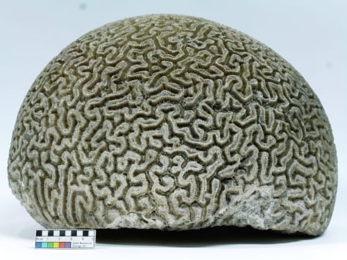
Dry specimen of a brain coral, Diploria labyrinthiformis LDUCZ-C1439
What is a coral?
Corals are marine invertebrates that belong to the Cnidaria phylum (which also includes jellyfish and sea anemones). These colonial animals consist of groups of genetically identical polyps which are sac-like in appearance and only a few centimetres long. Calcium carbonate is excreted near the polyp’s base to form an exoskeleton [1]. Corals can reproduce asexually (a polyp divides in two) or sexually (polyps release eggs and sperm simultaneously, allowing corals to spread) [2].

Brain coral spawning in the Flower Garden Banks National Marine Sanctuary (a protected site in the Gulf of Mexico). Image by NOAA, Emma Hickerson via Wikimedia Commons, Public Domain
The brainiest coral in the reef
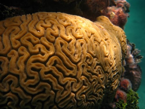
Brain corals (Diploria labyrinthiformis) are reef-building stony corals found in the tropical areas of the west Atlantic such as the Gulf of Mexico, the Caribbean Sea, Florida and the coasts of Central America [3]. They live anywhere from 1-43 metres below sea level but are most commonly found at a depth of 2-15 metres in lagoons or forereefs [4]. Looking at their maze-like patterns and dome shape, you can see why they are called brain coral (and why their species name is labyrinthiformis). Their characteristic pattern has featured on stamps for Belize and Mayotte. These corals only grow about 3.5 mm a year [3], but since they can live for up to 900 years, they can reach over two metres in diameter [5]! This picture of a scuba diver next to a brain coral shows how immense they can be.
The brain coral polyps sit within deep, interconnected valleys and eat zooplankton and bacteria by using the tentacles around their mouth. These tentacles have nematocysts (stinging cells) that use toxins to immobilise their prey before they consume them [3]. Brain corals display meandroid tissue integration which means that the polyps are highly associated with one another and are not separated by skeletal structures, allowing them to transfer nutrients, hormones and oxygen. Scientists believe that a high level of tissue integration is a sign of an advanced coral species [5].
Brain coral near the Island of Vieques, Puerto Rico. Image by Jaro Nemčok via Wikimedia Commons, CC BY-SA 3.0
Teamwork makes the dream work
Brain corals are hosts to Zooxanthellae which are photosynthetic dinoflagellates (a type of single-celled organism). Since their relationship is symbiotic, both of them gain benefits. The zooxanthellae live in a protected environment in the coral, and the coral receives some of the nutrients produced photosynthetically by the zooxanthellae. Brain corals also have a relationship with the black sea urchin because its grazing helps to prevent the overgrowth of macroalgae [3].
Threats
The IUCN lists brain corals as “least concern” because they are so common and have a widespread range. This, along with their highly connected and stable population, means that they are more resilient to habitat loss compared to other corals. Nevertheless, climate change is still a major threat to them so the conservation status of the species will need to be reassessed within 10 years. There are also localised impacts from bleaching, bioerosion and fishing [6]. Due to the high level of polyp integration, diseases such as black band disease, which is mainly caused by the Phormidium corallyticum cyanobacteria, can easily spread across the coral [5] [7].
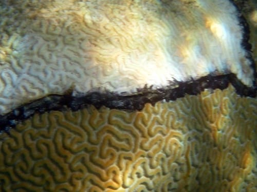
Brain coral showing signs of black band disease. The upper white portion is dead skeleton. Image by Jaro Nemčok via Wikimedia Commons, CC BY-SA 3.0
And finally, the most important thing to remember is “this is not a brain”!
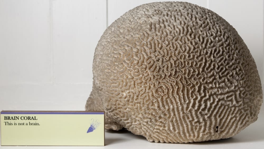
A brainy example of museum humour, LDUCZ-C449
Nadine Gabriel is the Museum Intern at the Grant Museum of Zoology
References
[1] Coral: https://en.wikipedia.org/wiki/Coral
[2] How Co Corals Reproduce?https://oceanservice.noaa.gov/education/kits/corals/coral06_reproduction.html
[3] Diploria labyrinthiformis https://en.wikipedia.org/wiki/Diploria_labyrinthiformis
[4] Goreau T. F. and Wells J. W., 1967. The Shallow-Water Scleractinia of Jamaica: Revised List of Species and Their Vertical Distribution Range. Bulletin of Marine Science, 17(2), 442-453
[5] What are brain corals? https://oceanservice.noaa.gov/facts/brain-coral.html
[6] IUCN: Diploria labyrinthiformis: http://www.iucnredlist.org/details/133257/0
[7] Black band disease: https://en.wikipedia.org/wiki/Black_band_disease