X Close

Researchers in Museums

Home

Engaging the public with research & collections

Menu

Myths in the Museum: The Iron-Eater and the Ostrich Egg

By Jen Datiles, on 4 July 2019

This is the fourth segment in the Myths in the Museum series; you can go back and read about the horseshoe crab, the dugong and mermaid, and the narwhal and unicorn.

The now-extinct Arabian ostrich, depicted in The Book of Animals, 1335. [Source: The Book of Animals of al-Jahiz, Syria]

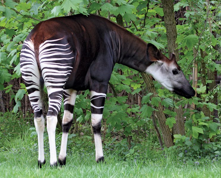
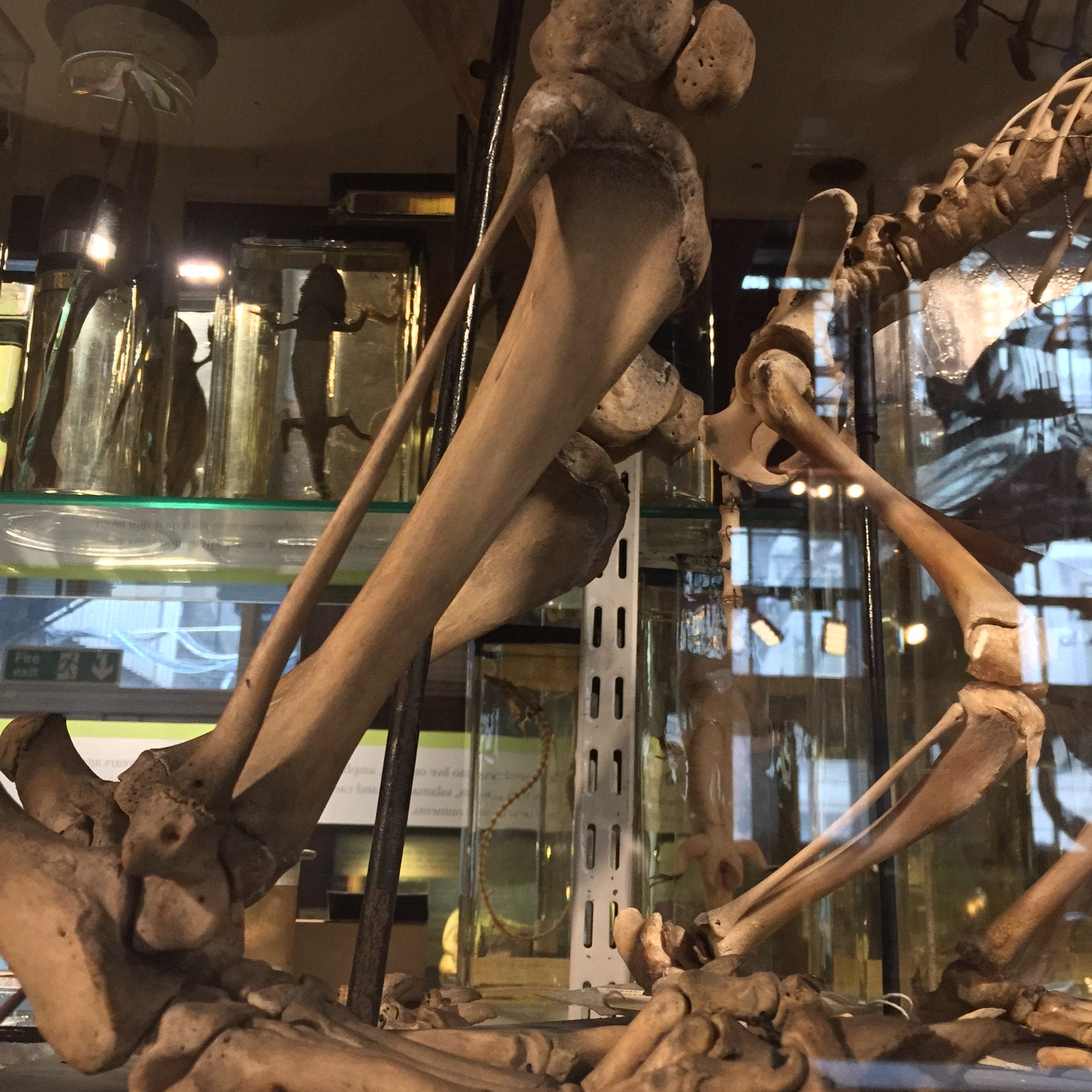
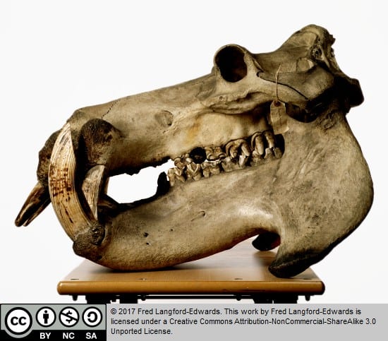
The UCL Grant Museum of Zoology is currently undergoing a significant restructuring of its displays. The Grant Museum is the last of London’s university natural history museums and has amassed a fascinating collection — but only a fraction is on display. From a set of warthog and domesticated pig skulls now placed over the entranceway as part of the expanded comparative anatomy section, to a lost set of dodo bones found in a drawer in 2011, to the world’s rarest skeleton, the extinct quagga (think zebra with fewer stripes), the museum’s collection is vast. In the newly reorganised avian section, some exceptionally large bird eggs are neatly lined up like a mini hall of fame. And the largest (non-extinct) egg of all, of course, belongs to that famous 9-foot-tall marvel with legs strong enough to kill a lion in one blow, the OG kickstarter, the Ostrich.

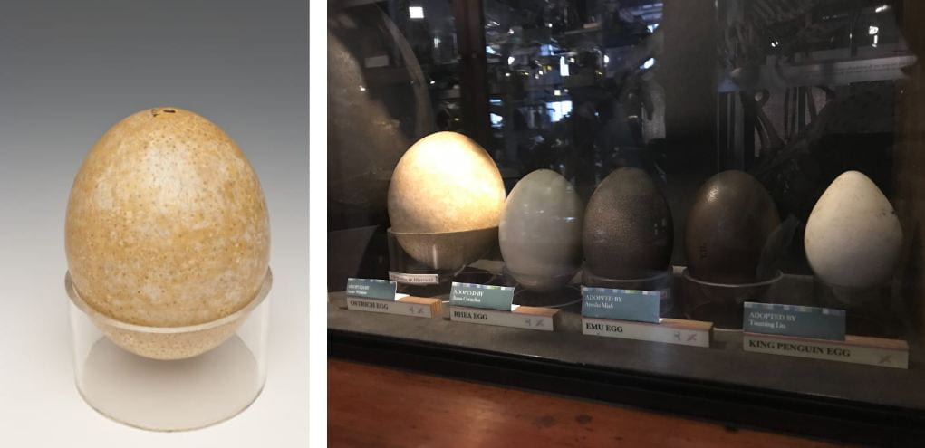
At the Grant Museum of Zoology, you can compare the pale-coloured ostrich egg with other large birds’ eggs. The photo on the left shows the individual ostrich egg; the photo on the right shows the museum’s display, with the now-extinct elephant egg dwarfing the rest, and the ostrich egg next to it. [Left: UCL Grant Museum, Y134; Right: image by Jen Datiles]

As the largest living bird species in the world, the ostrich unsurprisingly lays massive eggs that have been valued by humans for millennia. But their value goes beyond serving as a royal dish for ancient pharaohs; across cultures, the ostrich egg has long possessed symbolic significance and associations with prosperity, truth, life, and rebirth. Evidence as early as the 4th millennium BCE reveals eggs were hollowed and intricately carved, used as perfume containers and drinking cups, and buried as part of ancient Egyptian funerary rituals. Eggshells were found in sites of the early cultures of Mesopotamia and Crete, and the use of eggshells as drinking vessels was continued by desert peoples until at least the late 20thcentury. Ostrich eggs were also, of course, highly valued for their nutritional intake, with a whopping 120g or so of protein per egg.

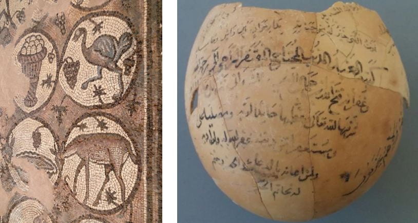
Left: A mosaic floor in the Byzantine Church of Petra, Jordan [Photo: Bernard Gagnon]; Right: A 15th-century ostrich egg with Arabic writing, describing the soul’s journey from death to life [Copyright University of Leeds. Source: Nature, 2002]

Ostrich eggs remained both spiritually and practically significant in the Greek and Roman worlds, where they were offered to deities and hung in temples as decorations or used as lamps. This association of ostrich eggs with sacred spaces carried over into Muslim and Christian practices. The ostrich, according to popular belief in the 2ndcentury BCE, the ostrich had the ability to make its eggs hatch by staring at them intensely rather than brooding, a trait that added to their significance as early Christian symbols of not only new life and rebirth, but also of single-mindedness, concentration, and determination. Pliny the Elder wrote on the ostrich’s mythical ability to eat iron and glass, which earned the bird a reputation as an iron-eater, and symbolized strength through resistance and hardship. In medieval and early modern Europe, the ostrich egg also came to symbolize the Immaculate Conception of the Virgin Mary.

Left: Ostrich eggs add both symbolism and splendor to the interior of the Lazarus Church in Cyprus. [Photo: Hannes Grobe/BHV]; Right: Piero della Francesca’s Brera Madona c. 1472, an altarpiece known for its pendant egg detail. [Source: Wiki Commons]

In the secular luxury trade of the 16-17th centuries, ostrich eggs became a subject of particular fascination for metalsmiths. Last week I oohed and ahhed my way through the famously fantastic Staatliche Kunstsammlungen Dresden, Germany. Amongst all the royal treasures of gems, ivory, gold, and crystal, a wall of the Grünes Gewölb (Historic Green Vault) was devoted to some seriously decorated ostrich eggs. These specimens had been fashioned with gilt-silver into figurines, goblets, and drinking vessels that once adorned the feast tables and halls of Saxon princes. Talk about egg-cellent conversation pieces!

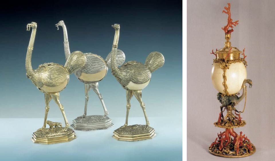
Left: Three ostriches, fashioned from eggs mounted in gilt-silver. Elias Geyer, c. 1595, in the Grünes Gewölbe, Staatliche Kunstsammlungen Dresden [Source: SKDresden Online Collection]; Right: An ostrich egg standing cup, c. 1570, from the Kunsthistorisches Museum, Vienna. Note the horseshoe in the ostrich’s beak — a reference to its mythical ability to eat iron [Source: Wiki Commons].

Ostrich eggs may also have use in modern medical research. Like all birds, ostriches pass on bacteria- and virus-fighting antibodies to their offspring through their yolk. Considering one ostrich egg contains as much yolk as about 24 chicken eggs, and one ostrich female can lay 50-100 eggs per year, a team of Japanese researchers have identified ostrich eggs as a promising source for developing drugs. Last October, they announced the commercial development of an ostrich antibody for dengue fever. The research is open to speculation, and still years away from clinical trials and regulatory approval, but our fascination large eggs continues!

Further reading:

Green, N. (2006) Ostrich Eggs and Peacock Feathers: Sacred Objects as Cultural Exchange between Christianity and Islam, Al-Masāq,18:1, 27-78, DOI: 10.1080/09503110500222328

When Plastics Saved Turtles

By Mark V Kearney, on 25 May 2019

As you may now know, UCL Culture has decided to defund our program next month and so this will be my final post. I thought I would take this opportunity to give a little back story to my PhD project and tell you all about how once upon a time plastic saved the fate of turtles!

Throughout history, natural materials such as tortoise shell and ivory have been coveted by the rich and famous. This led to two things happening – the price of these materials became very high (meaning it really was the rich and famous who could afford them) and the stock levels declined. By stock levels I mean the killing of thousands of animals such as the Hawksbill turtle to feed the needs of the bourgeoisie.

The Grant Museum has three fantastic examples of Hawksbill turtles on its back wall. I normally stand beside them so that I can kidnap engage with people about my work.

Figure 1 – Eretmochelys imbricata or known by its common name Hawksbill Turtle that is on display at the Grant Museum. (Object Number X1226)

By the mid-18th century, the farming of turtles for their shells had gotten to the point where we almost caused their extinction. A similar point had been reached with ivory where demand far outweighed supply. To give you an idea of the scale of what can only be described a mass slaughter, have a look at this doll’s house currently on display at the Rijksmuseum in Amsterdam – the outside is totally covered in tortoise shell!

Figure 2 – Dolls’ house of Petronella Oortman. Part of the collection at the Rijksmuseum. The object is totally covered with tortoise shell. It measures h 255.0cm × w 190cm × d 78cm which is a huge surface area to cover in shell!

These natural materials were being used for everything: shirt collars, corset boning, piano keys, knife handles, spectacle frames, combs and brush handles, to name the most common ones. The rising cost and demand for these materials lead to a prize of $10,000 being offered to anyone who could develop a material to replace one particular use of ivory: billiard balls. $10k in 1860 s was a huge sum of money — about $300k today!

Around this time, a material called cellulose nitrate had been discovered and was being used in England by Alexander Parkes. John Wesley Hyatt, an American scientist, added heat, pressure and camphor to this material and created what we consider the first successful plastic. This material was hugely popular and allowed the democratisation of many goods which up to that point had been exclusively in the literal hands of the 1%ers.

However, the only issue with cellulose nitrate was that it was HIGHLY flammable. Even today, it’s considered highly dangerous. Even in UCL’s chemistry department, where we have all the safety precautions, you could expect, when we asked to make some for our research we were turned down as the method carries so much risk. Storing this historic plastic is also a major issue. In 1929 a major fire at a hospital in Cleveland, where 123 people were killed, was caused by x-ray negatives made from cellulose nitrate igniting. Because of this danger, the material was changed and cellulose acetate was developed instead. Cellulose acetate is most noted for its ability to mimic tortoise shell and is highly prized in glasses frame manufacture.

While the development of plastic didn’t totally stop the culling, it did slow it enough so that these fantastic animals are still around today.

These two materials are known as semi-synthetic plastics because they are based on cellulose rather than petroleum. It’s not till the development of Bakelite in at the turn of the 20th century that we get fully synthetic plastics. But even at this point plastics held a privileged place within the hierarchy of materials. This changed after the second world war, where mass industrialisation and production of plastic altered its role from being a highly prized replacement for natural materials to what we unfortunately now know it to be – a mass-produced, often poorly made, single-use throwaway object.

Clearly this move away from small scale production of plastic has produced horrific results for the natural environment. But history is starting to repeat itself again and many of the new plastics being developed are based on cellulose, which naturally decays and can be composted.  So, in a way, cellulose acetate is again saving turtles!

Question of the Week: How do dogs recognise other dogs?

By Cerys R Jones, on 30 April 2019

From Great Danes and Dogue de Bordeauxs to miniature Dachshunds and Chihuahuas, man’s best friend comes in a variety of shapes and sizes, so how can they recognise fellow dogs even when they all look so different?

Dogs come in a variety of different shapes and sizes, featuring Jess the black Labrador, Jewell the miniature Dachshund, Percy the Bichon Frise, Luna the Dogue de Bordeaux, Scratch the Jack Russell Terrier, and Spud the mixed-breed. (Engager’s own photos)

The Kennel Club recognises 211 different breeds of dogs but with different coats and mixed-breeds, there are by no means 211 dog-shaped moulds. Despite this, your dog can decipher between a Bichon Frise and a lamb instantly. This is in part due to their impressive sense of smell which they use to smell the hormones secreted by other dogs. Not only do they have a large nose cavity, which contains a folded surface covered by the sensing organ that is up to 23 times larger than in humans, they also have a vomeronasal organ in the roof of their mouth for detecting smells [1]. This means dogs can smell up to 10,000 times better than humans [1].

Seven domestic dog skulls on display in the Grant Museum (Accession number: Z2909)

Dogs’ ability to recognise different chemicals through their sense of smell has been used by humans to sniff out drugs, explosives and even illnesses such as cancer and diabetes. But is this the only sense dogs rely on to recognise other canines? A study from 2013 tested nine dogs’ ability to correctly identify other dogs from pictures [2]. The dogs were shown two images: one of a dog (from a set of 3000 pictures of different breeds, including mixed-breeds) and one of a non-dog animal, which included cats, cows, rabbits, birds, reptiles and even humans. On command, the dog participant had to correctly distinguish between the images and place their paw on the picture of the dog. All nine dogs successfully chose the images of the dogs over the images of non-dogs the required 10 times out of 12. The study concluded that dogs could “form a visual category of “dog pattern”” ([2] page 647); however, it did not allow the researchers “to determine which dog morphotypes or which species were easier to discriminate” ([2] page 648). As the dogs were successful at distinguishing between dogs and other animals from photographs alone, it is clear that they don’t solely rely on a sense of smell.

Hair curlers with a hunting dog on from the Petrie Museum (Accession number: UC8529)

Although varying highly in appearance, from the colour of their coat to the length of their snout, dogs use both their senses of smell and sight to identify others. Exactly which visual cues are required is still unknown. One thing we know for certain is, regardless of how they look, they’re all good dogs!


Bibliography

[1] Miklosi, A., (2018) “The Dog: A Natural History” Ivy Press, Brighton

[2] Autier-Derian, D., Deputte, B.L., Chalvet-Monfray, K., Coulon, M., and Mounier, L., (2013) “Visual discrimination of species in dogs (Canis familiaris)” Anim Cong, 16, pp 637-651.

The case of the yellow T-Rex

By Mark V Kearney, on 24 January 2019

When I joined the engager team, we were given a great tour of all three UCL museums so as to help us relate our own research to their respective collections. In a simplified nutshell, my work focuses on trying to monitor plastic artworks as they fall apart, by smelling them (more on that in a future blog post). Natural History and Egyptology collections aren’t known for their large plastic collections, so I was naturally a little nervous about how I might relate my work – or at least I was until I spotted the amazing dinosaur collection the Grant Museum has!

Figure 1 – A selection of dinosaurs at the Grant Museum (Author’s own photo)

My earliest memory of a museum was when I was about 10 and I visited the Natural History Museum in London, for an exhibition which had animated robot dinosaurs. One clear memory I have of that trip was how they looked – the colours and patterns. They all looked like my own toy models, which to a kid is amazing but now as a scientist I find it all little sketchy.

Figure 2 – The museum label for the dino collection — notice the lovely yellow T-Rex in the background! (Author’s own photo)

The models in the Grant are accessioned objects, meaning they are formal museum objects with the same status as some of the most important objects in the collection. They were used to teach students about the form of the animals they were studying. It’s not clear from the label if the colour of the models was also used in their studies. But judging by the bright yellow T-Rex, one would hope not!

But if a T-Rex wasn’t yellow, is there any way that we could know what colour it — or any of the other dinosaurs — might have been? Unfortunately, 65 million years ago they didn’t have quite as active an Instagram as the quokka does.

Amazingly, scientists have found a way to view the colour patterns from fossilized feathers that adorned some dinosaurs. In fact, feathered dinosaurs were more common than initially thought. In a paper published in Science, in 2010, scientists based in China looked at the morphologies (aka the shapes) of melanosomes. These are a small subunit , around 500nm,  of a cell which are used for ‘synthesis, storage and transport of melanin’. Using a clever bit of statistics, they learnt that depending on density and length, they could figure out the creature’s colours. Basically, if these melanosomes are long and narrow or short and wide then you ended up with either black and grey colours or with reds and browns respectively.

The team in China looked at one dinosaur in particular, Anchiornis huxleyi, which had enough preserved feathers to recreate a full profile of what it would have looked like. What is most striking about their reconstruction is how closely it matches one of the models at the Grant. So while not as totally outrageous as the yellow T-Rex, basing patterns on animals alive now gets you pretty close to what their ancestors may have been like.

Figure 3 – Image of Anchiornis huxleyi showing the resulting colour pattern from the scientific analysis. Photo taken from Quanguo et al (2010)


Figure 4 – A model featured at the Grant, notice its tail whose pattern is similar to that found in the research by Quanguo et al. (2010 )(Author’s own photo)

However, there’s one clear area where the research falls down, and where it’s unlikely that we’ll ever know. Many animals have the ability to camouflage themselves, but given the uncertainty about their habitats millions of years ago (combined with a low number of preserved samples and the fact the only certain types of cells can be preserved), it is very unlikely that we will ever know if dinosaurs were able to blend into their environment.

Let’s end on this thought – through looking at the shape and size of certain parts of cells, and through some fairly understandable stats, scientists are basically able to do some paint-by-numbers on animals that lived over 65 million years ago. Sometimes I am left in utter awe of what science can tell us!

P.S – Plastic Dino figures are not only used in science but also in art – check these out at the new exhibition at the UCL Art Museum.

Figure 5 – A piece by Edward Allington which is currently on display at UCL Art Museum (Author’s own photo)

Question of the Week: How do you Describe the Jaw of a Crocodile?

By Cerys R Jones, on 11 January 2019

Like many of us, Leonardo da Vinci, the great polymath, wrote “to-do” lists. However, in true Leonardo form, his lists did not contain typical mundane tasks such as ‘pick up milk’ or ‘post mum’s birthday card’ but instead provide a fascinating insight into the mind of the Renaissance great. The entries on Leonardo’s list include ‘obtain a skull’, ‘describe the tongue of the woodpecker’ and ‘describe the jaw of a crocodile’. In the spirit of the New Year, with the motivation of completing tasks and resolutions, this blog post aims to tick off one of Leonardo’s 500-year-old objectives.

To start with, let’s return to a previous blog post by UCL museums that discussed the differences between crocodiles and alligators. It includes the location (alligators are typically found in North and South America, whereas crocodiles are typically found everywhere else), how porous the skin is (alligators only have pores around their jaws, whereas crocodiles have them everywhere),  and also the shape of the jaw. The blog post states that the crocodile’s jaw is narrower than the alligators: it is more of a V shape whereas the alligator’s is more rounded at the end, like a U. The jaw is also straighter in an alligator than a crocodile and crocodiles have bottom teeth that extrude from the bottom lip. This is enough information if you are simply looking to identify your crocodiles from your alligators but, for curiosity’s sake, we will continue.


The skull of a crocodile. (Grant Museum, X1224)

Walter Isaacson’s biography of Leonardo mentions the inventor’s interest in crocodile jaws. Isaacson states that “a crocodile, unlike any mammal, has a second jaw joint, which spreads out the force when it snaps shut its mouth. That gives the crocodile the most forceful bite of any animal. It can exert 3,700 pounds per square inch of force, which is more than thirty times that of a human bite” [1]. According to Science Daily, crocodiles have likely retained this ability since the Mesozoic Era, when dinosaurs roamed the earth].

A rather humorous experiment involving “ten gigantic crocodiles” was described in an article in Scientific American published February 25th 1882. The aim of the experiment was to calculate the strength of the muscles of the crocodile’s jaw, which they determined as 1540 lb, although noted that “this experiment was performed on a crocodile already weakened by cold and fatigue, its force when in its natural conditions of life must be enormous”. The text also mentions “how difficult it must be to manage such ferocious animals in a laboratory” and measures some of the crocodiles as ten feet long and 154 lb in weight! Leonardo was possibly interested in these creatures for their warfare potential. After all, he was hired as a military engineer and creatively designed weapons and armour.

Sketch of the experiment to determine the power of a crocodile’s jaw in Scientific American (Copyright: Universal History Archives, via Scientific American)


Although Leonardo has a bit of a reputation for not finishing his works (look at the Adoration of the Magi, the Battle of Anghiari, and Saint Jerome in the Wilderness to name a few), Leonardo did in fact complete this task. He wrote in one of his notebooks “[the crocodile] is found in the Nile, it has four feet and lives on land and in water. No other terrestrial creature but this is found to have no tongue, and it only bites by moving its upper jaw”. This actually isn’t entirely true. The crocodile does have a tongue – in fact, the female crocodile uses her tongue to help crack the eggshells of her young. There are also many scientific papers that discuss the tongue of a crocodile (for example, see [2]). Furthermore, ‘The British Cyclopaedia of Natural History’ published in 1837 mentions that the crocodile only moving its upper jaw was an “old belief” [3].

Leonardo’s inquisitive mind and thirst for knowledge is reflected on every page of his notebooks. He fills them almost entirely with his fervent list-keeping, avid note-taking, and intricate sketches. The child-like fascination with every aspect of the natural world is a quality that enabled him to become an expert in many areas of studies, including art, anatomy, optics, and geology.

As we enter the New Year, a time for reflections and new beginnings, we could all do with “being more Leonardo” and seeking the answers to life’s curiosities. What unconventional item will you add to your next “to-do” list?


References:

[1] Walter Isaacson, Leonardo da Vinci: The Biography (Simon & Schuster, 2017), 398.

[2] J.F. Putterill and J.T .Soley, “General morphology of the oral cavity of the Nile crocodile, Crocodylus niloticus (Laurenti, 1768). II. The tongue,”The Onderstepoort Journal of Veterinary Research71.4 (2004): 263-77.

[3] Charles Frederick Partington, The British Cyclopaedia of Natural History (Orr & Smith, 1835), 550.

Famous Butts of the Animal World: The Okapi

By Sarah M Gibbs, on 10 December 2018

Jungle-politan’s Senior Relationships and Lifestyle Correspondent, Sarah Serengeti, examines pressing posterior issues.

Hey there, all you sassy Jungle ladies! Sarah Serengeti here. Now, as you may have learned from a few little posts on my Instagram, Tumblr, Facebook, Twitter (retweeted thirty-seven times!), and Snapchat accounts, I was recently voted Best Lifestyle Columnist (Four-Legged and Flightless Bird Division) at the annual Savannah Magazine Awards. But I don’t want my readers to worry that my fame will make me rest on my laurels (or, you know, just eat a celebratory antelope and then sleep for three days). No, this award has spurred me on to pursue solutions to challenging reader dilemmas. Hence, my recent memorable columns: “So You’re Dating Your Natural Predator: Tips to Enjoy Times with the Bad Boys” and “Dying Your Pelt: How to Find the Best Spots and Stripes Stylists.” This month, I take on an even more pressing issue: butts.

Ever since Pippa Tiger-ton slunk her way into the jungle, the watering hole chatter has been all about generous backsides. How to get them? How to maintain them? Will they throw off your balance so much that you nosedive trying to swing through the canopy? To find answers, I’ve started a new series, “Famous Butts of the Animal World.” These interviews will get the facts direct from the horse’s (or baboon’s or thylacine’s) mouth. First up, we’ll be talking to a fierce four-legger: the Okapi.

The Okapi (Encyclopedia Britannica)

Sarah: Welcome, Miss Okapi.

Okapi: Uh, thanks. You can call me “Oki.”

Sarah: Okie-dokie, Oki! Can you tell me a bit about yourself?

Okapi: Um, I guess, but I’m a bit of a shy animal.

Sarah: Well, we all feel a little invisible sometimes.

Okapi: Actually, I’m way invisible. I live deep in the Ituri rainforest in the Democratic Republic of Congo, and have keen hearing that lets me detect any stumbling two-footers (humans) long before they see me. I wasn’t even known to science until 1900.

Sarah: Wow! You’re like a hoofed ninja!

Okapi: True dat. And I’m really not a people person. Okapis are solitary animals.

Sarah: Well, I don’t want to get too personal, but I hear you have a famous relative: the giraffe.

Okapi: Yeah, he’s pretty popular. The ladies love a tall guy.

Sarah: Was it difficult to grow up with such a well-known family member?

Okapi: Living in his shadow wasn’t easy. I mean, it’s huge. The dude is two stories tall. It doesn’t help that we have similar heads and ears, and the same long, prehensile tongues. I’ve been asked a lot of times whether I’m a giraffe standing in a hole.

Okapi Calf at the San Diego Zoo.

Sarah: Oki, let’s talk brass tacks. What about that butt?

Okapi: Well, you know, I was really self-conscious about it growing up. I felt that people were staring at it. Which they were, because it’s covered with stripes. The rest of my fur is dark purple or reddish brown, and feels like velvet. And it’s oily to allow water to roll off. Then suddenly, BAM! Butt stripes! One day my mom finally said to me, “It’s unique. It’s you. It’s time you owned that booty!” And she was right. That day, I strutted through the Ituri.

Sarah: Work it, girl!

Okapi: My butt is actually the reason I survive. The markings are great camouflage in the diffuse light of the rainforest, and they help okapis find each other as well. That, and the scent glands. Each of our feet secrets a tar-like substance that marks where we’ve walked. It means if you’re lost in the rainforest department store, you can always find your mom.

Sarah: Any parting words for our readers, Oki?

Okapi: Make sure you love that junk in your trunk!

Sarah: Oh, what a lovely—she gone! She really is a hoofed ninja! Well, until next time, readers, keep it furry and fabulous!

Come see the Okapi at UCL’s Grant Museum of Zoology!

Myths in the Museum: Horseshoe Crabs, Blue Blood, and Modern Medicine

By Jen Datiles, on 7 December 2018

This is the third segment in the Myths in the Museum series; you can go back and read about the dugong and mermaid, and the narwhal and unicorn.


With Halloween now behind us and the golden days of autumn getting shorter and shorter, a new time of year is fast coming upon us…one filled with tissues, stuffy noses, and general misery. Flu season.

Yes, it’s that time again, when the cold frost that heralds winter comes nipping at our toes at night to suck the warmth from our bodies like the vampire that it is. Feverishly we brew our teas, cling to those hankies and wrap ourselves in our best woollies and Jon Snow faux furs in an attempt to fend off illness. Yet we ourselves are guilty of our own vampiric methods in this War of the Wheezing. Our flu shots, and basically most drugs and medical injections today, are possible because we harvest another species’ blood: Horseshoe crab blood.


Still from the PBS Documentary Crash (Source: The Atlantic, 2014)


The horseshoe crab, Limulus Polyphemus, is actually more closely related to scorpions, spiders, and mites than to crabs. Its common name is obvious; its exoskeleton is a large shell shaped like—you guessed it—a horseshoe. These strange-looking creatures have 10 eyes distributed around the shell to help them navigate their way. Don’t be fooled by the tail that looks like a stinger; it serves as a rudder while swimming, and can help the crab reorient itself when it gets flipped over. The horseshoe crab is the only species within its family, Merostomata, which means “legs attached to mouth”. Take a look at the 6 pairs of appendages on its underside, and you’ll see why.


Horseshoe crabs, our ‘living fossils’ (Source: PBS)


The blood of horseshoe crabs produces limulus amebocyte lysate (LAL), a protein that can detect the presence of endotoxins, bacteria, and other sources of contamination, which we use to render our medicines safe. This protein is found nowhere else on earth. It’s no wonder that this marvellous miracle protein would be found in the blood of horseshoe crabs; they’ve have remained virtually unchanged in the 450 million years they’ve existed. They’re literally living fossils, and yet another example of the strange mysteries of ocean life.

In the 1960s humans discovered the amazing LAL and soon after put it to use in pharmaceutical laboratories around the world. Horseshoe crabs were gathered from their native Atlantic habitats, taken to facilities, drained of up to 40% of their blood, and returned to the ocean. The problem, however, is that this method does little to track what happens to the crabs after they’ve returned to the wild, starved and injured. It is estimated that 50,000 die in the process each year; this, sadly, may be a gross underestimation.


Crabs collected from Delaware Bay, 1928 (Source: Delaware Public Archives)


Since the 1850s, Atlantic fishermen have harvested about 1.1-2 million horseshoe crabs annually to use as eel and fish bait. Once the medical industry got involved, however, horseshoe crab populations have drastically reduced, and by 2016 the species was added to the IUCN Red List.

A recent publication in June 2018 claims to have found a synthetic alternative to LAL; if true, this could mean a total turnaround for the species. And, possibly, humans may not have to rely on draining these ocean species’ blood and threaten their existence to protect ours.


The leg bones connected to the hip bone…

By Mark V Kearney, on 28 November 2018

One of the nice benefits of working as a student engager is that during the down times when there aren’t that many people in the museum — the last 20-30 min before closing gets pretty calm — I have a little time to explore the collection. Despite my lack of formal study in biology (a strange feature of the Irish school system which allows one to obtain a degree in science without having to study biology), I have always really enjoyed my explorations in the Grant Museum of Zoology.

I’ve spent the majority of my time as an engager working at the Grant rather than the other museums and I keep coming back to the same observation when I look into the display cases: Why would animals evolve to have fibulas that seem so incredibly fragile? It’s a weird observation I know, but I’m a Physicist at heart and these mechanical aspects jump out at me.

The fibula is a very slender bone that is found in the lower leg and sits behind the tibia.

Figure 1 – Where the fibula is located in humans (Source: Wikimedia)

Now the issue I have with the fibula is this: our leg bones need to be strong enough to run on, how else would we evade some other animal that’s trying to eat us… and that goes for most other animals too (FIG.1). But why then is it so slender? What is it actually doing? Why don’t we break it all the time? And if it’s more of a hindrance to use, why haven’t we evolved to remove it?

The back legs of a Brown Bear (Author’s own photo)

The Tiger on display at the grant has a tiny fibula compared to its tibia. (Author’s own photo)

Let’s start by talking about what it does – from the reading I found for this blog it would seem that it doesn’t do very much. The fibula only takes about 10% of body weight and that is likely the answer to why it’s so slender. It doesn’t need to take as much weight as you might think; the tibia does that and that bone is multiple times the fibula’s diameter. The fibula also connects some muscles together and in the case of humans, it helps keep our ankle stable when we move.

More tiny fibula, this time from a Macaque on display at the Grant (Author’s own photo)

This Tree Shrew on display has a broken fibula… kinda proving my point that they break easily… (Author’s own photo)


Trying to find out about how often we fracture the fibula has proven more difficult than I thought. The studies that I have found don’t separate it from the tibia or sometimes they lump it in with the ankle making it hard for me to judge if it’s the tibia or the fibula that break first – though I did find one study suggesting the ‘fibula made up 12% of the tibia/fibula fracture cohort’. But that was only one reference so we won’t base everything off of that! At about 10% of fractures, that area seems to be one of the more common ones to get broken.

If the fibula doesn’t do much or if other things could do it better why do animals still have it and why hasn’t it been removed over the years via evolution? Well, it turns out that some animals are doing just that! First off, humans still have it because all tetrapods have inherited this basic form; we all ‘share the same pattern of bones in [our] limbs’. But some animals have started to evolve to do without it. For example, scientists have studied chickens and by looking their ancestors, early theropod dinosaurs, they’ve found that the fibula is now shorter and ‘splinterlike toward its distal end’.  This idea of the reduction of the fibula isn’t new at all… in fact, one reference I found is dated to 1918!

Through noticing the peculiarities of fragile fibulas my research has led me to learn more about the function and form of the skeleton, evolution, and finally landing on dinosaurs (which is always a great place to end up after a days research!) . That’s the beauty of museums, you can go in to kill some time and end up learning something fantastic!

The Plagues of Egypt

By Hannah B Page, on 23 October 2018

For my blog post this week I am starting a new series based loosely on the Plagues of Egypt. The idea came to me while I was working in the Grant Museum and was thinking about possible connections between the Grant and the Petrie Museum of Egyptian Archaeology. For some reason as I was stood next to the insect cabinet, the plague of locusts was the first thing that came to mind.. and conveniently, I have already written a blog post about the 2nd plague of frogs. Before I launch in I must note briefly that I don’t particularly wish to talk about religion or religious texts. Instead I will use the 10 plagues to discuss some (hopefully) interesting zoological and sociocultural phenomena that link the two museums.

So, what are the 10 Plagues of Egypt?

  1. Water turning into blood
  2. Frogs
  3. Lice
  4. Wild animals
  5. Diseased livestock
  6. Boils
  7. Thunderstorms of hail and fire
  8. Locusts
  9. Darkness for three days
  10. Death of the firstborn

The first plague of water turning into blood is an interesting one to start with, but the topic of the two liquids is very pertinent to both collections. Water has an incredibly important role in the ideological and cultural landscape of ancient Egypt. The waters of the Nile were the lifeblood of ancient Egyptian society. It provided vital irrigation for farming, transport through the kingdom, and was linked closely with ideology and religion in Egypt. The Greek Herodotus is recorded as calling Egypt the “gift of the Nile”, implying that Egypt itself was born from the river—this further develops an idea I have discussed in a previous blog post: that the Nile is deeply connected with fertility. With this in mind it is not difficult to see how devastating the idea of water turning into blood would be for Egyptian society.

One papyrus from the twelfth dynasty (c.1991-1803 BCE) interestingly states that the “river is blood“, which has caused some debate over the occurrence of the plagues in Egyptian history. However, the most probable explanation is that during the harsh flooding of the Nile the disturbed red river silt would create this phenomena.

Blood as well as water was also symbolically significant to the Ancient Egyptians. Wine was given as “blood of the Gods” during certain religious offerings, something akin to the Christian symbolism of using wine as the blood of Christ, and the deity Shesmu is also linked with blood, being the lord of wine and the “great slaughterer of the gods”.

It is also not difficult to connect the Grant Museum with water and blood as they are both vital components to many living creatures on earth. For this post I wish to focus in on one of my favourite water dwellers in the museum and one that has a deep connection with ancient Egypt. This mammal can certainly displace a lot of water and coincidently produces a fluid over its skin that is often called blood sweat. The hippopotamus, known as a “river horse” by the ancient Greeks secretes a substance called hipposudoric acid. The liquid is red, which gives it its colloquial name, but it is neither sweat nor blood. In fact the secretion is an example of an evolutionary masterpiece—a natural sunscreen! This fluid is very much needed due to their skin being exposed in blistering high UV environments (and being a redhead who works in sub-Saharan Africa- I can fully appreciate this)! As well as the blood sweat creating UV protection it is also a very good antiseptic, which is useful as hippos can be extremely aggressive animals.

Fig 2. Hippo skull in the Grant Museum of Zoology (Catalogue no. Z32)

Sadly, the hippo is no longer found in Egypt but in dynastic times it was a hazard to boat travellers along the Nile and was present in ideological and cultural symbolism.  The deity Taweret was often depicted in the form of a pregnant hippo as she represented fertility (like frogs!). Hippo figurines are also found on ancient Egyptian sites (Fig 3) and hippo tusk ivory was used to make pendants, amulets and sculptural pieces.

Fig 3. Blue glazed faience hippopotamus (Petrie Museum Catalogue No. UC45074)

As you can see, water and blood were and still are incredibly important cultural symbols, most probably due to their inescapable connection to the natural world and to life and death. It really is no wonder that that these themes come up time and time again all over the world.

I hope you have enjoyed my first foray into the Plagues of Egypt as much as I have… I’m quite excited about what direction they might take my research in next!

Question of the Week: Why does the Kingfisher look blue?

By Cerys R Jones, on 25 September 2018

The Common Kingfisher is one of Britain’s most colourful native birds and a personal favourite of mine. Despite the name, the Common Kingfisher isn’t actually all that common. I’ve only been lucky enough to see one in the wild and it was a brief encounter; I still vividly remember the bright blue flash of its feathers. Although these creatures are known for their striking colours, the blue feathers down the back of the Kingfisher are actually brown.

The bright blue colour you perceive is due to a phenomenon called structural colouration. Structural  colouration is seen throughout the animal kingdom and makes creatures appear much more colourful than they actually are. So while the coloured pigments in the kingfisher’s feathers are brown, you actually view them as a brilliant blue.


The brightly coloured Common Kingfisher (Image: Avijan saha)

Structural colouration, first described by Robert Hooke and Isaac Newton, is when the observed colour of an object is not due to the pigment but rather caused by some interference effects instead. The structure of the object itself causes a different colour to be perceived than what would typically be observed by the pigment. Structural colouration can result in iridescent colours – i.e. colours that are dependent on the viewing angle – or non-iridescent colours, when the colour remains constant regardless of the viewing angle. Examples of iridescent colours are the feathers of a peacock, which are also pigmented brown but appear blue due to the structural colouration, and the setea (or spines) of the sea mouse. The nanostructures of the setea of the sea mouse and peacock feathers are regular and so reflect the light in the same direction. This means that the bright colour is only perceived at a certain angle.

The setea of the sea mouse appear red, green and blue to act as a warning to potential predators. The sea mice in the Grant Museum are some of my favourite specimens in the museum and are often unfortunately overlooked by visitors. Their interesting name likely derives from the fact that they look like drowned mice when washed up on shore, but their Latin name, Aphrodita, comes from the Ancient Greek goddess of love, Aphrodite, supposedly due to their resemblance to female anatomy…

The Sea Mouse specimen in the Grant Museum, G15 (Author’s own photo)

In contrast, the kingfisher’s feathers are an example of non-iridescent structural colouration. The blue stripe appears blue regardless of the angle of the viewer. This is because the structures are randomly oriented and so the reflections of the light are not angled in the same direction. The blue-and-yellow macaw similarly displays bright blue feathers that are due to non-iridescent structural coloration. These feathers also contain the brown-black pigment melanin that is present in those of the kingfisher.

Let that be a lesson that you can never trust your eyes – at least, not when it comes to structural colouration! Next time you visit the Grant Museum, look out for our kingfisher taxidermy specimen, the sea mice and any other brightly coloured creatures that may be cleverly appearing more colourful than their pigments might suggest!

To read more about this phenomenon, check out this paper.