X Close

Researchers in Museums

Home

Engaging the public with research & collections

Menu

Question of the Week: Why do brains have wrinkles?

By ucbtch1, on 27 April 2017

The brains displayed at the entrance of the Grant Museum are mostly mammal’s brains but we can observe differences in sizes and in how smooth or wrinkly they are. The folds of a brain are called gyri and the grooves are called sulci. These morphological features are produced by the folding of the cortex, the part of our brain responsible for higher cognitive processes like memories, language and consciousness. During development, all brains start off with a smooth surface and as they grow, gyrification (the development of the gyri and sulci) occurs. It is interesting to note that the major folds are very consistent amongst individuals, meaning that development is similar sometimes even amongst species.

c_ucl_gmz_matt_clayton020

The brain collection on display at the Grant Museum of Zoology (Image credit Grant Museum of Zoology).


It has been assumed that the wrinkles in brains correlate with an animal’s intelligence. The reasoning behind this is that a bigger brain, and hence more neurons, need more space. The folds allow the cortex to increase its area while being packed in a confined space like our cranium. There are several factors and hypothesis of how gyrification occurs. Recently, researchers at Harvard developed a 3D gel model based on MRI (magnetic resonance imaging) images to understand how this process occurs. They found out that it all boils down to the mechanical properties of the cortex. While neuronal cells grow and divide, the increasingly bigger brain leads to a compression of the cortex and to the formation of the folds. The researchers were able to mimic the folds of the cortex and were stunned at how similar their gel model looked to a real human brain.

tumblr_o21mv85Nt01t5fphqo1_1280

Gel model of a foetal brain (Image credit: Mahadevan Lab/Harvard SEAS).


Even though most of the brains on display in the Grant museum have gyri and sulci, in nature, most animals have smooth brains. In general, larger brains have folds while smaller brains do not, even small mammals like rats or mice have smooth brains. In humans, a lissencephalic brain is one without gyri and sulci and is a result of a rare disorder that is characterised by mental abnormalities. From the collection of brains in the Grant Museum, there is only one lissencephalic brain—next time you visit the museum see if you can spot it. Additionally, try to find the brain coral. Because of its intricate maze–like pattern, Diploria labyrinthiformis has very similar ridges and grooves as a brain, and so is referred to as brain coral. Overall, I find looking at brains and their grooves fascinating, each species with their own pattern and each groove in a specific place. Makes me wonder how brain coral gets its patterns.

brain coral 3

Diploria labyrinthiformis also known as brain coral(Grant Museum C1084).


References:

Roth, G. and Dicke, U., 2005. Evolution of the brain and intelligence. Trends in cognitive sciences9(5), pp.250-257.

Ronan, L. and Fletcher, P.C., 2015. From genes to folds: a review of cortical gyrification theory. Brain Structure and Function220(5), pp.2475-2483.

Manger, P.R., Prowse, M., Haagensen, M. and Hemingway, J., 2012. Quantitative analysis of neocortical gyrencephaly in African elephants (Loxodonta africana) and six species of cetaceans: comparison with other mammals. Journal of Comparative Neurology520(11), pp.2430-2439.


Question of the Week: What’s that zigzag on your skull?

By Arendse I Lund, on 25 April 2017

Stan hangs out in a corner of the Grant Museum amid cases filled with insect exoskeletons and bisected animal heads. Standing at around two meters, he keeps watch through empty sockets over the animal bones, taxidermy, and jar specimens.

“Can I hold his hand?” I’ve been asked more than once. “Is he real?” comes the hesitant question. As a matter of fact, Stan is a model skeleton, the likes of which you’ve probably seen in any biology classroom. Although he’s resin and missing a joint or two he’s still a remarkably good way to explain what we’re made of once you strip all our clothes, skin, and muscles away.

One of Stan’s characteristics is a zigzagging line arching its way across his skull. Surprised by the mark, a visitor wanted to know why Stan bears this line. She might have been surprised to know that she has one too. It’s actually a feature all human skulls have. Known as the coronal suture, it’s an immovable joint that runs transverse across the skull, separating the frontal bone from the parietal bones.

Top view of a skull with coronal suture extending from ear to ear (Image: Stanford's Children Health Hospital)

Top view of a skull with coronal suture extending from ear to ear (Image: Stanford’s Children Health Hospital)

At birth, the various bones of the skull don’t quite join up, making it easier for the infant to fit through the birth canal; following the birth, the gap persists for a while and the coronal suture reflects where that separation once was. There can be “premature closing” of the suture if the bones fuse too soon and people will develop conditions such as oxycephaly—where the skull is lengthened—or plagiocephaly—where the skull is flattened.

Top view of skull casts, the left found in Beijing and commonly referred to as the "Peking man" but is actually thought to be female (Grant Museum Z2681); and the right of a Rhodesian Man found in Kabwe and known as the Broken Hill 1 skull (Grant Museum Z2684).

Top view of skull casts, the left found in Beijing and commonly referred to as the “Peking man” but is actually thought to be female (Grant Museum Z2681); and the right of a Rhodesian Man found in Kabwe and known as the Broken Hill 1 skull (Grant Museum Z2684).

If you take an “exploded skull” view then you can see how the various parts of your head all join up. We can see these sutures in other skulls than just modern humans as these skulls are formed in similar ways.

Chimpanzee skull (left, Grant Museum Z461) and Neanderthal skull (right, Grant Museum Z2020) both showing coronal sutures.

Chimpanzee skull (left, Grant Museum Z461) and Neanderthal skull (right, Grant Museum Z2020) both showing coronal sutures.

Stan has a few friends at the Grant Museum. There’s a Neanderthal skull alongside Homo erectus, Homo habilis, and Australopithecus afarensis. There’s also a human skeleton that oversees the museum up on a balcony accompanied by an orangutan, gorilla, and chimpanzee—all bearing these sutures.

Next time you see a human skull in a museum, see if you can spot the coronal suture. While knowing its name may not win you any prizes in a pub quiz, it’ll certainly impress Stan. He’ll be waiting to say hi.

Follow @Arendse on Twitter or read more of her blog posts here.

A History of Legs in 5 Objects

By Stacy Hackner, on 11 April 2017

DSC_0745by Stacy Hackner

My research focuses on the tibia, the largest bone in the lower leg. You probably know it as the shin bone, or the one that makes frequent contact with your coffee table resulting in lots of yelling and hopping around; that’s why footballers often wear shinguards. The intense pain is because the front of the tibia is a sharp crest that sits directly beneath the skin. There are a lot of leg-related objects in UCL Museums, so here’s a whirlwind tour of a few of them!

One of the few places you can see a human tibia is the Petrie’s pot burial. This skeleton from the site of Badari in Egypt has rather long tibiae, indicating that the individual was quite tall. The last estimation of his height was made in 1985, probably using regression equations based on the lengths of the tibia and femur (thigh bone): these indicated that he was almost 2 meters tall. However, the equations used in the 80s were based on a paper from 1958, which used bone lengths from Americans who died in the Korean War. There are two problems that we now know of with this calculation: height is related to genetics and diet, and different populations have differing limb length ratios.

Pot burial from Hemamieh, near the village of Badari UC14856-8

Pot burial from Hemamieh, near the village of Badari UC14856-8

The Americans born in the 1930s-40s had a vastly different diet from predynastic Egyptians, and the formulae were developed for (and thus work best when testing) white Americans. This is where limb length ratios come into play. Some people have short torsos and long legs, while others have long torsos and short legs. East Africans tend to have long legs and short torsos, and an equation developed for the inverse would result in a height much taller than he actually was! Another thing to notice is the cartilage around the knee joint. At this point in time, the Egyptians didn’t practice artificial mummification – but the dry conditions of the desert preserved some soft tissue in a process called natural mummification. Thus you can see the ligaments and muscles connecting the tibia to the patella (knee cap) and femur.

The Petrie also has a collection of ancient shoes and sandals. I think the sandals are fascinating because they show a design that has obviously been perfected: the flip flop. One of my favorites is an Amarna-period child’s woven reed sandal featuring two straps which meet at a toe thong. The flip flop is a utilitarian design, ideal for keeping the foot cool in the heat and protecting the sole of the foot from sharp objects and hot ground surfaces. These are actually some of the earliest attested flip flops in the world, making their appearance in the 18th Dynasty (around 1300 BCE).

An Egyptian flip-flop. UC769.

An Egyptian flip-flop. UC769.

Another shoe, this time from the site of Hawara, is a closed-toe right leather shoe. Dating to the Roman period, this shows that flip flops were not the only kind of shoe worn in Egypt. This shoe has evidence of wear and even has some mud on the sole from the last time it was worn.  This shoe could have been worn with knit wool socks, one of which has been preserved. However, the Petrie Collection’s sock has a separate big toe, potentially indicating that ancient Egyptians did not have a problem wearing socks and sandals together, a trend abhorrent to modern followers of fashion (except to fans of Birkenstocks).

Ancient Egyptian shoe (UC28271) and sock (UC16767.

Ancient Egyptian shoe (UC28271).

sock UC16767

Ancient Egyptian sock (UC16767).

The Grant Museum contains a huge number of legs, but only one set belonging to a human. For instructive purposes, I prefer to show visitors the tibiae of the tiger (Panthera tigris) on display in the southwest corner of the museum. These tibiae show a pronounced muscle attachment on the rear side where the soleus muscle connects to the bone. In bioarchaeology, we score this attachment on a scale of 1-5, where 5 indicates a really robust attachment. The more robust  – attachment, the bigger the muscle; this means that either the individual had more testosterone, which increases muscle size, or they performed a large amount of activity using that muscle. (We wouldn’t score this one because it doesn’t belong to a human.) In humans, this could be walking, running, jumping, or squatting. Practice doing some of these to increase your soleal line attachment site!

The posterior tibia of a tiger.

The posterior tibia of a tiger.

Moving to the Art Museum, we can see legs from an aesthetic rather than practical perspective. A statue featuring an interesting leg posture the legs is “Spinario or Boy With Thorn”, a bronze statue produced by Sabatino de Angelis & Fils of Naples in the 19th century. It is a copy of a famous Greco-Roman bronze, one of very few that has not been lost (bronze was frequently melted down and reused). The position of the boy is rather interesting: he is seated with one foot on the ground and the opposite foot on his knee as he examines his sole to remove a thorn. This is a very human position, and shows the versatility of the joints of the hip, knee, and ankle. The hip is adducted and outwardly rotated, the knee is flexed, and the ankle is everted. It’s rare for the leg to be shown in such a bent position in art, as statues usually depict humans standing or walking.

Spinario, or Boy With Thorn.

Spinario, or Boy With Thorn.

Bipedalism, or walking on two legs, is one of the traits we associate with being human. It’s rare in the animal world. Hopefully next time you look at a statue, slip on your flip flops, or go for a jog, you’ll think of all the work your tibiae are doing for you – and keep them out of the way of the coffee table.

(OK, I know that was six objects… but imagine the sock inside the shoe!)

Question of the Week:

Was Using Human Remains for Science Taboo?

By ucramew, on 20 January 2016

Misha Ewen

By Misha Ewen


During a shift in the Grant Museum of Zoology recently, an American high school student asked me about the history of the collection and how it has been (and still is) used to teach students about anatomy. We got on to talking about museum collections that have specimens of human remains, like the Hunterian Museum in London. His next question was, when did we stop feeling that studying human remains through dissection, for the purposes of science, was taboo?

Nowadays, it’s commonplace for students studying anatomy to encounter human remains as part of their university degree, but this wasn’t always the case. In the early nineteenth century, there was a dire shortage in Britain of bodies for the purpose of medical research. For instance, the Edinburgh Medical College received fewer than five cadavers a year [1]. This was because only the remains of executed criminals could legally be used. The limitations put on scientific research because of this policy gave oxygen to the criminal business of ‘body-snatching’. When it began, the ‘snatchers’ invented a method to remove bodies from graves without detection: they used to dig holes, some distance away, and tunnel down into the graves before pulling bodies out by rope or hooks. Those who could afford it soon began to invest in mausoleums, vaults and table tombstones to ensure the safekeeping of their eternal resting places [2].

Medical students? Body-snatchers? Or both?

Medical students? Body-snatchers? Or both?

The business of bodysnatching, that fuelled medical research, soon turned even more sinister… In 1831 three men were arrested in London for the murder of vagrants, individuals whose deaths they thought would go unnoticed. On the day they were arrested, they had tried to sell the body of a fourteen year old boy to the lecturers of King’s College for twelve guineas [3]. There was also the famous case of William Burke and William Hare in Edinburgh, who murdered seventeen victims between 1827 and 1829, before selling the corpses to Dr Robert Knox at the Edinburgh Medical College. Unfortunately, this grisly business was inherently tied up in the advancement of medical knowledge.

The dissection of bodies was problematic, in both religious and moral terms, for contemporaries. In the first instance, many believed that their bodies had to remain intact for the afterlife, and dissection was also widely considered to be a punishment for the worst type of criminal. Take the fate of the Edinburgh bodysnatcher William Burke, for instance: he was executed by hanging in 1829 and his body was then publicly dissected at the Edinburgh Medical College [4]. And yet, in this period, recognition of the need for medical students to learn from human subjects was growing.

Courtesy of the Edinburgh City of Literature

Courtesy of the Edinburgh City of Literature

Public outcry, because of the black-market that had developed around medical research, helped the passing of a new bill: the 1832 Anatomy Act, which recognised that more bodies were needed for research and teaching. University College London’s Jeremy Bentham, who donated his own body to science (his auto-icon remains in the UCL South Cloisters), helped prepare the bill before his death in 1832. The act significantly extended access to cadavers, by allowing anatomists to dissect ‘unclaimed bodies’, individuals who died without anyone coming forward to pay for their burial. This was mostly people who died destitute in hospitals, workhouses and prisons. Dissection was no longer solely associated with individuals who were executed for murder, it was now also associated with the shame of dying in poverty [5].

It was really only in the mid-twentieth century that the donation of bodies to science became commonplace. Yet even now, we often feel squeamish about donating our bodies to science after we die. Attitudes certainly have changed, however, since 1832. From December 2015, individuals living in Wales will now have to opt-out if they don’t want their organs donated when they die, and legislation will certainly change soon in the rest of the United Kingdom.


[1] http://www.edinburgh-history.co.uk/burke-hare.html

[2] http://www.history.co.uk/study-topics/history-of-death/the-rise-of-the-body-snatchers

[3] http://www.exclassics.com/newgate/ng609.htm

[4] http://www.edinburgh-history.co.uk/burke-hare.html

[5] http://www.kingscollections.org/exhibitions/specialcollections/charles-dickens-2/italian-boy/anatomy-act

Further reading:

Colin Blakemore & Sheila Jennett, ‘body snatchers’, The Oxford Companion to the Body (2001). Encyclopedia.com. <http://www.encyclopedia.com>.

Question of the week: Do other animals have belly buttons?

By Stacy Hackner, on 19 March 2014

Stacy Hackner_Thumbnail

by Stacy Hackner

This question was thrown at me at the end of a conversation about juvenile bone growth, and I was completely blindsided. I know my cat definitely has a bump in the place his navel should be, and I assumed all placental mammals have them.

Further research shows that indeed, all placental mammals start with a belly button (or navel, or umbilicus if you’re scientific). The navel is the remnant of the umbilical cord, which attaches a fetus to the mother’s placenta to deliver nutrients in utero. Thus animals that hatch from eggs don’t have them – this includes marsupials like kangaroos and wombats, which have not evolved a placental structure and instead incubate their young in a pouch. However, in most other mammals (and certain humans) they’re obscured by fur, and in some species they are a thin scar rather than a small bump, and fade over the course of the animal’s lifetime.

beluga

Umbilicus evident on a Grant Museum specimen of a fetal beluga whale.


The Biomechanics of Breasts

By Stacy Hackner, on 24 February 2014

Stacy Hackner_ThumbnailBy Stacy Hackner

Have you ever wondered what biomechanics has ever done for you? Well, if you’re a runner, it can tell you a lot about your gait and efficiency. It tells us why people with long legs are good at running and people with long arms are good at swimming, and the forces they use per stride or stroke. It can teach us proper runner techniques. If you’re a female runner, you may have encountered a problem biomechanical researchers are actively working to solve: bouncing breasts.

I’ve only been a runner since I started my PhD. As you may remember from my last post, I learned from my research that it’s very important for your bone strength to practice weight-bearing activity (sorry, astronauts), which includes running. As professional running goes, for some reason marathon organizers decided to exclude women from participating until the mid 1980s, when just a few women snuck into the Boston and London marathons and achieved quite good times (see Heminsley’s book for an exciting run-down of the sneaking). Since then, women have been participating in most major sports, including (very recently) American football; 36.5% of 2012 London Marathon finishers were female (Brown et al 2013). But sports equipment for women is still catching up, and biomechanics – long applied to gait and stride, torso and head movements – is now being brought in to design a better bra. Most biomechanical studies of breasts involve attaching markers to women on treadmills in clothed and unclothed conditions and filming them with an infrared camera – a slightly awkward study for the volunteers, but it’s worth it for the results.

The breast in three dimensions. Zhou et al 2012.

The breast in three dimensions. Zhou et al 2012.

First, let’s look at a breast from an anatomical perspective. Breasts are composed of milk glands and ducts, fat, connective tissue, and Cooper’s ligaments. The latter are fibers that attach the breast to the underlying fascia and pectoral muscles; throughout life, and in vigorous exercise, they can stretch or break and cause sagging and breast pain. Imagine a laundry line with wet clothes hanging on it – now shake it: that’s what happens during intense exercise. Of course, these forces have been measured, which can be difficult as unlike bones and muscles, they squish and deform, and each of the above types of tissue reacts to running forces differently. During the beginning of a running stride, the breasts are found to accelerate at up to 3 G (where G is the force of gravity – at that point in time, it’s like the breast weighs 3 times as much). This is considerably more than the rest of the trunk, and puts strain on Cooper’s ligaments. You can then imagine that each breast goes through a cycle of acceleration, stasis, and deceleration for each stride, like your head when stopping and starting in a car. For each stride, the breasts move forward into the air and backward into the ribcage, in what is called anterior displacement.

The figure-8 movement of breasts in a digital reconstruction. Image via ShockAbsorber website.

The figure-8 movement of breasts
in a digital reconstruction.
Image via ShockAbsorber website.

Now it gets more serious. In addition to being displaced anteriorly, breasts move in three dimensions. When running, the goal is generally to move forward. In order to do this, you need to move up as well. And with each step, you also sway side to side. Breasts respond to this combination of forces by actually moving in a figure-8, experiencing additional vertical and horizontal displacement. Studies show that it’s these three directions of displacement rather than acceleration that cause breast discomfort and pain; the worst seems to be vertical displacement, which peaks at mid-flight. At this point, the Cooper’s ligaments are basically floating upwards and then being tugged back down during deceleration (not to mention the fat and glands moving about internally). This is important to know for bra design, as many sports bras take the approach of “flattening everything is best” – however, as we’ve now learned, flattening will only reduce anterior displacement! Flattening bras can also cause breast pain, so it’s lose-lose situation.

Now let’s discuss what a bra actually does. The everyday padded bra is an attempt to compromise comfort, sexuality, and stabilization, often emphasizing one to the detriment of the other two. The goal is to hold the breasts in an uplifted position so they appear firm and don’t jiggle around too much while walking or climbing stairs. Sports bras, on the other hand, prioritize stabilization, as they’re to be worn in high-impact activities. Many sports bras take the approach that flatter is better, which as I’ve shown is not quite the case, but they do prevent one kind of displacement. Regular, everyday support bras lift the breasts up, reducing strain on the Cooper’s ligaments, but in tests of treadmill running do little to prevent any kind of displacement. Running bare-chested causes the most displacement, and – in the case of marathons – can lead to a breast injury experienced by men as well, where the nipples chafe against the fabric of the shirt. (This is actually very common, and there are marathoner web forums devoted to sharing prevention tips.) A newer kind of sports bra attempts to encapsulate rather than flatten, and researchers from biomechanics and textile manufacturing have been collaborating on new design. This bra holds each breast separately and matches the figure-8 to the overall movement of the torso, reducing displacement in all three directions.

As more women get into sports (which is particularly important for the prevention of osteoporosis), making us comfortable and keeping us engaged should be a high priority for sports equipment manufacturers. Most runners can find shoes that fit, as shoes have been tested and re-designed for the last thirty years, and have a high profile in the press. Despite the increase in women running, 75% of female London marathoners still reported a problem with their sports bra, with the prevalence higher among larger-breasted women. However, proper fitting technical sports bras receive significantly lower press coverage than proper running shoes. Clearly, there is more work to be done!


Sources

Brown, N., J. White, A. Brasher, and J. Scurr. 2013. An investigation into breast support and sports bra use in female runners of the 2012 London Marathon. Journal of Sports Sciences 2013:1-9.

Heminsley, A. 2013. Running Like a Girl. London: Huntchinson.

Scurr, J., J. White, and W. Hedger. 2010. The effect of breast support on the kinematics of the breast during the running gait cycle.  Journal of Sports Sciences 28(10): 1103-1109.

Zhou, J., W. Yu, and S.P. Ng. 2012. Studies of three-dimensional trajectories of breast movement for better bra design. Textile Research Journal 82(3): 242-254.

Update: The post originally stated that Cooper’s ligaments connect breast glands to the clavicle; this was incorrect. 

How are Ancient Nubians Like Astronauts?

By Stacy Hackner, on 6 January 2014

Stacy Hackner_Thumbnail By Stacy Hackner

Some respected individuals (supervisors, mentors, parents) have advised me to not get distracted by the primrose paths that crop up during a PhD. These primrose paths are always deliciously exciting, offering opportunities to study wonderful new topics that one can justify as marginally related to one’s thesis and therefore potentially of use. Of all the primrose paths I’ve followed, I never expected the most relevant one to be about astronauts.

Credit: Wikipedia Commons

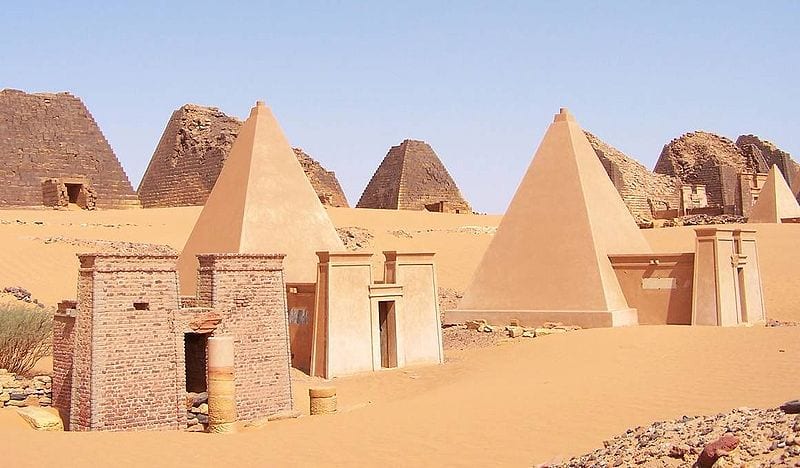
Sudanese pyramids. (Wikimedia commons.)

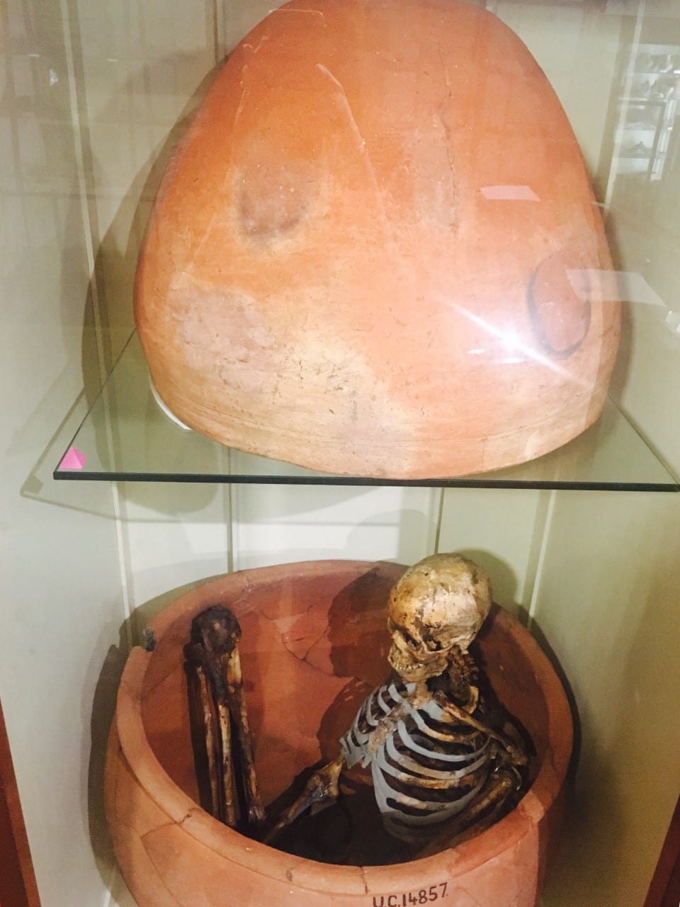
My thesis explores ancient Nubia, the region that is now northern Sudan, from roughly 3000 years ago to medieval times. Unlike their contemporaries, the Egyptians, the Nubians didn’t have a system of writing until the Meroitic period (300 BCE-400 CE), a time of Egyptianizing influence. They built small pyramids and imported Egyptian goods, attesting to the influence of their famous northern neighbors. In the absence of writing (and even in the presence of texts, as humans tend to play with the truth), archaeologists try to build a picture of the ancient society using physical evidence, including human remains. Fortunately, the dry climate and sandy soil usually result in excellent bone preservation, allowing me to identify differences in bone shape. But wait – let me back up a little.

We aren’t entirely sure how bone works. There are two types of cell responsible for bone maintenance – osteoblasts and osteoclasts. Osteoblasts build bone, and osteoclasts take it away. The body is highly responsive to changes in activity, and bone is constantly updating itself accordingly. The general principle is that your body thinks what’s happening now will happen forever. Think about when you’re running a race: it’s hard to start because your body’s been used to standing still and needs some time to amp up your heart rate and muscle contractions. When you finish the race, your heart keeps pounding for a minute or two because it hasn’t quite got the signal to stop running yet. Bone works in a similar way. In response to physical stress, bone will accumulate more osteoblasts to strengthen itself. Each step makes tiny microfractures, which tells the bone “Come on, I’m breakin’ here! Give me more strength!” and the osteoblasts pile on. In the absence of activity – during periods of prolonged sitting or lying down – the osteoclasts come in to take away unnecessary bone. “You’re not using this one, right? Then we can send the calcium somewhere else.” The thing is, scientists don’t know all the signals involved in this process. We know what happens, but not the channels of communication. I like to imagine bone cells having little conversations with each other, but clearly it’s all on a neurochemical level we haven’t yet discovered.

The concept of bone building in necessary areas is keenly presented in studies of elite athletes. In a study by Haapsalo et al (1998) of young female tennis players, the players gained significant bone mineral content in the bones of their dominant (forehand and serving) arm. When the authors looked at a control sample (girls who did not play tennis), there was minimal or no difference between their arms; there was also minimal difference between the nondominant arms of the tennis players and the controls.

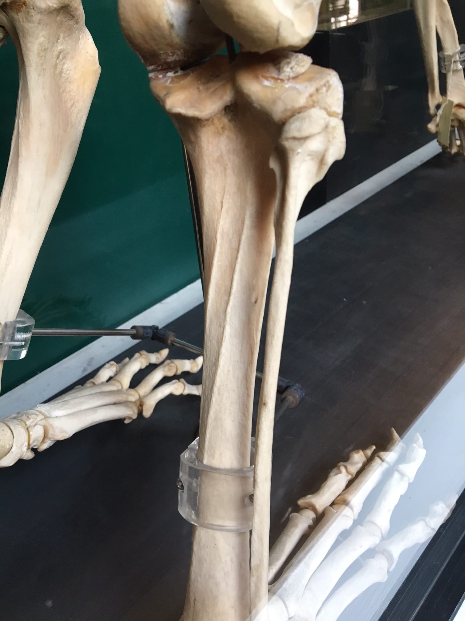
Another study, by Shaw & Stock (2009), examined differences between university athletes who competed in either hockey or long-distance running. They found significant differences in the actual shape of the tibia (shin bone) due to the physical stress of these activities. The tibias of the long-distance runners were more elongated front to back while the tibias of the hockey players were more even side-to-side, showing a distinct difference in the direction of activity in these sports. Clearly, osteoclasts were being sent to the bone locations these athletes needed them most: for runners, the front, and for hockey players, the sides. It is important to point out that many of the studies investigating activity and bone growth look at adolescents, since their bones develop until the end of puberty. After that, it seems to take a lot more effort to alter bone shape and density.

Credit: wikipedia

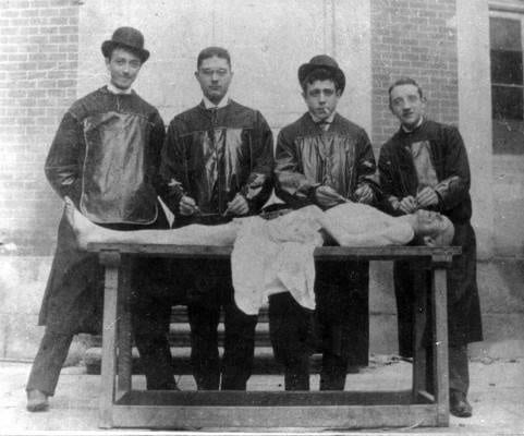
Her bones are losing mineral content by the minute! (Wikimedia commons.)

And what about the other side of the cycle? The osteoclasts? That’s where the astronauts (and cosmonauts) come in. The constant pounding of our feet against the floor keeps our bones as strong and dense as they need to be for everyday use. In zero gravity, though, there’s no pounding, just the occasional soft push off the wall of the space station. The osteoblasts don’t have any stress to react to, and the osteoclasts assume the extra bone is useless, so it starts to be resorbed. It helps that astronauts are some of the most-studied individuals on our planet (and definitely the most studied off the planet!). During spaceflight, urine calcium output is found to increase, indicating that bone is being sapped of minerals, and post-spaceflight bone scans reveal a condition called “spaceflight osteoporosis”, similar to the osteoporosis experienced on earth – but the bone density is only lost from the legs, feet, and hips, all weight-bearing regions, including an 8% loss in four months (compared to 1% loss per year for earth-bound sufferers of osteoporosis). The upper body and head generally remain unaffected (unless one of the astronauts was a tennis player, of course). One study found that after a “long-duration” spaceflight of 4-6 months, it took up to three years for astronauts to recover the bone density they’d lost in space (Sibonga et al, 2007). It makes one really appreciate gravity.

So how do I apply this to ancient populations? The data from astronauts indicates that most of the density lost is from trabecular bone, from the internal core, rather than from the outside. This means that even if ancient bones have lost density due to age, either before death or after burial, it’s likely to have happened from the inside out and thus the external shape should remain intact. This gives me more confidence in figuring out what kinds of activities they performed during adolescence, which in most cultures was when young people started to take up adult cultural roles. I hope to compare the shape of the bones of Nubians to those of athletes and to other populations whose activities are known in order to draw a better picture of their society.

Sources

For an amusing (but factual) look at the craziness that is astronaut and cosmonaut research, check out Mary Roach’s “Packing for Mars: The Curious Science of Life in the Void” (Norton & Co, 2010).

Haapasalo, H, P Kannus, H Sievänen, M Pasanen, K Uusi-Rasi, A Heinonen, P Oja, and I Vuori. 1998. Effect of long-term unilateral activity on bone mineral density of female junior tennis players. Journal of Bone and Mineral Research 13/2, 310-319.

Shaw, CN and JT Stock. 2009. Intensity, Repetitiveness, and Directionality of Habitual Adolescent Mobility Patterns Influence the Tibial Diaphysis Morphology of Athletes. AJPA 140, 149-159.

Sibonga, JD, HJ Evans, HG Sung, ER Spector, TF Lang, VS Oganov, AV Baulkin, LC Shackelford, and AD LeBlanc. 2007. Recovery of spaceflight-induced bone loss: bone mineral density after long-duration missions as fitted with an exponential function. Bone 41, 973-978.


Sexual Conflict in Nature and Museums: Specimen Ratios and Duck Genitalia

By Suzanne M Harvey, on 18 November 2013

Suzanne Harvey #2by Suzanne Harvey


The Duck Penis Controversy of 2013 is well known amongst science bloggers, evolutionary anthropologists and Fox News viewers alike [1]. Now, the time has come for the worlds of museum collections and duck genitalia to collide.

There are some interesting facts about duck penises. For example, they measure a third of the length of the duck’s body, and they cannot become erect outside of the female duck’s vagina (or, as we will find out later, a man made substitute created in the name of science)[2]. Probably most surprising of all, duck penises are corkscrew shaped. However, in March 2013, Fox News conducted a poll in which 89.14% of respondents agreed that the research that brought us these fascinating facts was a waste of public money. At a time when funding for basic science research is becoming more and more difficult to obtain, I disagree with 89.14% of Fox News respondents. And as is so often the case, by clicking on links that come up in a search for ‘penis’, we miss the fact that the most interesting findings of this research come from the vagina. The duck penis controversy not only gives us the opportunity to talk about research, but also the curious bias towards male specimens in museums.

Specimen Ratios and Sexual Dimorphism

Duckling

Duckling preserved in fluid.
Research sugests 97% are of
ducklings are voluntarily
conceived despite
forced copulations.
Photograph: Grant Museum
of Zoology. 


On first arrival at the Grant Museum of Zoology, or indeed most natural history museums, it’s not obvious that the vast majority of specimens on display are male. But why is this the case? One possible explanation is the sexual dimorphism present in many species – the fact that males and females often look different, either in colouring or size [3]. Specifically, males are often larger than females due to competition for mates and sexual selection, and thus make more impressive specimens for display. Perhaps the most obvious example of sexual dimorphism at the Grant is the giant deer at the entrance to the museum, with his imposing 3.6m wide antlers.

It’s also been suggested that male animals were seen as a greater challenge and a more impressive trophy for the Victorian hunters who collected zoological specimens [4] – an acquisition policy that would not be used by the modern day Grant Museum! As well as this unavoidable bias in the specimens on display, some of the most popular blogs on this site have focused on the penis. With the onset of the duck penis controversy, we now have an opportunity to redress this balance, and assess the value of duck genitalia research from a more feminine perspective…

Corkscrews, Angles and Dead Ends: Welcome to the Duck Vagina

That ducks have corkscrew shaped penises is obviously a fact worth knowing, but surely the more interesting question is why do ducks have corkscrew shaped penises? The answer comes from sexual conflict. Forced copulations are common in ducks, presenting an evolutionary problem for females who only want to mate with high quality males of their choice. Females are rarely able to physically resist forced copulations, so in order to control the father of their offspring, their genitalia have evolved an elaborate structure that effectively prevents unwanted suitors from fathering offspring.

Here’s where the research comes in. By creating four substitute duck vaginas from glass tubes (one straight, one twisting in the same direction as a penis, one twisting in the opposite direction from the penis, and one with a sharp bend) researchers were able to assess which shape effectively prevents ducks from depositing semen at the site of fertilisation. The actual duck vagina is a combination of a sharp angle, and a anti clockwise spiral that twists in the opposite direction to the penis. As confirmed by the experiment, this makes it very difficult for males to inseminate females. The female must solicit males with a particular posture in order to make fertilisation likely, therefore gaining control over which males they breed with. In fact, while forced copulations are common, only 3% result in fertilisation.

Duck Vaginas

Glass substitute duck vaginas. A combination of
the two examples on the right most closely represents
an actual duck vagina. Photograph: adapted from
Brennan et al. 2009.

Ducks then are an example of the males and females of a species evolving equally elaborate genital anatomy under the pressures of sexual conflict and sexual selection. There are certainly some impressive male specimens in the Grant Museum, but those giant antlers and corkscrew penises did not evolve without the female of the species.


Suzanne Harvey is a PhD student in Biological Anthropology, working on social interactions and communication in wild olive baboons. She is also a teaching assistant on the UCL Arts and Sciences BASc, a new interdisciplinary degree, and can be found on twitter @suzemonkey.


References

[1] Yong, Ed. (2009). Ballistic penises and corkscrew vaginas – the sexual battles of ducks. Not Exactly Rocket Science. http://scienceblogs.com/notrocketscience/2009/12/22/ballistic-penises-and-corkscrew-vaginas-the-sexual-battles/

[2] Brennan, P., Clark, C., & Prum, R. (2009). Explosive eversion and functional morphology of the duck penis supports sexual conflict in waterfowl genitalia. Proceedings of the Royal Society B: Biological Sciences DOI: 10.1098/rspb.2009.2139

[3] Machin, R. (2008). Gender Representation in the Natural History Galleries at the Manchester Museum. Museum and Society 6(1) 54-67. ISSN 1479-8360.

[4] Shamloul, R., El-Sakka, A., & Bella, A. J. (2010). Sexual selection and genital evolution: an overview. Journal of Sexual Medicine (7): 1734–1740.


Painted Skins & Butterfly Wings

By Gemma Angel, on 1 April 2013

Gemma Angelby Gemma Angel


When I first began my doctoral research into tattoo preservation three and a half years ago, I assumed that tattoo collections such as those held by the Science Museum in London were rare. Whilst collections of inked human skin are most definitely unusual, I was soon surprised and intrigued to discover that such objects exist in almost every museum archive, university anatomy department, or pathology collection that I have visited over the course of my research. The largest collections, of which the Wellcome Collection is the major exemplar, are dry-preserved and date from the 19th century – similar collections can be found across Europe, and the MNHN in Paris has a collection of 56 tattoos which are very similar to those in the Wellcome collection. Historically, these collections may be medical, anthropological, or criminological in origin.

In anatomy and pathology collections, tattoo specimens tend to be wet-preserved and date more recently, usually from the early part of the 20th century anywhere up to around the 1980s. But why are these objects preserved in anatomy and pathology departments at all? There is of course nothing pathological about tattooed skin in itself – so it seems strange that specimens like the one pictured below are displayed alongside other pathological skin specimens such as cutaneous anthrax, fibromas, keloids and glanders. What, if anything, can be learned from these tattoos in medical terms? Or are these striking collections of decorated human skin merely objects of curiosity? Often, the simple answer is that they are a little bit of both…

Tattooed human skin specimen. UCL Pathology Collections. Photograph © Gemma Angel.

Tattooed human skin specimen. UCL Pathology Collections.
Photograph © Gemma Angel.

The collection of tattoos pictured here are a case in point. These particular tattoos belonged to one individual, whose very brief case notes have been recorded and retained along with the specimen in UCL Pathology Collections. The notes provide an intriguing glimpse into the life of the individual to whom the tattoos belonged, as well as revealing something of the clinical interests and collecting practices of the doctor who preserved them:

From a man aged 79 years who had earned his living for many years as the Tattooed Man in a circus. His entire body, except for the head and neck, hands and soles of his feet, was covered with elaborate tattoo designs. He died of peritonitis due to a perforation of an anastomatic ulcer … In tattooing, fine particles of pigment are introduced through the skin, taken up by histiocytes and become lodged in the tissue spaces of the dermis. Pigment also passes to the regional lymph glands via the lymphatics. In this case, all the superficial lymph nodes were heavily pigmented.

It is clear from these brief comments that the nature and extent of this man’s tattoos were indeed of anatomical interest to the medical practitioner: The tattooed man had been so extensively tattooed that gradual migration of ink particles resulted in the collection of pigment in the lymph glands. This demonstrates that although tattoo ink is trapped permanently under the skin following healing, it does actually travel within the body over time, filtering into the body’s tissue drainage system, and collecting in the lymph glands. Whilst this is certainly an interesting anatomical observation, it is not the pigmented lymph glands that the doctor has chosen to preserve, but rather the tattooed skin itself. Without these accompanying case notes, we would never have known that this man’s tattoos had exerted any effect on another of the body’s organs and systems at all.

Reverse panel of tattooed human skin specimen Z6,  showing tattoos of a butterfly and a flying fish.UCL Pathology Collections.Photograph © Gemma Angel.

Reverse panel of tattooed human skin
specimen Z6, showing tattoos of a
butterfly and a flying fish.
UCL Pathology Collections.
Photograph © Gemma Angel.

It would be equally impossible to know whether or not these were the only tattoos he possessed – or indeed, if they all necessarily belonged to the same person. There are strong stylistic similarities between the butterfly motifs, suggesting the work of a single tattooist, or perhaps that the individual motifs were part of a larger design. But just how large or complex the design may have been, we certainly cannot tell just by looking at these 5 small tattoos. We know that they belonged to a 79-year-old man, who made his living as a Tattooed Man, only because the doctor tells us so. He or she also tells us that his body was covered in tattoos – yet only 5 small pieces have been preserved. Five carefully selected motifs, chosen by the doctor from an already complete collection, which provided the livelihood and told the life story of one unnamed man. What selection criteria did the pathologist adopt when deciding which tattoos to preserve, and which to consign to the grave? The manner in which the specimens have been excised and mounted are strikingly reminiscent of a lepidopterist‘s collection of butterflies – could this reflect the personal collecting interests of the pathologist, or perhaps even the Tattooed Man himself? Both the pathologist and Tattooed Man alike chose these butterflies – did they also share a passion for lepidoptery?

Many people will be familiar with the kind of insect specimen displays that are a staple of natural history collections – the old 19th century museum cases containing neat rows of pinned and mounted moths and butterflies, neatly organised according to subspecies and morphological characteristics. The tattooed butterflies share some remarkable similarities with these entomology collections; they are arranged one above another, and “pinned” to a support with small surgical stitches. Unusually for specimens found in pathology collections, this support is a slightly translucent black. This appears to be a deliberate choice on the part of the pathologist – the black perspex provides a contrasting ground for the display of tattoos on opposite sides of the vitrine, such that they do not visually detract from one another. These aesthetic choices suggest a nuanced interest in the collection and display of these specimens, which goes far beyond a straightforward medical interest in the anatomy of the tattoo. From the limited case notes and analysis of the specimen itself, we can learn something about the pathologist’s interest in the tattoo, but we are still no closer to being able to answer the fundamental question – why collect tattoos at all?

Butterfly display at UCL's Grant Museum. Photograph © UCL, Grant Museum of Zoology.

Lepidoptera display at UCL’s Grant Museum. Photograph © UCL, Grant Museum of Zoology.

There is no part of the body able to register the history of a life lived so much as the skin: wrinkles, scars and lines all map out our lives on the surface of our bodies as we age. The tattoo reinforces this unique capacity of the skin to record the traces of our experience, in the conscious act of permanently inscribing memory in skin. The pathologist, uniquely acquainted with death by virtue of their specialism, is perhaps best positioned amongst medical professionals to appreciate the peculiar relationship of the tattoo with mortality. It is a trace of the subjectivity of the deceased that is capable of outliving them, akin to a photograph or written memoir. From this point of view, it no longer seems surprising that tattoos are so often found in pathology collections; perhaps the pathologist who collected the Tattooed Man’s butterflies simply wished to preserve a small part of a colourful and remarkable life.


[analytics-counter]

Doctors, Dissection & UCL

By Gemma Angel, on 21 January 2013

  by Sarah Chaney


A visit to the current Museum of London exhibition, Doctors, Dissection and Resurrection Men (on until 14 April 2013), brought to mind the recent Buried on Campus exhibition in the Grant Museum. Several of us have previously blogged about reinstating the stories of the forgotten dead, as well as the issues around the display and interpretation of human remains in a museum context. As I myself wrote, the disinterrment of human remains is not unusual during building work: the Museum of London exhibition focuses on the excavation of the former Royal London Hospital burial site, during recent improvement works. The bones found showed traces of a variety of practices, including dissection for autopsy, as well as marks made during surgical practice and articulation for the creation of teaching specimens.

Dissection, particularly in the case of medical teaching, was often linked to artistic practice. Doctors, Dissection and Resurrection Men opens with the grisly plaster cast of James Legg, hanged for murder in 1801. Legg was subsequently flayed and posed as if crucified: a collaborative project between artists Benjamin West and Richard Crossway, and sculptor Thomas Banks, who believed that most depictions of Christ’s crucifixion were anatomically incorrect (for more on the Anatomical Crucifixion see Gemma Angel’s post). Rather less theatrically, anatomical drawings and textbooks were also created directly from dissection practice. During a recent session in the Art Museum, I discussed with visitors the way in which anatomy textbooks create stylised images, removing certain body parts in order to emphasise others. Students re-created these images for themselves: first with the corpse, then in their own sketches, re-interpreting the body in a way that made sense for their practice.

Joseph Lister – Side of the Neck and Floor of the Mouth (1850), UCL Art Museum #4801

Amongst the UCL Art Collections are a number of student sketches of the famous surgeon Joseph Lister (1827 – 1912), well-known for his introduction of antiseptic techniques into surgical practice. Born in Essex, Lister came to UCL in 1844, initially as a student of the arts. After graduating, however, he subsequently turned his attention to medical studies, continuing at UCL until he gained his M.B. in 1852. The sketches in the collection mainly date from 1849-50, produced as part of Lister’s studies. The techniques used indicate some of the interesting artistic choices available to anatomical illustrators: perhaps also the influence of Lister’s varied education and interests. The sketch above, for example, was made on tinted paper, which enabled the young Lister to highlight structures using white chalk. This emphasis, along with the effective use of colour (in this instance, major blood vessels are depicted in red, standing out clearly in an otherwise monochrome drawing), enables quick and easy recognition of bodily structures, adding depth to the sketch. For an un-trained eye, the mass of tissues within the human body could not be read in such a manner. The ability to render the three-dimensional body in a series of recognisable images – and then understand the physical body through such images – was as important as surgical skill.

Box Viewer from the UCL
Physiology Collections (080:RFH)

The huge variety of techniques for anatomically representing the human body is also evident elsewhere in the UCL Collections. The Physiology Collection includes a volume of the Edinburgh Stereoscopic Atlas of Anatomy, published in 1905. Stereoscopy became a popular technique of representing three-dimensional structures from its inception in the 1840s. Two offset photographs or other images are presented to the viewer which, when viewed through the stereoscope, are seen separately by the left and right eye. As occurs in ordinary vision, the brain combines the images perceived by both eyes; in the case of stereoscopy, giving the illusion of three-dimensional depth. The Edinburgh Atlas aimed to use this technique to represent photographs of dissections in a manner closer to that seen in the three-dimensional human body than simple sketches. Bulky and expensive, the success of the Atlas was relatively limited. It still serves, however, as an unusual reminder of the way in which the human body has continued to require anatomical translation.