X Close

UCL Culture Blog

Home

News and musings from the UCL Culture team

Menu

Hypsilophodon foxii cast part 3

By ucwehlc, on 3 November 2023

Previously on the Grant Museum blog we introduced our biggest dinosaur, along with several mysteries. How did we get it? Who made it? Why does its head look so strange? Then in an exciting turn of events 4 years later we discovered a picture of our cast being made at the Natural History Museum. Now another 4 years on the story continues with Museum Studies students Imogen Geoghegan, Nadia Adal, Lauren Brown Phelan and Yanning Huang who have written this blog about their dinosaur research project.

**The Hypsilophodon foxii cast (Part 3)**

We took part in a research project with the Grant Museum of Zoology (GMZ) to uncover the truth behind this mystery object in the GMZ’s collection. It started with a few photographs and texts from the GMZ which led us to discover the cast’s background and even the species of the dinosaur (Hypsilophodon foxii), both of which were in doubt and unconfirmed before our project began.

Image of LDUCZ-X185 cast of skeleton of Hypsilophodon foxii from the Grant Museum of Zoology UCL

LDUCZ-X185 a one armed cast of skeleton of Hypsilophodon foxii

(more…)

The Giant Thorny-Headed Worm of Swine

By ucwehlc, on 20 October 2023

The Grant Museum is currently closed for refurbishment works until the new year, but we still have plenty of exciting stories to tell from the collection.

Today’s blog is by visiting researchers Dr Andrew McCarthy and Dr Jennie C. Litten-Brown from Canterbury College, UK, who have been looking into some of the parasites in our collection. This week we meet the Giant Thorny-Headed Worm of Swine, Macracanthorhynchus hirudinaceus (Acanthocephala: Oligacanthorhynchidae), a parasite that causes disease in both pigs and humans, that you will be able to see on display when we re-open.

The Giant Thorny-Headed Worm of Swine

Parasitism is probably the most common life strategy on Earth. Parasites are known to be important factors in maintaining ecosystem health, and the relatively new discipline of Parasite Conservation is devoted to preventing the extinction of endangered parasite species. However, several parasites cause serious disease in both humans and livestock. With increasing trends in more ethical farming methods such as Wild Farming, (often associated with rare breeds conservation), where livestock come into close contact with the natural environment, an awareness of potential pathogenic parasites is important. Specimens of one such parasite, an acanthocephalan, may be found in the Grant Museum of Zoology.

Acanthocephalans, commonly known as the “Thorny-Headed Worms” are a group of parasites typically under-represented in museum collections. Members of the group are known to cause a disease, acanthocephaliasis, in their vertebrate hosts, and some are of both veterinary and medical importance. The Grant Museum of Zoology has in its collection both fluid preserved adult worms (specimens LDUCZ-F21, F22 & F23), and Rudolf Weisker wax teaching model specimens c.1884 (LDCUZ-F26 & F84), of one of the largest acanthocephalans known to science. It goes by the rather dramatic common name of the “Giant Thorny-Headed Worm of Swine”, Macracanthorhynchus hirudinaceus. It may be described as a neglected parasite upon which relatively little research has been carried out. Its scientific name refers to the large, thorned proboscis (see Fig. 1), and the leech-like (“hirudo”) nature of its body (see Fig.2 ).

Black and white electron microscope image of the head of a thorny-headed worm showing rows of hooked spines

Fig. 1. The Parasite: Macracanthorhynchus hirudinaceus.
Scanning Electron Micrograph of the proboscis bearing thorns.
(© Migliore et al. (2021) )


Adult thorny-headed worm on a black background. The worm is a pale pink-beige colour and is curled in loops. It lies next to a ruler that shows it is 10cm long while curled up.

Fig. 2. The Parasite: Macracanthorhynchus hirudinaceus. Entire worm from intestine of pig.
(CDC, Public Domain via Wiki Commons)

(more…)

The Unsung Heroes of UCL Museums and Collections

By ucwehlc, on 6 July 2021

During 2020 and 2021 while UCL was closed due to the coronavirus pandemic, UCL Culture’s curatorial team worked with students from the Institute of Archaeology’s MA Museum Studies on our first-ever virtual work placements. These projects, which included archive transcription, documentation and object label writing, provided opportunities for the students to gain practical curatorial skills to prepare them for their future careers while undertaking valuable work towards better understanding the collections.

This blog was written by Nicky Stitchman, UCL MA Museum Studies

As part of my MA in Museum Studies, I undertook a work placement with Hannah Cornish, Science Curator at UCL. My brief was to discover the different locations of UCL’s museums and teaching collections from the university’s origins in 1828. Ferreting out information from primary and secondary sources and finding maps that showed the movement of the various museums as the university expanded was fascinating, but I found myself drawn to people behind the museums. I am not talking here about the headliners – the Flinders Petries or Robert Grants of this world – but rather the curators, assistant curators and demonstrators who would have done most of the day-to-day tasks such as cataloguing, labelling, teaching, and physically moving the artefacts and objects within the collections. 

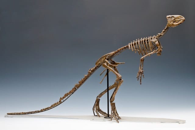
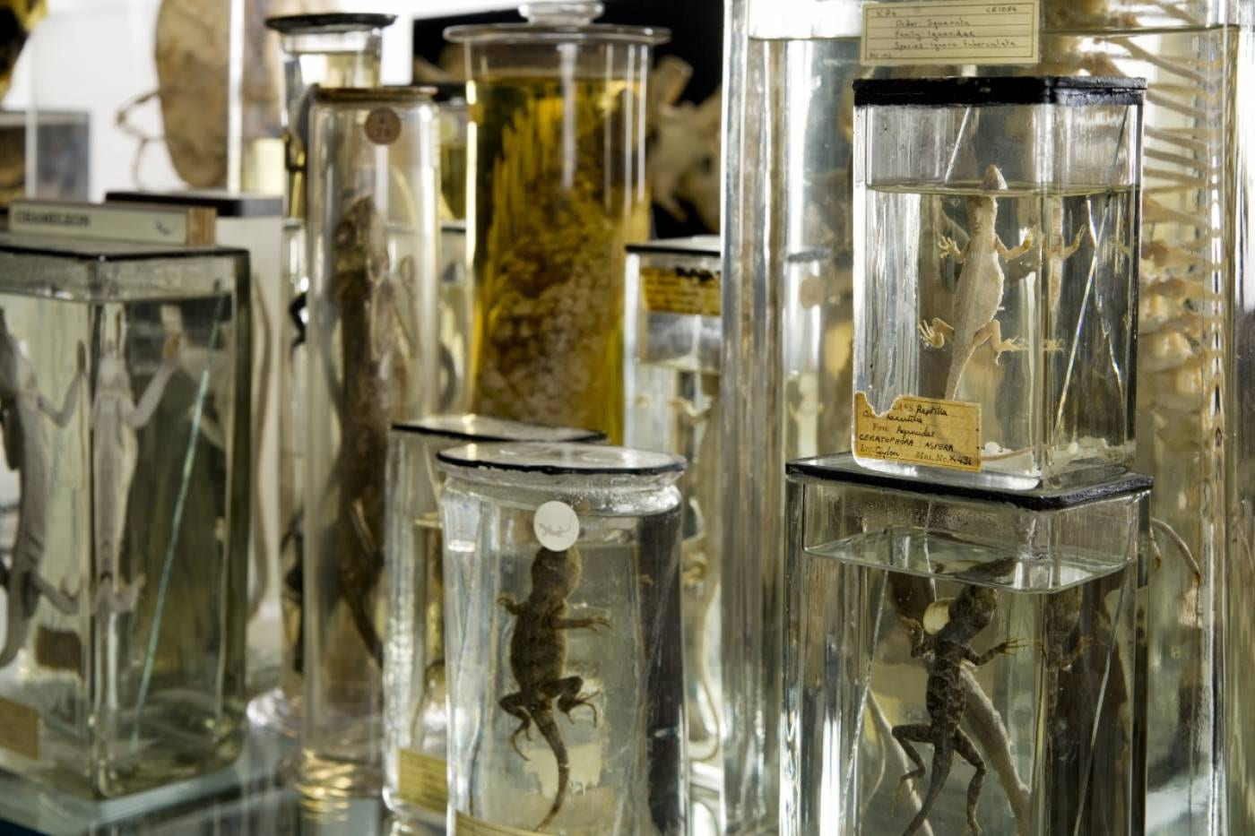
James Cossar Ewart at the Grant Museum 

J Cossar Ewart was the first professional, rather than professorial, curator of both the Anatomical Museum and Comparative Anatomy/Zoology Museum between 1875-1878. He was appointed after the retirement of William Sharpey (curator of the Anatomical Museum) and the death of Robert Grant (professor of comparative anatomy). In the official records, it was Professor Lankester, Grant’s successor, who refitted and rearranged the Museum of Zoology over this period but Ewart was instrumental in making the zoological preparations and was also known to have helped organise and take the subsequent practical classes that Lankester introduced to UCL. 

Black and white image of the head and shoulders of James Cossar Ewart

James Cossar Ewart worked at the Grant Museum 1875-1878. Image in public domain

After Ewart, there was a change in the running of the two largest museums at UCL at that time, with a separate curator appointed for the Museum of Anatomy, while the Zoological Museum (now the Grant Museum of Zoology) was titularly run by the Head of Department with a curatorial assistant.

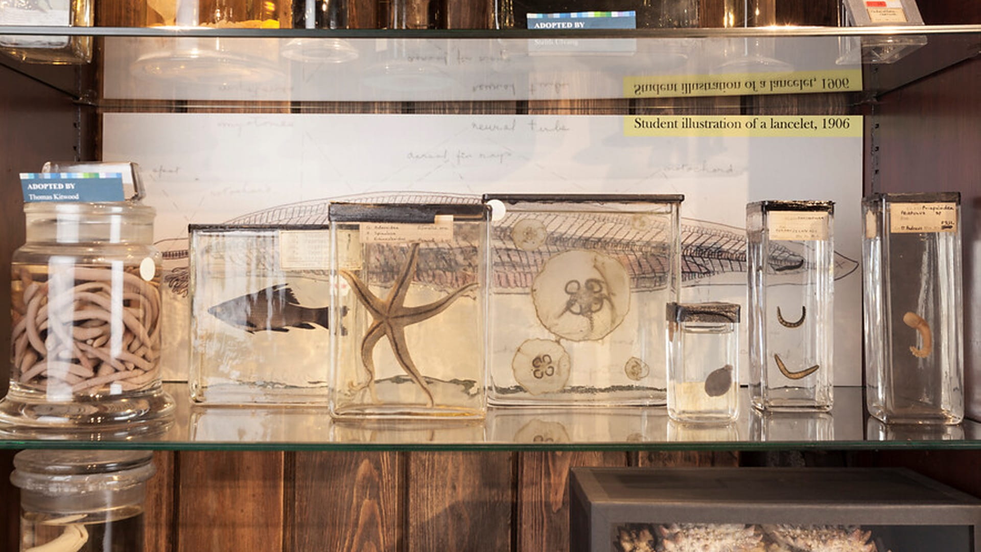
Shattock and Stonham: Anatomy and Pathology

Mr Samuel Shattock succeeded Ewart as the Curator of the Anatomical and Pathological Museum. He had originally shown up in the records as a Mr Betty which caused me some confusion at the time, until I discovered that he had decided to change his name to prevent the extinction of the Shattock family name!  Shattock never qualified as a physician but dedicated his life to pathological medicine. He was responsible, alongside Dr Marcus Beck, for a descriptive catalogue of the surgical pathology preparations at UCL. His successor Charles Stonham also worked with Marcus Beck on Part II of this catalogue and then in 1890 produced another medical pathology catalogue, which can be found online at the Wellcome Collection. In the preface to this catalogue, Dr Barlow and Dr Money acknowledge the work of Charles Stonham, and state that it is ‘to him the preparation of this work is almost entirely due’.

Black and white image of Charles Stoneham in military uniform sitting on a wooden chair holding a riding crop

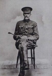
Charles Stonham Curator of the UCL Anatomical and Pathological Museum (copyright UCL CC By 3.0)

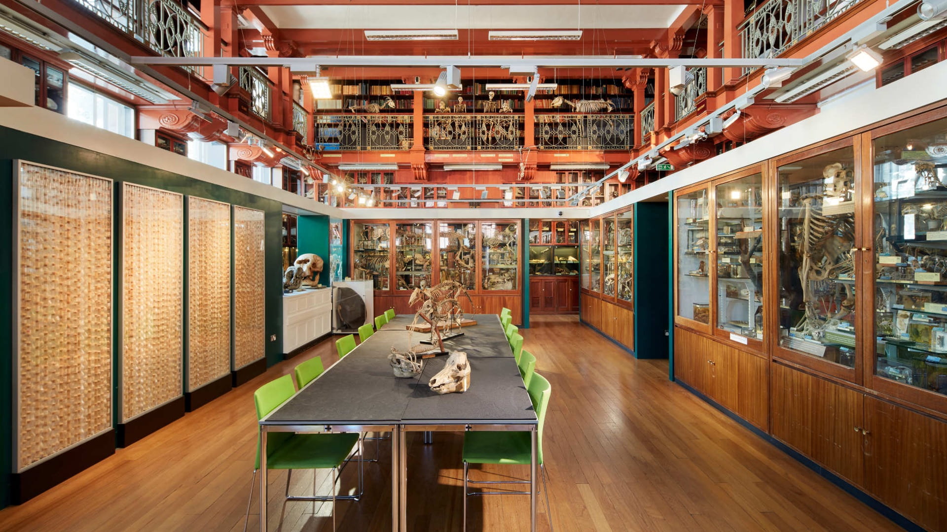
Stonham was also responsible for the division of the pathology from the anatomy collection and its rehousing within the museum. He is also remembered on the UCL Roll of Honour as not only was he an instrumental figure in the London Mounted Brigade Field Ambulance, but he volunteered as a member of the Royal Army Medical Corps during WWI and died on service in January 1916 aged 58. 

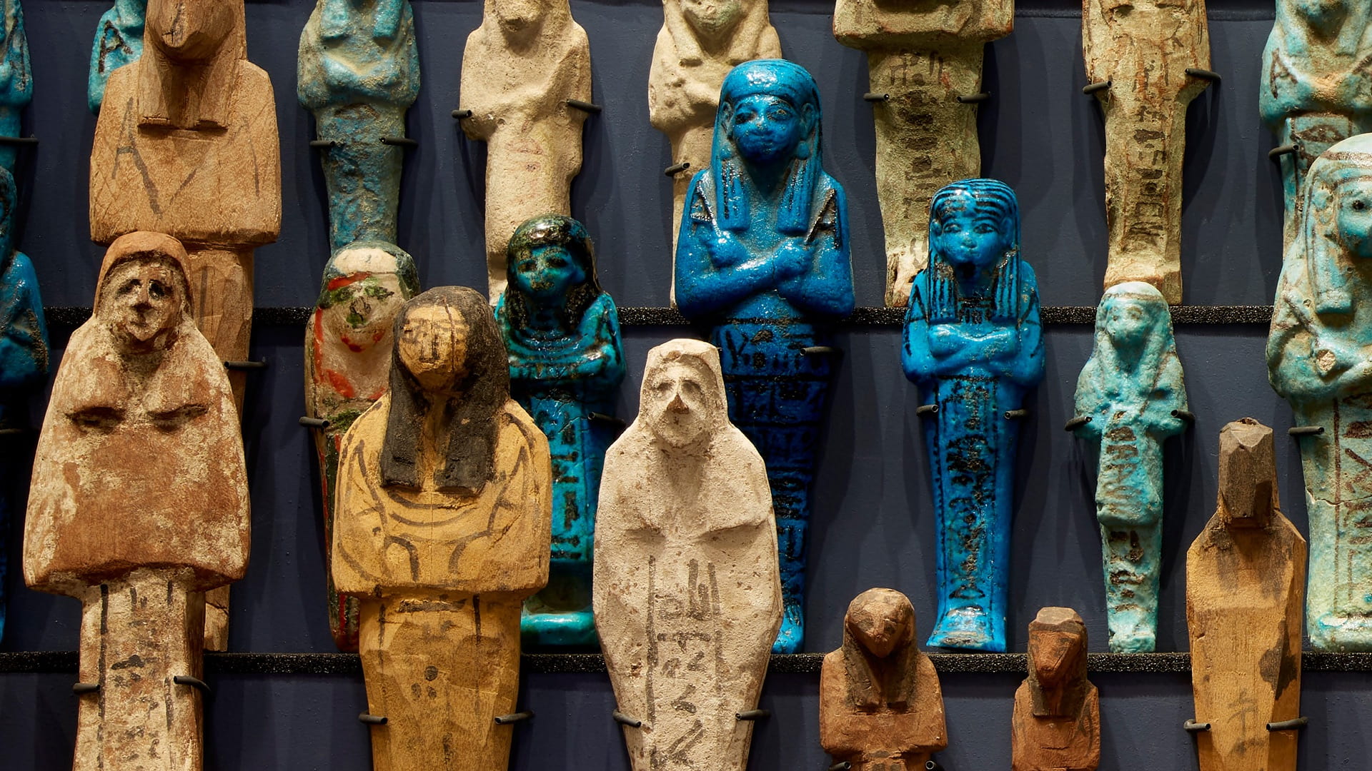
Margaret Murray and the Petrie Museum

The dedication of many assistant curators at UCL is very clear. The redoubtable Margaret Murray (pictured here), who worked alongside Flinders Petrie for many years, was effectively in charge of the museum’s collections during Petrie’s many excavations in Egypt and the Levant. It should also be remembered that the Petrie Museum of Egyptology and the Department itself was founded on the collection and endowment of Mrs Amelia Edwards, who favoured UCL due to the early admission of women into the College.

Black and white image of Margret Murray wearing a lace shawl, seated and holding a book

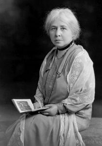
Egyptologist Margaret Murray (Image in public domain)


Edith Goodyear and the Geology Museum

One of the first women to be involved in UCL’s museums was Edith Goodyear who was appointed as the Assistant in the Geology Museum in 1904 and subsequently remained in the department until the Second World War. Edith worked alongside Professor Edmund Garwood, reorganising the museum, teaching, and researching papers. A room in the Lewis Building was named for her, along with the First Year Student prize within Earth Sciences. It is also worth noting that in an age of inequality, the 1916/17 council minutes show Edith was paid £150, the same as her male colleague Dr J Elsden. 

Keeping it in the family: Mary and Geoffrey Hett

1917 also saw the appointment of Mary L Hett as Assistant in the Zoology Dept on the same salary of £150, where she remained until she took up the post as Professor of Biology at the Hardinge Medical College, Delhi.  She had followed her brother Geoffrey S Hett to UCL where he held the post of Curator of the Anatomical Museum from 1907-1910. Like his sister, he had a great interest in the natural world and was an authority on both British birds and on bats. Geoffrey became an ENT Specialist and during his time at UCL completed valuable research on the anatomy of the tonsils. Like his predecessor, Charles Stonham, he also served in World War I and used the skills learnt at UCL to treat head, and in particular, nasal injuries during this period. 

The stories of the men and women who studied and worked at UCL museums over the years are many and various, and these are just a sample of those whom I have met in my research for the Mapping UCL Museums Project. We may never be able to give the Curators and Assistant Curators the recognition that their work and dedication deserve but in introducing these few to you, I hope to have redressed the balance very slightly in their favour!  

Download our free virtual meeting backgrounds from UCL Culture

By f.taylor, on 22 June 2020

Like many people around the world, the UCL Culture team has spent the last few months collaborating with colleagues via Microsoft Teams. Now you can bring some of our amazing collections into your meetings. Click on the images below to see them at full size and then download them to your computer. You can then upload them to the virtual meeting platform of your choice. Enjoy!

Grant Museum of Zoology

The Grant Museum of Zoology is one of the oldest natural history collections in the UK and is the last remaining university natural history museum in London. Home to 68,000 zoological specimens, the collection is a unique window on the entire animal kingdom. The final image below is from our Micrarium, a beautiful back-lit cave of 2,300 microscope slides.

Grant Museum of Zoology

Grant Museum of Zoology

The Petrie Museum of Egyptian Archaeology

The Petrie Museum contains over 80,000 objects and ranks among some of the world’s leading collections of Egyptian and Sudanese material. Below you can see our collection of Shabtis. They are small figures in adult male or female form created to carry out tasks in the afterlife.

Petrie Museum

Petrie Museum

UCL Art Museum

UCL Art Museum holds over 9,000 paintings, drawings, prints and sculptures dating from the 1490s to the present day. One of the the most important parts of the collection is the unique archive of works by staff and students from the Slade School of Fine Art.

UCL Art Museum


Bloomsbury Theatre

Bloomsbury Theatre and Studio is the home of home of cutting-edge performance in the heart of London. You can see our upcoming shows here.

Bloomsbury Theatre


Specimen of the Week 391: The Domestic Cat Skeleton

By Katie Davenport-Mackey, on 17 January 2020

This blog was written by UCL Culture volunteer Jingyuan Zou.

The Grant Museum not only has many fascinating specimens in its collection such as the subfossils of extinct giant deer and dodo bones, skeletons of lions and dugongs, but also many common domestic animals that we may see in everyday life. Many people may be familiar with the appearance of an extinct animal such as saber-toothed cat, however often the skeletons of more common animals are the most unfamiliar specimens viewed from a museum. This week’s Specimen of the Week features one such ‘common’ animal that looks quite different in its Grant Museum guise…

**The Domestic Cat Skeleton**


LDUCZ-Z2602 Felis silvestris catus Domestic cat skeleton

(more…)

Specimen of the Week 389: The Monarch Butterfly

By Lisa Randisi, on 16 August 2019

This blog was written by UCL Culture volunteer Melissa Wooding.

Today’s specimen of the week highlights one of the world’s longest animal migrations at 6,000 miles1– completed by an insect!

This beautiful insect has an internal biology including a sundial 2 compass 3, and a gene enabling it to suppress its own ageing and increase its own lifespan 8 times4… all inside a brain the size of a single sesame seed5.

It’s time to give this mind-boggling butterfly its due moment in the spotlight:

Pair of monarch butterflies

(more…)

Specimen of the Week 386: A Cocktail of Spirits: Fluid Preservation at the Grant Museum

By Katie Davenport-Mackey, on 19 July 2019

The Grant Museum contains a staggering number of specimens – the estimate is more than 68,000 – and approximately 10% are fluid preserved specimens. Fluid preservation describes specimens that are preserved in chemicals that protect them from deterioration. There are three components to a fluid preserved specimen:

  1. The fixed specimen: The specimen is injected or ‘fixed’ with a chemical that prevents decomposition by stabilising the protein molecules. The most common fixative is formaldehyde.
  2. The preservative fluid: Most fluid preserved specimens are preserved in solutions of alcohol or formalin, a mixture of formaldehyde and water.
  3. The container: Containers are typically glass jars or bottles sealed with a closure.

Below is a brief description of some of the different preservative fluids that can be found in the Grant Museum.

(more…)

Specimen of the Week 381: What Lies Beneath

By Tannis Davidson, on 17 May 2019

Mystery ceramic pots

Mystery ceramic pots

The fluid store room for the Grant Museum is perhaps an unlikely setting for a hair-raising tale. In it, rows of metal shelves are neatly arranged holding jars of preserved zoology specimens arranged by taxonomy. Order and classification dictate the placement of specimens, and as a whole, the contents of the store are visible, documented and accessible.

Apart from the bottom shelf of the last row. Although a numbered (legitimate) location, it is a wildcard area which houses several large ceramic pots of unknown content. No one knows exactly how and when the pots came to be a part of the Grant Museum collection and because it is impossible to see into them, no one knows what they hold. A few clues exist – the occasional faded label or a more modern post-it note – but as the pots have not been opened and investigated in living memory, their contents are a mystery.

What follows is the first-hand account of the opening of two of the pots. This is a true story.

**WARNING  Graphic images below **

(more…)

Plural Animal Wednesdays

By ucwehlc, on 24 April 2019

This blog is about a centuries-old quirk of the English language that has become a Grant Museum tradition.

If you follow us on twitter (@GrantMuseum) you may have come across Plural Animal Wednesday (#PAW), our weekly tweet about collective animal nouns. These are the words used to describe groups of animals, you are probably familiar with a herd of sheep, a flock of birds and a swarm of insects. There are, however, an astonishing number of obscure and wonderful plural animal names, enough to keep us in tweets for years and years.

#PAW was the brainchild of former Grant Museum Curatorial Assistant Emma Louise Nicholls. It all began on 16th November 2011 with a crash of rhinos (because rhinos are Emma’s favourite), and has continued every week for 7 years. All our plural animal discoveries are kept in a big spreadsheet and we are now approaching 400 entries. So why are there so many? Where do they come from? How long can we keep finding them to boost our social media content? Read on to find out.

A 'crash' of white rhinos Ceratotherium simum by Chris Eason CC-BY 2.0

A crash of white rhinos Ceratotherium simum by Chris Eason CC-BY 2.0

(more…)

Specimen of the Week 377: The Lobster Claw

By Graham Isted, on 15 March 2019

Hello and welcome to the next instalment of Specimen of the Week. This week’s specimen is a mighty claw (LDUCZ-H671) from the lobster species Homarus gammarus, also known as the European or common lobster. Lobsters are great, whether you like them to be freely going about their lives at the bottom of the sea or perhaps prepared by a chef on a dish with some butter. Either way, I am fairly certain there are aspects of their lives you are unaware of and this blog will hopefully either make you hungry for more knowledge or perhaps just dinner.

Lobster claw LDUCZ-H671 Homarus gammarus

(more…)